|
|
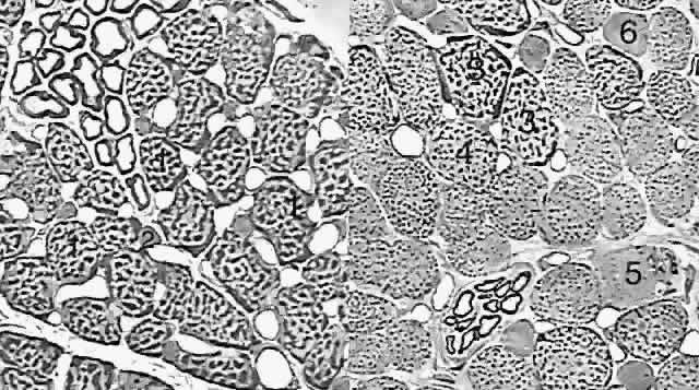
| Fig. 3. Thin section (1-μm) light micrographs of the fiber types in human medial rectus muscle. (Left) Orbital layer. Singly innervated fibers (SIFs, 1) are large and densely packed with dark-staining aggregates of mitochondria, especially in the periphery of each fiber. Multiply innervated fibers (MIFs, 2) are much smaller and contain fewer mitochondria. (Right) Global layer. The three types of SIFs really form a continuum distinguished by their density of mitochondria. The largest and most granular fiber (3) is very similar to the orbital SIF; the other two SIFs (4 and 5) have fewer mitochondria. Global MIFs (6) are still smaller, contain fewer mitochondria, and resemble orbital MIFs. (Courtesy of Dr. John D. Porter.) |