
|
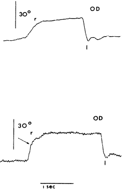
| Fig. 17. Electro-oculographic recording in a patient before and after transposition surgery for lateral rectus paralysis. Upper tracing: eye position showing slow saccades to the right (r) and rapid saccades to the left (l). Lower tracing: horizontal saccades after muscle transposition show an increase in saccadic velocity to the right. (Metz HS, Jampolsky A: Change in saccadic velocity following rectus muscle transposition. J Pediatr Ophthalmol 11:129, 1974) |