|
|
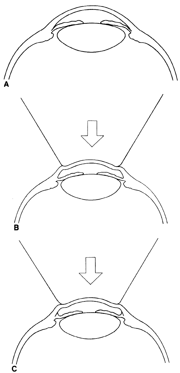
| Fig 9. Use of indentation gonioscopy to differentiate between appositional and synechial angle closure. (A) Eye with angle closure. (B) With indentation gonioscopy, aqueous is forced peripherally, opening the appositional closure. (C) The synechial closure is not affected by the indentation even though the peripheral iris is pushed posteriorly. |