|
|
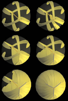
| Fig. 5. 3D-computer assisted drawings (CADs) showing key structural elements in the formation of Y sutures during fetal development. Upper left. After the primary fiber mass (dark gold embryonic nucleus) has been formed, six straight fibers normally positioned equidistantly around the equator, separate growth shells into equal sextants composed of S-shaped fibers (upper right and middle left). The ends of the S-shaped fibers abut and overlap to form suture branches (blue lines) that extend to confluence at the poles (middle right and lower right and left). Note that the length and location of suture branches are defined by the ends of straight fibers. The end result is an upright Y anterior suture (lower right) and an inverted posterior Y suture (partially shown in lower left). |