|
|
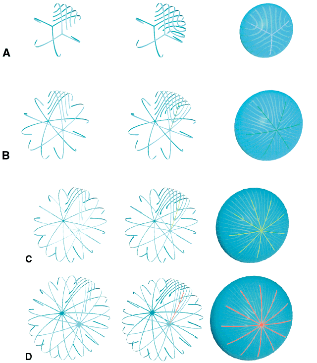
| Fig. 4. A series of scale (approximately 3:1) three-dimensional computer-assisted drawings (3D-CADs) showing normal human lens sutural anatomy as a function of fetal development (A), infantile and juvenile growth (B and C) and adulthood (D). Throughout fetal development, six straight fibers are positioned precisely to divide growth shells into six equal segments. Three of the straight fibers, oriented at 120° to one another, have one end that extends to confluence at the anterior pole, whereas the other three comparably oriented straight fibers have one end that extends to confluence at the posterior pole. All other fibers in any shell are S-shaped fibers, or fibers with opposite end curvature. Between any two straight fibers, are groups of S-shaped fibers with progressively variable degrees of opposite end curvature. Neither end of an S-shaped fiber reaches a pole, and because of the variable degree of opposite end curvature, the anterior and posterior ends of these fibers become aligned as offset (60°), anterior, and posterior latitudinal arc lengths. The ends of S-shaped fibers in neighboring groups overlap precisely to form suture branches. Although the anterior ends of S-shaped fibers in neighboring groups overlap to form an anterior suture branch because of opposite-end curvature, the posterior ends of these same fibers do not overlap with one another to form a posterior suture branch. In the fetal nucleus, the anterior and posterior ends of S-shaped fibers from all neighboring groups overlap to form three anterior and three offset (60°) posterior branches arranged respectively as Y and as inverted Y suture patterns. The key parameters of the progressively more complex generations of human lens suture patterns formed throughout life are shown in the second through fourth rows. From birth through infancy, 12 groups of S-shaped fibers become symmetrically positioned between 12 straight fibers to form a simple star. From juvenile through sexual maturation, 18 groups of S-shaped fibers become symmetrically positioned between 18 straight fibers to form a star suture. Finally, throughout adulthood, 24 groups of S-shaped fibers become symmetrically positioned between 24 straight fibers to form a complex star suture. As more suture branches are formed as a function of development, growth, and age, the degree of opposite end curvature and variation in intrashell fiber length decreases. |