|
|
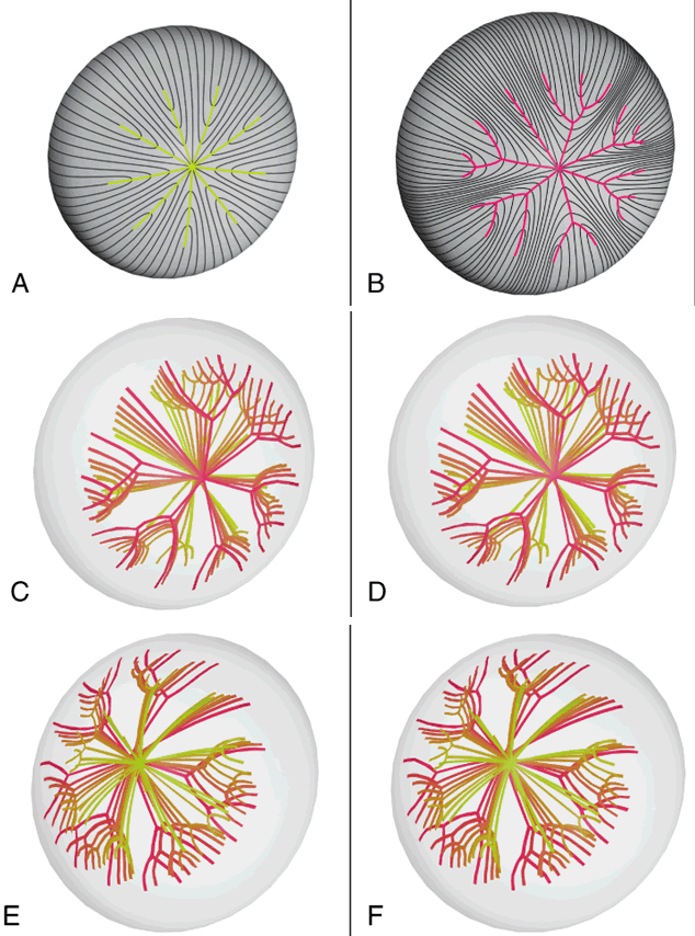
| Fig. 6. Scale (3.5:1) schematic 3D-CADs depicting a normal young adult anterior suture pattern (A) and an abnormal aged anterior suture pattern from a surgically removed (intracapsular technique) cortical cataract (67-year-old man; B). The aged cortical cataract had 13 nonidentical and asymmetrically positioned suture subbranches extending from an irregular (7-branch) star suture. Stereo scale schematic 3D-CADs depicting the three-dimensional anterior (C and D) and posterior sutural anatomy (E and F) of the aged cortical cataract shown in B. In this case, it clearly can be seen that abnormal suture formation throughout adulthood resulted in continuous abnormal suture planes of comparable shape (conical or cuneiform) and size (progressively larger as a function of depth or age), and in analogous locations as typical aged cortical opacities seen by slit-lamp biomicroscopy. |