|
|
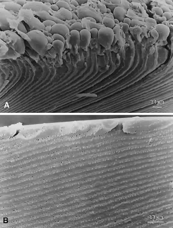
| Fig. 8. Higher-magnification SEM micrographs of the RCS PSC shown in Figure 7. A. At this magnification, it can be seen that the PSC was the result of enlarged posterior ends of fibers turning up and away from the polar axis rather than overlapping within and between growth shells to form suture branches. It also can be seen that many of the posterior ends had blebbed off from the rest of the fibers. In any plane of section, these blebs would appear to be Wedl cells. B. A comparative view of posterior fiber ends beneath the posterior capsule of a normal adult rat lens. |