|
|
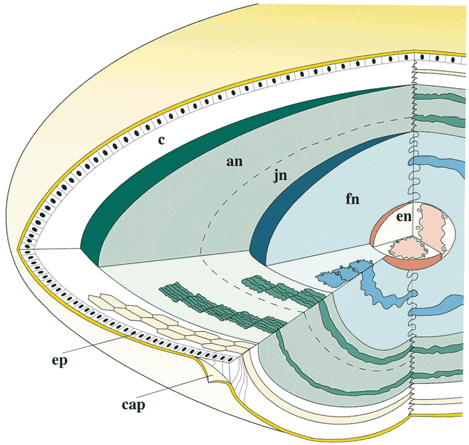
| Fig. 9. Schematic scale (15:1) diagram of a 60-year-old aged noncataractous human lens. The embryonic nucleus (en) is enlarged 4× , and the epithelium (ep) and capsule (cap) are enlarged for clarity. Fibers are approximately to scale relative to each other (but not to the region thickness). The sutures are not shown. The fetal nucleus (fn), juvenile nucleus (jn), adult nucleus (an), and cortex (c) are composed of secondary fibers formed during specific periods of development and growth. (Taylor VL, Al-Ghoul KJ, Lane CW et al: Morphology of the normal human lens. Invest Ophthalmol Vis Sci 37:1396, 1996) |