|
|
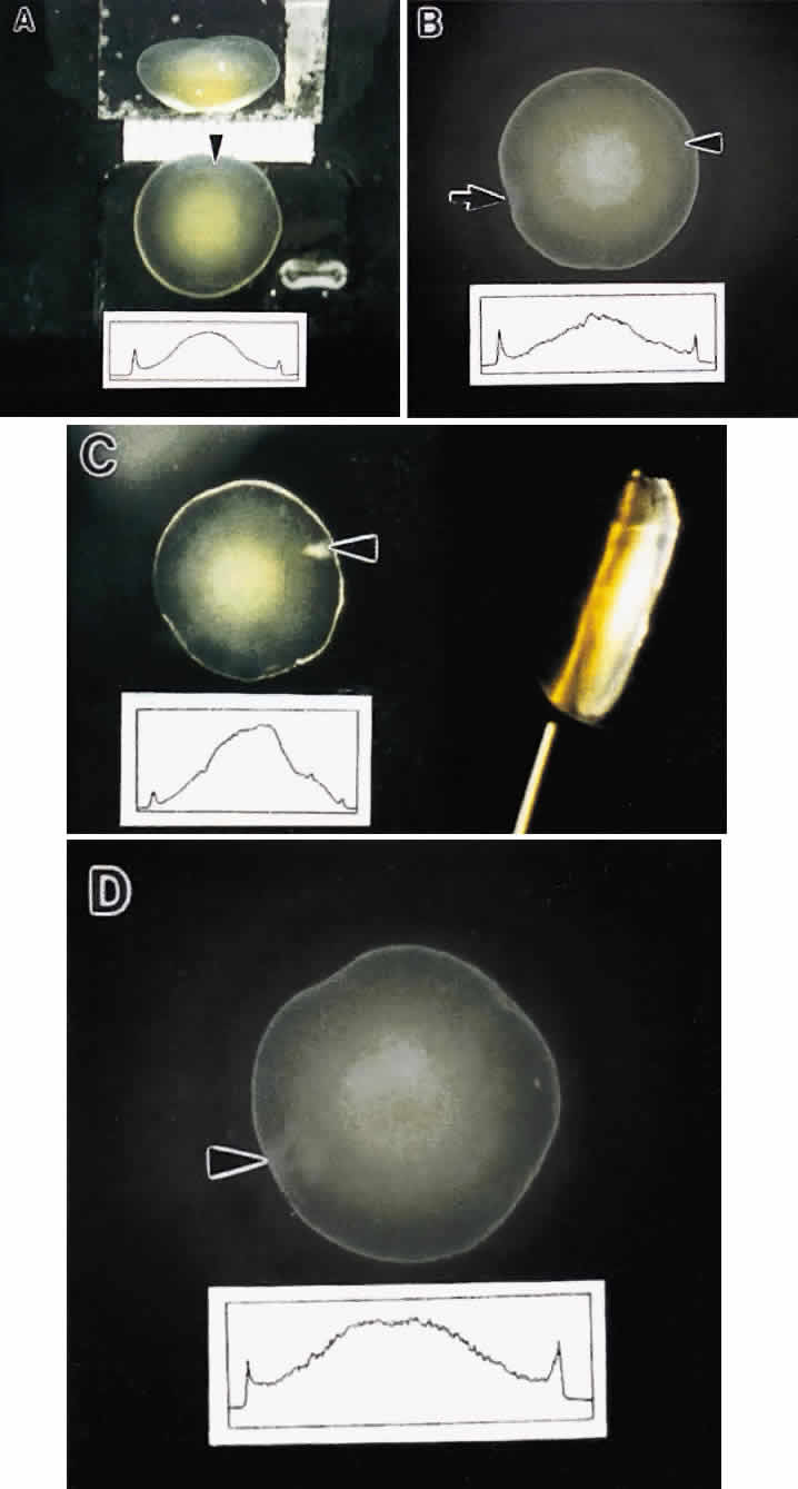
| Fig. 10. Representative in vitro dark-field photomicrographs of surgically removed (intracapsular technique) age-related nuclear cataracts. Each lens has pronounced nuclear scattering and localized cortical opacities (arrowheads). The optical density scans (insets) demonstrate that the central regions of the lenses scatter the greatest amount of light. A. Anterior and side (prism) views from a 61-year-old cataractous lens. A calibration bar in millimeters is included. B. Anterior view of a 73-year-old cataractous lens with an asymmetric distribution of the whitish central scattering also indicated by the fluctuations at the apex of the density scan. The arrow indicates the probable attachment site of the cryoprobe used to extract the lens. C. Anterior (left) and in vitro slit-lamp (right) views of an 81-year-old cataractous lens. Note the markedly higher scattering from the central region corresponding to the embryonic and fetal nuclei. D. Anterior view of a 67-year-old cataractous lens with a very slight decrease in scattering at the lens center. (Al-Ghoul KJ, Lane CW, Taylor VL et al: Distribution and type of morphological damage in human nuclear age-related cataracts. Exp Eye Res 62:237, 1996) |