|
|
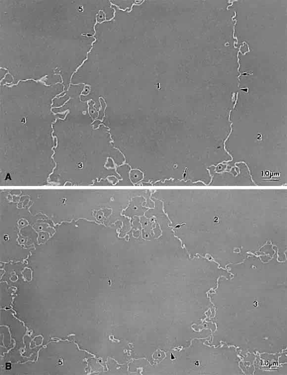
| Fig. 12. Transmission electron micrographs of embryonic nuclear fiber cross-sectional profiles from an aged transparent 62-year-old lens (A) and a nuclear cataractous 83-year-old lens (B). In several numbered fibers, gap junctions (space indicated by arrowheads), undulating membrane pairs (arrows), and interlocking edge processes (circular profiles marked with asterisks) are apparent. Cataractous embryonic nuclear fibers have quantifiably more numerous edge processes (asterisks) than aged embryonic nuclear fibers. (A, Al-Ghoul KJ, Costello MJ: Fiber cell morphology and cytoplasmic texture in cataractous and normal human lens nuclei. Curr Eye Res 15:533, 1996; B, Al-Ghoul KJ, Lane CW, Taylor VL et al: Distribution and type of morphological damage in human nuclear age-related cataracts. Exp Eye Res 62:237, 1996) |