Chapter 12
Concomitant Esodeviations
MARSHALL M. PARKS, PAUL R. MITCHELL and MAYNARD B. WHEELER
Main Menu   Table Of Contents

Search

ACCOMMODATIVE ESODEVIATIONS
NONACCOMMODATIVE ESODEVIATIONS
PRISM ADAPTATION TESTING
BOTULINUM A TOXIN (BOTOX)
DIVERGENCE PARALYSIS
REFERENCES

In concomitant esodeviations, the convergent angle of the eyes remains unchanged regardless of the direction of gaze: that is, the alignment is the same in the primary position, lateral gaze, and vertical gaze. Concomitant esodeviations are caused by or associated with various unrelated factors, such as the near reflex, a congenital hypertonus of the medial rectus muscles, an acquired hypotonus of the lateral rectus muscles, and unilateral reduced visual function in infants or young children.

Risk factors for esotropia and exotropia were studied in the Collaborative Project of the National Institutes of Neurological Disorders and Stroke, from 1959 through 1965.1 To evaluate the developmental consequences in children of complications during pregnancy and the perinatal period, the study of socioeconomic, perinatal, and neonatal characteristics of 39,227 children and their mothers was compared at birth, 4 months, 8 months, 12 months, and 7 years. Esotropia developed in 1187 (3%) and exotropia in 490 (1.2%). Esotropia was more common in white patients (3.9%) compared with nonwhite patients (2.2%). The incidence rates of exotropia were similar (1.2% and 1.3%, respectively). Maternal cigarette smoking during pregnancy and low birth weight increased the risk of both esotropia and exotropia. There was also an increased risk of esotropia with increasing maternal age up to age 34.

Back to Top
ACCOMMODATIVE ESODEVIATIONS
A convergent deviation of the eyes associated with activation of the accommodation reflex is an accommodative esodeviation. If the accommodative esodeviation is within the fusional divergence amplitude, it is termed accommodative esophoria; however, if the accommodative esodeviation is beyond the scope of fusion, it is termed accommodative esotropia.

ETIOLOGY

Accommodative esotropia is precipitated by the following hereditary disorders: (1) hypermetropia; and/or (2) a high accommodative convergence to accommodation (AC/A) ratio. Patients may have both hypermetropia and a high AC/A ratio.

As Donders2 described, the first cause is the convergence associated with accommodation applied to clear the blurred retinal image caused by hypermetropia. If the retinal image is allowed to remain blurred, the hypermetropic patient is not accommodating and the eyes remain straight. However, clearing the blurred hypermetropic image is accomplished by accommodation, and there is also a synkinetic accommodative esodeviation.

A high AC/A ratio occurs in patients with normal refractions, but with an abnormal relationship between accommodation and its synkinetically associated accommodative convergence, which can cause an esodeviation at near. The amount of convergence associated with every 1 diopter (D) of accommodation may vary from slight to marked.

CHARACTERISTICS

In children, accommodative esophoria is usually asymptomatic. If a symptom appears, it is usually asthenopia, which occurs after prolonged near work. As the fusional divergence fatigues, controlling the esophoria by maintaining fusion becomes increasingly difficult. Eventually, esotropia momentarily replaces esophoria, and the patient experiences diplopia. The diplopia causes the patient to react by reducing the accommodation, hence lessening the associated accommodative convergence, which reduces the esophoria to the level at which the fatigued fusional divergence can regain fusion. Esotropia returns to esophoria, but the retinal image is blurred as a result of underaccommodation and, as a consequence, visual acuity is reduced. The reduced acuity stimulates the patient to increase the accommodation, which, in turn, causes the diplopia to return. This recurrent cycle of diplopia vacillating with blurred vision is often experienced by the fatigued esophoric patient. Patients with accommodative esophoria caused by a high AC/A ratio have these symptoms at near, whereas those with accommodative esophoria caused by hypermetropia have symptoms at both distance and near.

The onset of accommodative esotropia may occur at any age between 6 months and 7 years; the average age of the patient at onset is 2½ years, regardless of the cause. Apparently, this is the usual age at which the youngster first accommodates sufficiently to appreciate the visual gain, although diplopia is experienced. Accommodation is not sustained, but the recurring esotropia produces the clinical pattern of intermittent esotropia. Attention to fine visual detail causes momentary esotropia at both distance and near if hypermetropia is the cause, and only at near if a high AC/A ratio is the cause. Some patients maintain the intermittent esotropia pattern without manifestations of an increase toward constant esotropia; however, others increase the frequency and duration of esotropia and rapidly convert to constant esotropia.

CLINICAL INVESTIGATION

The clinical investigation of these patients demands an evaluation of the cycloplegic refraction, the AC/A ratio, and the fusional divergence amplitude because these three factors determine the cause of esodeviation and the patient's ability to contain the accommodative esodeviation and maintain fusion.

Cycloplegic Refraction

An adequate cycloplegic refraction need not be atropine refraction. One drop of 2% cyclopentolate (Cyclogyl) within 40 minutes provides within 0.25 D of the plus refractive error produced by one drop of 1% atropine three times daily for 3 days in white children. Deeply pigmented irides require more than one drop of 2% cyclopentolate: namely, the addition of one drop of 2.5% phenylephrine hydrochloride (Neo-Synephrine), and 40 minutes later one drop of 1% tropicamide (Mydriacyl). This produces superb cycloplegia 1 hour after the first drops are instilled in the most deeply pigmented eyes. Ideally, evaluation of the cycloplegic refraction should be included in the examination conducted during the initial appointment, and if needed, glasses should be prescribed at the conclusion of the examination. Also, regardless of the cycloplegic drugs used in children, a certain residual hypermetropia remains. Repeat cycloplegic refraction within weeks after prescribing the first glasses usually discloses more hypermetropia than was detected at the initial refraction.

Accommodative Convergence to Accommodation (AC/A) Ratio

The AC/A ratio is determined by comparing the distance prism, near prism, and alternate cover accommodation-controlled measurements. A near esodeviation measurement within 10Δ of the distance measurement is considered within the normal range. An excess of 10Δ difference between distance and near constitutes a high AC/A measurement. Since the severity of the high AC/A ratio varies from patient to patient, it is graded as follows:

  Grade 1: The difference from 11Δ to 20Δ between the distance and near measurements
  Grade 2: The difference from 21Δ through 30Δ
  Grade 3: The difference in excess of 30Δ

Infants and children fixate toys at distance and near as the prism and alternate cover measurements are performed. Children who submit to a visual acuity test with Snellen letters or the Snellen illiterate E fixate the same targets for their distance-near testing. The lines of columns of letters are read as the cover test is performed on literate children, and illiterate children point out the direction of the E, which are continuously presented during cover testing.

Any combination of refraction and AC/A ratio can be found, but statistically there is a definite relationship. Patients with a normal AC/A ratio have relatively more hypermetropia, and those with a high AC/A ratio have relatively less hypermetropia. One study3 revealed the relationships shown in Table 1.3 These findings are in accordance with expectations, since patients with a high AC/A ratio should have to accommodate less than patients with a normal AC/A ratio to produce equal angles of accommodative esodeviation.


TABLE 1. Relationship Between AC/A Ratio and Refraction in Patients with Accommodative Esotropia


PatientsAC/ARefractions
378High+ 2.25
289Normal+ 4.75


The evidence for altering the AC/A ratio after surgery is inconclusive. In a prospective study of 38 patients with concomitant strabismus, the effect of strabismus surgery was investigated.4 In both esotropia and exotropia, the AC/A ratio was found to decrease after surgery. A trend became evident, suggesting an increased risk of overcorrection in those children with esotropia with a high preoperative AC/A ratio equal to or greater than 7:1.

Fusional Divergence Amplitude

The fusional divergence amplitude usually ranges between 12Δ and 20Δ; when the accommodative esodeviations exceed this range, esotropia prevails. By trial and error, the fusional divergence amplitude can be determined indirectly by doing the prism and alternate cover test while the patient is wearing the minimal hypermetropic lenses that permit fusion. Patients with accommodative esodeviation who remain intermittently esotropic for several years tend to maintain the largest fusional divergence amplitudes. The patient who lapses into constant esotropia soon loses the large fusional divergence amplitude previously possessed when the strabismus was intermittent.

Sensory and Motor Complications

Sensory and motor complications soon evolve if esotropia repeatedly vacillates with esophoria. Initially, diplopia is experienced when esotropia first appears; however, the young child soon learns suppression and adopts anomalous retinal correspondence (ARC) peripheral fusion, removing any sensory annoyances that occur during the esotropic phase. Until this happens, the child often manifests diplopia and visual confusion by expressing it verbally, by closing or covering one eye, and by awkwardness. As soon as these sensory annoyances are eliminated by development of the sensorial adaptations, the patient is happier and more willing to tolerate the tropia. Eventually, constant esotropia may replace intermittent esotropia. After developing suppression and ARC and while still intermittently esotropic, these patients—when their eyes are straight—have normal retinal correspondence (NRC) and central and peripheral fusion. Thus, these patients instantly adjust their sensory status to conform to the alignment of the eyes.

A motor complication also occurs in intermittent esotropia; it is presumably a change in the medial rectus muscles secondary to their increased and more frequent contraction. Whatever the change—hypertrophy or contracture—a gradual increase in nonaccommodative esodeviation appears. Eventually, the nonaccommodative esodeviation buildup exceeds the fusional divergence amplitude in most patients, and the intermittent esotropia is replaced by constant esotropia. As the angle of esotropia increases, the ARC values and localization of the suppression scotoma change accordingly to conform to the larger angle.

With constant esotropia comes an opportunity for amblyopia to develop. In contrast to those with congenital esotropia, most patients with acquired esotropia select one eye for fixation to the exclusion of the other eye; this soon results in amblyopia of the unused eye. If, by chance, alternate fixation is chosen, amblyopia is prevented. Amblyopia is unrelated to suppression and ARC. Although the intermittent phase of the accommodative esotropia prevails, suppression and ARC may begin; in some patients, however, amblyopia does not develop because there is sufficient bifoveal fixation to prevent it. Only when constant esotropia replaces intermittent esotropia is the strabismus capable of causing amblyopia to develop in the patient with nonalternate fixation. However, suppression and ARC are always present in constant acquired esotropia, regardless of alternate or nonalternate fixation; they also occur during the intermittent esotropia phase.

TREATMENT

Early recognition of the disorder and early initiation of treatment are mandatory if the sensory and motor complications are to be prevented. The basic treatment involves some technique that curbs accommodation. This is accomplished either by an optical method (e.g., glasses) or by instillation of a parasympathomimetic drug (miotic) into the eyes.

The treatment varies according to the patient's age at the time of initial ophthalmologic consultation. For convenience, the treatment is described for three age groups: younger than 4 years, 4 to 8 years, and older than 8 years.

Children Younger Than 4 Years of Age

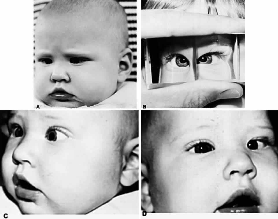
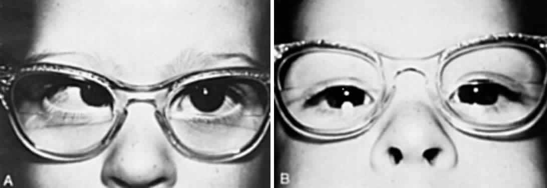
In all children younger than 4 years of age, the full cycloplegic refraction spectacle correction is prescribed (Fig. 1). Occasionally, the patient's history is not helpful, the findings are questionable, and the ophthalmologist wishes to gain evidence regarding improvement in the angle of esotropia by curbing the accommodation, but may have reason to doubt whether glasses are necessary. Alternatively, glasses may be rejected despite all efforts to encourage eventual acceptance, such as prescribing instillation of 1% atropine each morning for 1 month. If glasses cannot be used, it is helpful to prescribe instillation of a miotic each morning. The miotic should be considered temporary therapy: it is to be used to determine the antiaccommodation effect, or until the child has matured and accepts the needed glasses. Miotics should not be considered as alternative permanent antiaccommodation therapy equivalent to glasses.

Fig. 1. (A) Straight eyes while large hypermetropic spectacle correction is worn. (B) Accommodative esotropia when spectacles are removed.

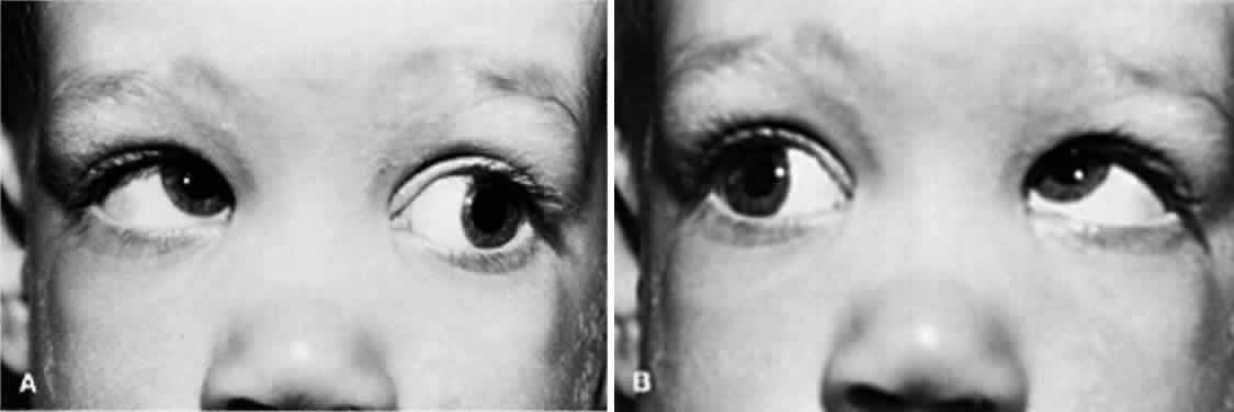
SPECTACLE CORRECTION. The patient is reexamined at monthly intervals until the ophthalmologist is certain that the glasses are controlling the accommodative esotropia. If the esotropia remains at near but the eyes are straight at distance, an additional + 2.5 D bifocal segment is prescribed (Fig. 2). Care is taken to inform the optician that the top of the lower segment must be higher for children than for adults. Ideally, the top of the segment must be 3 mm above the 6-o'clock limbus position. Segments that are too low are either viewed over, or the child must raise the chin too high in order to depress the eyes to see through the segments. If the optician places the top of the segment too high, the bifocals will be higher than midpupil, causing the chin to be depressed while viewing at distance through the upper portion of the bifocals. Children who do not readily accept the lower segment for near viewing and continue to cross their eyes for near viewing while looking through the upper portions of the lenses are started on 1% atropine instillation daily. This therapy is continued for up to 1 month. After the atropine is discontinued, the child usually continues to use the lower segment correctly. The ophthalmologist judges the correct acceptance of the lower segment by observing whether or not the child invariably raises the chin and depresses the eyes to look through the lower segment when small target material is presented at eye level or slightly above at a distance of 0.33 meter. The most satisfactory bifocal is a flat-top segment that traverses the entire width of the lens, such as the executive bifocal. With this type of lens, regardless of the position in which the child depresses the eyes in straight down gaze, left and down gaze, or right and down gaze, the lower segment is simultaneously engaged by each eye. The parents should make sure the glasses are in proper adjustment, since tilted or crooked glasses cause one eye to look through the lower bifocal segment and the other eye to look above it. Some patients obviously need bifocals, and these are prescribed as were the initial glasses. For example, the child who, without glasses, has straight eyes for distance but 35Δ of esotropia for near and ±1 D cycloplegic refractive error needs bifocals. Progressive addition lenses, such as the Varilux 2 lenses (Essilor Canada, Ltd., Ville Saint-Laurent, PQ) have been advocated because of the lack of image jump without the demarcation line, and because of the cosmetic improvement of the lenses without lines.5 There is difficulty, however, in determining the proper height of the progressive zone, and the lenses are more expensive.

Fig. 2. (A) High AC/A ratio, causing near esotropia despite fully corrective distance hypermetropic spectacles. (B) Bifocal segment adds compensation for the high AC/A ratio, permitting straight eyes for near viewing.

Substituting a miotic for bifocals when the patient needs glasses because of a refractive error usually does not succeed, since the child tends to peer over the glasses and abandons them. Miotics work best in lieu of glasses, not in conjunction with them.

MIOTICS. Two miotics have been used extensively in North America for accommodative esotropia: isoflurophate (Floropryl 0.025% ointment)6–8 and echothiophate iodide (Phospholine Iodide 0.03% to 0.25%). These are long-acting cholinesterase inhibitors that facilitate neuromuscular transmission.9 Isoflurophate is inactivated by water and once was dispensed in USP anhydrous peanut oil. Currently it is not available in the United States. If control of the esodeviation is achieved during this time, the frequency of administration is diminished to every other day for 2 months and finally to twice a week (and in rare cases, once a week) for as long as therapy is required. Echothiophate is stable in a water solution if refrigerated; four concentrations are available (0.03%, 0.06%, 0.125%, and 0.25%). One drop is instilled in each eye each morning. Both drugs cause miosis, but more severe miosis is caused by isoflurophate than by echothiophate. Sustained miosis is often associated with pigmented hypertrophic epithelial cysts at the pupillary border. These cysts project into the pupillary space and may seal the space shut if they are allowed to develop further by continuing the medication. This occurs more readily with isoflurophate than with echothiophate. After discontinuing the miotic, the cysts diminish in size but remain for years as an obvious pigmented hyperplastic mass (“tags”) on the pupillary border as viewed in the slit lamp. The cysts can be prevented by instilling 2.5% phenylephrine hydrochloride twice daily.10 This diminishes the miosis slightly, but does not interfere with the function of potentiating the transmission of acetylcholine at the myoneural junction of the ciliary musculature. Miotics have a residual action for several days to a few weeks, making cycloplegia and mydriasis less than thorough during this period.

Although miotics have a residual action for several days to a few weeks, it appears to be unnecessary to discontinue echothiophate in order to obtain a reliable cycloplegic refraction. Raab11 compared the results of cycloplegic refraction in 18 patients on echothiophate, and then off echothiophate for 4 weeks. The average interval was 9 weeks (range, 2 to 39 weeks). The results were calculated to account for the various strengths of echothiophate (0.06%, 0.125%, or 0.25%), and for the interval off echothiophate. From the time that echothiophate was stopped, refraction was 4 weeks or less, the change in refraction was + 0.08 ± 0.48 D; at greater intervals, the change in refraction was -0.12 ± 0.29 D.

Although isoflurophate causes greater miosis, it performs better than echothiophate in controlling accommodative esodeviation. It cannot be titrated as easily, however, because of the single strength. Echothiophate has a systemic effect: it destroys the cholinesterase in plasma and erythrocytes. This constitutes an additional risk for the patient undergoing anesthesia, particularly if other anticholinesterase drugs, such as succinylcholine, are used during intubation to relax the pharyngeal muscles. As a result, the muscles used in respiration may be rendered functionless for a few hours, and the anesthesiologist may have to supply artificial ventilation for the patient until the muscles recover. Finally, subcapsular vacuoles, and even cataracts, have been reported in the eyes of patients receiving echothiophate. Neither drug can be considered innocuous, and the indication for their use must be evaluated carefully. No patient should receive either of these medications without being observed at least every 3 months. After the patient's deviation is controlled, either the concentration may be reduced or the frequency of instillation diminished progressively to every other day and then to every third day. The minimum dosage should control the deviation and allow fusion. The medication deteriorates with time, and the patient develops some degree of tolerance to it; these variables must also be considered. The medical approach to controlling accommodative esodeviation is more precarious and difficult to manage than the optical approach. Since accommodative esotropia requires long-term control, miotics are not the agents of choice for prolonged therapy. The prime importance of glasses in the management of accommodative esotropia has not been displaced by miotics.

CLINICAL COURSE. The cause of accommodative esotropia determines the clinical course. Accommodative esotropia due to hypermetropia and a normal AC/A ratio remains well controlled once glasses straighten the eyes. This is not true, however, for accommodative esotropia due to a high AC/A ratio. Despite the use of glasses to immediately control the esotropia, there is a significant possibility that a gradual, nonaccommodative esotropia component will appear. Table 2 shows that the more severe the high AC/A ratio, the greater the possibility of the patient's deteriorating and developing nonaccommodative esotropia, thus escaping the control that glasses rendered originally.12


TABLE 2. Relationship Between the Deterioration Rate and the Severity of the High AC/A Ratio


PatientsAC/ADeterioration Rate (%)
40Normal5
19Slightly high11
20Moderately high35
21Severely high43


In another study, Ludwig and associates13 analyzed the rate of deterioration in accommodative esotropia in relationship to the AC/A ratio. The AC/A relationships were graded according to the difference between the distance and near measurements:

  Normal: 0Δ to 9Δ
  Grade 1: 10Δ to 19Δ
  Grade 2: 20Δ to 29 Δ
  Grade 3: 30Δ and higher

Deterioration occurred in 7.7% of patients with a normal AC/A ratio, in 25% with grade 1 high AC/A ratio, 44% with grade 2 high AC/A ratio, and 52% with grade 3 high AC/A ratio, confirming that accommodative esotropia tends to deteriorate with greater frequency if the AC/A ratio is high.

Patients with a high AC/A ratio and a need for bifocals do not usually manifest improvement in the severity of their abnormal ratio until after 7 years of age. The hypermetropia disclosed by subsequent cycloplegic refraction usually increases until 6 years of age, levels off for 2 or more years, and after 8 years of age frequently decreases until the teens. Patients with accommodative esotropia controlled by glasses, including bifocals, are reexamined at 6-month intervals until 6 years of age and annually thereafter.

In a retrospective study, Wilson and colleagues14 reviewed the records of 127 patients with accommodative esotropia, who were within 10Δ of orthophoria, and who had stereopsis and other binocular sensory test measurements at their latest examination. With bifixation defined as 50 arc-seconds of stereopsis or better, 31 (24%) met this criterion; average follow-up was 89 months. Monofixation, or peripheral fusion, was present in the remaining 96 patients (76%); average follow-up was 84 months. In comparing the two groups, the patients with bifixation were less likely to have presented with constant esotropia (19% vs 39%), were more likely to be aligned within 8Δ of orthophoria with their first eyeglasses (84% vs 21%), were less likely to have worn bifocals (39% vs 59%), and less likely to have undergone surgery (23% vs 62%). Wilson and colleagues suggest that the maintenance of bifixation is possible in accommodative esotropia if the eyes can be straightened before the deviation becomes constant, or shortly thereafter. With the early therapy, amblyopia and deterioration of ocular alignment are less likely to occur. None of the patients with bifixation had constant esotropia lasting longer than 4 months.

Children who became constantly esotropic before receiving treatment and whose glasses did not reduce their angle to straight, or children whose angle was originally straight with glasses but escaped the control supplied by glasses and who are now esotropic with glasses, require surgery for the quantity of esotropia measured at distance with their full corrective lenses. Empirically, we have learned that patients with a high AC/A ratio who require surgery should receive more than the customary amount of surgery done for that angle of esotropia. For example, if the angle of deviation would normally require a 4-mm recession of the medial rectus muscles, we would do a 5-mm bilateral medial rectus recession for the patient with a high AC/A ratio. Any child on whom surgery is performed should have amblyopia eliminated by the appropriate occlusion therapy before the surgery. If, after wearing glasses for 1 month, the eyes remain esotropic and there is no amblyopia, surgery is performed. We have not noticed improvement in the angle of esotropia in patients wearing glasses for several months. The constantly esotropic eyes either straighten promptly with glasses or assume an angle of esotropia that remains approximately the same until surgery is performed.

A high rate of undercorrection has occurred when performing standard surgery for the nonaccommodative component, when full hypermetropic correction is worn.15–18 Because of these undercorrections, Wright and Bruce-Lyle18 have recommended augmented surgery for esotropia with high hypermetropia. In a retrospective study, they reviewed 70 patients with the following criteria: acquired esotropia, hypermetropia of 3 D or more, surgery for the residual esotropia with recession of the medial rectus muscle both eyes, full optical correction, surgery after 6 months of age, follow-up of at least 1 year, normal neurologic testing, and absence of eye muscle palsy. Based on the average of the deviation at distance and near, 30 patients received standard surgery; the other 40 patients had augmented surgery, based on the average of the near deviation, with and without correction. Correction to 10 D or less of orthophoria was achieved in 22 of the 30 patients (74%) in the standard surgery group; undercorrection was noted in the remaining 8 patients (26%). Correction to 10 D or less of orthophoria was accomplished in 35 of the 40 patients (88%) in the augmented surgery group; however, 10 D or more of consecutive exotropia developed in the remaining 5 patients (12%) with their full optical correction. After reduction of the hypermetropic power, or in some patients, removal of the eyeglasses, success was achieved in 39 of the 40 patients (98%). One patient had intermittent exotropia even after removing the eyeglasses.

Children 4 to 8 Years of Age

The treatment of accommodative esotropia in children between 4 and 8 years of age contrasts with that described for children younger than 4 years old in that the minimum power lenses required to maintain fusion and provide maximum visual acuity can be determined and prescribed. With accommodative targets at distance and near, as well as the cover-uncover test, the least power plus lenses that keep the eyes straight are prescribed, including bifocals if necessary. Ideally, esophoria is demonstrable by an alternate cover test with these minimal plus lenses, since it maintains a high degree of tone in the fusional divergence. This satisfies the objective of accommodative esotropia therapy, which is to reduce the esodeviation with an antiaccommodation measure just enough to allow accommodative esophoria to replace accommodative esotropia. The object is not to convert accommodative esotropia to orthophoria. Maintaining orthophoria with glasses over several years, never putting a load on the patient's fusional divergence, decreases the probability of eventual withdrawal of glasses without symptoms of asthenopia, blurred vision, and diplopia. It is generally impossible to apply this objective of therapy for accommodative esotropia to children younger than 4 years of age because the examiner is unable to evoke a response in this age group that is indicative of the visual acuity.

Children Older Than 8 Years of Age

In the treatment of accommodative esotropia in children older than 8 years of age, the ophthalmologist must realize that this is the earliest age at which improvement should be expected. The hypermetropia usually decreases, and the severity of the high AC/A ratio diminishes from this age into the early teens. The power of the spectacle plus lenses can usually be decreased and the bifocals reduced and frequently withdrawn. The prognosis for withdrawal of the spectacles must be related to the severity of the hypermetropia, the astigmatism, the anisometropia, and the AC/A ratio. Some patients who should have a good prognosis for part- or full-time spectacle withdrawal but show no spontaneous improvement may be induced to remove their glasses after expanding their fusional divergence amplitude, or at least encouraged to become more reliant on using their fusional divergence to its maximal potential. Patients are taught to limit their accommodation to that quantity that does not evoke more accommodative esodeviation than can be contained by the fusional divergence amplitude, even though the retinal image remains blurred, since suboptimal accommodation was applied to gain maximal visual acuity. Either an orthoptist or an ophthalmologist can teach this with the aid of diminishing-strength miotics.

The orthoptist teaches dissociation exercises to enable the patient with accommodative esotropia to remove the glasses. First, the patient is taught to maintain straight eyes without glasses in the unaccommodative state, accepting blurred vision. Next, small increments of accommodation that are associated with increasing amounts of esophoria are allowed. Repeating this maneuver increases the fusional divergence amplitude. Gradually, larger amounts of esodeviation are withstood until, eventually, clear vision and straight eyes are maintained. Physiologic diplopia (framing) and bar reading should be practiced during this routine to prevent the patient from lapsing into esotropia with suppression and to guarantee maintenance of fusion with clear vision. Training to increase the fusional divergence amplitude may be done at the same time by using gradually stronger base-in prisms while watching television and reading or by employing stereoscopic or Polaroid vectographic techniques (Orthofusor).

Miotics can be withdrawn gradually while preserving fusion. By gradually diminishing the concentration or frequency of the instillation of the miotic, the fusional divergence may be expanded. This requires the patient to accommodate more in order to obtain clear vision. This causes greater esophoria, which in turn puts more stress on the fusional divergence amplitude if binocularity is to be maintained. By gradually expanding the fusional divergence amplitude, some patients can eventually discontinue all medication, keeping the eyes straight and seeing well. The program ideally follows this plan:

  1. Remove glasses and start 0.125% echothiophate daily for 1 week.
  2. Decrease to 0.06% echothiophate daily for 1 week.
  3. Decrease to 0.06% echothiophate every other day for four doses.
  4. Decrease to 0.06% echothiophate every third day for three doses.
  5. Decrease to 0.06% echothiophate every fourth day for two doses.
  6. Decrease to 0.06% echothiophate every week for two doses.
  7. Discontinue all medication.

The patient is examined each time the dosage is changed, and further decrease is not made unless the eyes are straight. This routine can be combined with orthoptic dissociation exercises and fusional divergence amplitude exercises.

These procedures are successful in patients 8 years of age or older with moderate refractive errors and in those with moderately severe high AC/A ratios. If, however, the policy of providing no lens power above that necessary to keep the eyes straight is pursued from 4 years of age, most of those who required medication or orthoptics to rid themselves of glasses at 8 years of age will have achieved the same result by 10 to 12 years of age.

Deterioration of accommodative esotropia to nonaccommodative esotropia is a well-recognized complication requiring surgical intervention.19 Although most cases of accommodative esotropia subside by 10 to 12 years of age, there are many cases of accommodative esotropia, with or without deterioration, persisting into the later teen years and occasionally into adulthood.

Ludwig and colleagues20 studied 65 patients with accommodative esotropia who required bifocals to maintain near alignment. The average follow-up was 10.5 years. Forty patients (61.5%) were able to discontinue bifocals after an average of 5.5 years. Twenty-five patients (38.5%) continued to wear bifocals or reading glasses after an average follow-up of 9.7 years. Twenty patients in the first group (50%) and 9 patients in the second group (36%) required surgery for deterioration of accommodative esotropia. The average age of bifocal discontinuation was 9.3 years in the nonsurgical patients and 9.7 years in the surgical patients. The surgical patients had lower hyperopia (average, + 2.4 D) than the nonsurgical group (+ 3.5 D) and an earlier age of onset of bifocal wear (3.3 vs 4.6 years). Although bifocals may be discontinued successfully in a majority of patients at an average age of 9.5 years, a significant percentage require long-term wear, even among those who have had surgery. The only predictive factor for long-term bifocal wear was a relatively high AC/A ratio.

Back to Top
NONACCOMMODATIVE ESODEVIATIONS
Nonaccommodative esodeviations are the various types of convergent strabismus not associated with accommodation. Whether or not the fusional vergences can control the esodeviation determines if the deviation is an esophoria or an esotropia.

The cause of nonaccommodative esodeviations is neurogenic and anatomic. The innervating factor that causes convergence other than accommodative and fusional convergence is known as tonic convergence. Accordingly, the innervational cause of nonaccommodative esotropia is reasoned to be some vague disturbance within the tonic vergences, resulting in either excessive tonic convergence or deficient tonic divergence. It is convenient, although unproved, to use this speculation to explain both congenital esotropia and the esotropia that gradually develops secondary to an anomaly that impairs the sight in one or both eyes of infants and young children.

The anatomic factors that probably account for some of the nonaccommodative esodeviation problems are primary anomalies, such as abnormal medial rectus muscles; and secondary anomalies, such as the change that occurs in the medial rectus muscles with excessive innervation, as is encountered in deteriorated accommodative esotropia, abducens palsy, and Duane syndrome.

CONGENITAL ESOTROPIA

The term congenital esotropia has been criticized as improper by some authors, who argue that these patients cannot be proved to have been born with esotropia.21,22 The basis for this criticism is that the deviation is not present at birth. Based on clinical observations, however, the term congenital esotropia is valid, and it can be easily distinguished from other forms of acquired esotropia.23

Congenital esotropia is usually apparent to the parents shortly after the child's birth. After the first few days of life, when the eyes are open sufficiently for study, the esotropia is detected. Some parents claim the esotropia was not obvious until 1 or 2 months later, but the onset of esodeviation is open to subjective analysis, and it is difficult for the physician to procure good objective analysis.

Nixon and co-workers24 observed 1219 alert infants in a newborn nursery to learn whether esotropia is present at birth or develops later in infancy. Orthophoria was found in 593 (48.6%), exotropia in 398 (32.7%), intermittent esodeviations in 17 (1.4%), varying between esodeviations and exodeviations in 14 (1.1%), and variable esodeviations in 9 (0.7%). None of the infants had findings diagnostic of congenital/infantile esotropia. The authors concluded that congenital/infantile esotropia is not present at birth, but develops in the first few weeks to months after birth.

Maumenee and associates25 analyzed 173 pedigrees involving 1589 people in an effort to determine the etiology of congenital esotropia. Their results were compatible with a mendelian codominant model, but the estimated transmission probability was not consistent with the mendelian expectation. This suggested that these families had etiologic heterogeneity, the majority being autosomal recessive and some dominant; there may have also been an aggregation of some nongenetic cases.

The prevalence of the monofixation syndrome in the general population is approximately 1%. Scott and colleagues26 studied 90 children with congenital esotropia, and 129 of the biological parents. Twelve parents had secondary monofixation syndrome, which reduced the numbers studied to 117 parents and 78 patients. Of the 117 parents, 7 had primary monofixation syndrome, yielding a prevalence of 6% for the parents and 9% (7/78) for the families. Because congenital esotropia is most likely inherited in a multifactorial fashion, the authors believe that the increase in prevalence of primary monofixation syndrome compared with the general population supports the hypothesis that primary monofixation syndrome may be a mild subthreshold effect of the gene or genes responsible for congenital esotropia.

Characteristically, the angle of convergence is large and constant, showing little sign of change with age in children without brain damage. A high percentage of brain-damaged infants have congenital esotropia, but their angle of strabismus is usually more variable, frequently diminishing with age; occasionally, the esotropia is spontaneously replaced with exotropia between 6 months and 1 year of age.

In a study of patients with infantile esotropia, Holman and Merritt27 found neurologic problems, both general and ocular, in 29 of 47 patients (62%). Problems included prematurity, hydrocephalus, mental retardation, cerebral palsy, meningomyelocele, intraventricular hemorrhage, neonatal and postnatal seizures, and abducens palsy. The authors believed that the high incidence of neurologic problems was, in part, due to the fact that the study was conducted at a university medical center. Nelson and co-workers28 found a prevalence of strabismus of 24% in 29 infants who were prenatally exposed to various psychoactive drugs: 14% had esotropia and 10% had exotropia, which is high compared with the incidence of strabismus in the general population (2.8% to 5.3%).

Patients with congenital esotropia must be differentiated from those with very early onset accommodative esotropia who present with a large-angle esodeviation or variable deviation combined with hypermetropia in the range of + 3.75 D to + 7.5.D.29

Diagnosis

The majority of patients with congenital esotropia alternate fixation in the primary position and cross-fixate on side gaze, using the right eye in left gaze and the left eye in right gaze, as illustrated in Figure 3. The minority who happen not to alternate their fixation have amblyopia in the nonpreferred eye, which may be profound, as manifest by eccentric fixation. The cross-fixation pattern can be confusing and misleading to the examiner who is not experienced in examining infants with congenital esotropia. The examiner must learn that the absence of the abduction so frequently encountered in these infants is secondary to the cross-fixation habit. The infant who cross-fixates has never abducted an eye, and the examiner finds it impossible to evoke an abduction response on either version or duction studies. The inexperienced examiner may assume that the patient has bilateral abducens paralysis. Similarly, the inexperienced examiner may mistakenly suspect unilateral abducens paralysis in the eccentrically fixating eye of a patient with a large angle of congenital esotropia who does not cross-fixate and has amblyopia. To help avoid this pitfall, the examiner should be alert to this possibility and also realize that unilateral and bilateral congenital abducens paralyses are rare disorders; there is also the possibility of confusing abducens paralysis with Duane and Möbius syndromes.

Fig. 3. (A) Congenital estropia, primary position. (B) Krimsky measurement. (C) Cross fixation, using right-eye fixation for left gaze and (D) left-eye fixation for right gaze.

Two diagnostic maneuvers can assist the examiner in differentiating congenital esotropia and its associated abduction limitation from abducens paralysis, Duane syndrome, and Möbius syndrome:

  1. By spinning the child with the head fixed in an upright position, clasping the head with his or her hands so it cannot be turned and holding the child tightly against his or her chest, the examiner swiftly accelerates and decelerates the child's head horizontally through space. This labyrinthine stimulation, specifically of the horizontal semicircular canals, causes a subtle abducting movement of the eye on the side opposite the direction of head acceleration that lasts for only 1 second. By closely concentrating on the eye, a favorably positioned second examiner can detect the abduction.
  2. The examiner can produce abduction in the cross-fixating congenital esotrope by occluding one eye for several days. Abduction will appear in the other eye.

The traction test performed in an anesthetized cross-fixating congenitally esotropic child, who manifests no abduction, is universally normal. No passive resistance to abduction is encountered. Also, with the patient under surgical plane anesthesia, the large angle of esotropia disappears, and often the eyes are divergent.

The esotropia angle is measured by the Krimsky prism technique in infants and young children (Fig. 3B) until the prism and alternate cover measurements are determinable. In infants and young children, usually only a horizontal deviation is present; however, the majority of children 2 to 3 years of age manifest a dissociated vertical deviation (DVD).30–32 Early surgical correction of the congenital esotropia does not reduce the incidence of this interesting cyclovertical disorder. The DVD is manifest by the nonfixating eye being elevated and extorted. Alternate cover testing reveals that the eye behind the cover elevates and extorts, and the eye assuming fixation depresses and intorts. As the cover moves from eye to eye, this process repeats itself and results in a positive vertical vergence with the cover before the right eye, and a negative vertical vergence with the cover before the left eye, with simultaneous movements being dextrocycloversion with the cover before the right eye, and levocycloversion with the cover before the left eye. The fact that these cyclovertical movements occur simultaneously with the bilateral abducting movements during the alternate cover testing in a congenital esotrope makes them difficult to detect. They are easily detected, however, after the horizontal deviation is eliminated by surgery. The presence of DVD is not pathognomonic of congenital esotropia, although the majority of congenital esotropes have it. DVD is encountered infrequently in patients with exotropia and in patients having horizontally straight eyes. Approximately one third of patients with DVD will require surgical treatment.31

Version studies in infants younger than 1 year of age are usually normal, but overacting inferior oblique muscles (Fig. 4) appear in the majority of the older congenitally esotropic children. This problem also occurs with the same incidence regardless of whether or not early surgery was performed and the horizontal deviation eliminated.32,33

Fig. 4. Overaction of inferior oblique muscles. (A) Right inferior oblique overaction with left eye fixing in left gaze. (B) Left inferior oblique overaction with right eye fixing in right gaze.

Hiles and associates33 noted overactive inferior oblique muscles in 42 of 54 infants (78%) with congenital esotropia, the onset most frequently occurring during the second year of life. Surgery was performed on 21 of the 54 infants (39%). Wilson and Parks32 found overactive inferior oblique muscles in 72% of congenital esotropes with an average age of 3.6 years. The incidence was not related to the age of onset of strabismus, time from onset of strabismus to surgery, age at first surgery, or decompensation of ocular alignment. The incidence was positively related to the number of surgeries. The incidence of inferior oblique overaction in accommodative esotropia was 34% (average age, 5.2 years).

The cycloplegic refraction can be hypermetropic or myopic, but it is usually equal in the two eyes and hypermetropic by the usual amount for the chronologic age.

In older children in whom sensory testing is reliable, the absence of binocular vision is the invariable finding in a patient who has not received treatment to straighten the congenitally esotropic eyes. Children whose eyes are straight as a result of early surgery often manifest peripheral fusion with NRC, but they rarely have central fusion.22,23,33–43 Children whose eyes were straightened at a young age and then either had a return of esotropia or developed secondary exotropia may have suppression and ARC.

The monofixation syndrome35 or subnormal binocular vision is usually the best attainable sensory result in the treatment of congenital esotropia.22,23,33–43

Management

The management of congenital esotropia essentially involves surgical straightening of the eyes. The controversial aspects of this need are when and how the surgery is best accomplished. The need for eliminating amblyopia, if present, before surgery is not controversial. Alternate occlusion therapy may benefit the minority of patients having a propensity for amblyopia until the surgery is performed, but it is of no other value. Alternate occlusion therapy for congenitally esotropic patients to prevent suppression and ARC is completely unfounded. Suppression and ARC are adaptations to NRC fusion; they never develop unless binocular vision, which is lacking in congenitally deviated eyes, is already present. Apparently, a requisite for the conditioned reflex of single binocular vision development is either straight eyes or eyes that are at least within 10Δ of being horizontally straight during infancy or early childhood. Therefore, treating congenital esotropia with alternate occlusion for a few years, until the child is sufficiently mature to be taught by orthoptics how to fuse, and then performing the necessary surgery illustrates a gross lack of knowledge about the disorder.

SURGICAL APPROACH. Surgery is performed ideally at 6 months of age in children who do not have brain damage. By this age, the examiner can assess the Krimsky prism measurement of the esotropia angle and the patient's fixation ability. All children undergo refraction, and their fundi are viewed before the child is committed to surgery. Amblyopia, if present, is overcome with occlusion therapy before surgery. Early surgery is safe and rewarding. The surgical objective in children younger than 4 years of age is to place the eyes within 10Δ of orthophoria, not simply to reduce the angle of esotropia. The response to surgery in terms of the quantity of improvement in the angle of esotropia is variable in infants; therefore, it is justifiable to operate on only two muscles and then assess the result. As the initial procedure, we routinely recess the media rectus muscles 7 mm in infants having 60Δ of deviation or more, and performing the quantities listed in Table 3 otherwise.


TABLE 3. Quantity of Surgery for Various Esotropia Angles*


ETRecess MROUResect LROU
15Δ3 mm4 mm
20Δ3.5 mm5 mm
25Δ4 mm6 mm
30Δ4.5 mm7 mm
35Δ5 mm8 mm
40Δ5.5 mm9 mm
50Δ6 mm10 mm
60Δ7 mm10 mm
ETRecess MR andResect LR

* Read from top down for MROU and LROU. Read horizontally from the bottom up for MR and LR.
ET, esotropia; MROU, medial rectus muscles of both eyes; LROU, lateral rectus muscles of both eyes; MR, medial rectus of one eye; LR, lateral rectus of one eye.


With large-angle infantile esotropia, averaging 74Δ, Weakley and associates44 performed 7-mm medial rectus muscle recessions, in both eyes, in a series of 36 patients. Of the 36 patients, 27 (75%) achieved successful horizontal alignment, 5 (14%) had undercorrection, and 4 (11%) had overcorrection. Even in very large angle congenital esotropia, the advantages over three- and four-muscle procedures are speed, simplicity, and less trauma, which leaves the lateral rectus muscles available for future surgery when necessary. In a subsequent article, however, Stager and colleagues45 reported delayed consecutive exotropia after the 7-mm procedure for congenital esotropia. Of a total of 88 patients, 25 (28%) had overcorrection, which was larger than initially reported. The overcorrection rates according to age group were as follows:

  Younger than 7 months of age: 8 of 21 patients (38%)
  7 to 12 months of age: 10 of 49 patients (20%)
  13 months or older: 6 of 18 patients (33%)

The consecutive exotropia became evident at an average of 26.8 months after surgery. Others have reported excellent results with medial rectus muscle recessions of 6 to 8 mm, without compromising adduction or convergence.46,47 Recession of the medial rectus muscles 8 mm has been advocated for infantile esotropia of 80 to 90 D.48 In a series of 16 patients, 12 (75%) achieved successful alignment and 4 (25%) had undercorrection. None had overcorrection, although the follow-up of 6 to 48 months postoperatively may not have been long enough. The authors recommended this procedure in place of three- and four-muscle procedures.

Patients with residual esotropia angles of 15Δ or more 6 weeks after surgery receive secondary surgery on the lateral rectus muscles, the quantity of resection depending on the magnitude of the angle. Some surgeons routinely recess and resect the horizontal rectus muscles of one eye, rather than performing bilateral medial rectus recession, and they have reported similarly good results. It is a mistake to state that one approach is better than another, since each surgeon has developed confidence in certain surgical methods, and these techniques should be followed. The important issue is that the eyes be surgically straightened; ideally, this should be performed when the patient is young. If the surgeon performs recession-resection on the horizontal rectus muscles of one eye and residual esotropia persists at 6 weeks postoperatively, a recessionresection quantitated to the angle of deviation should be performed on the other eye. A guide to the quantity of surgery performed for various esotropia angles is presented in Table 3. The number of millimeters of surgical recession or resection is not varied according to the age of the patient. Children 4 years of age or younger usually receive surgery simultaneously on only two horizontal rectus muscles, but older children may have surgery simultaneously on as many as four horizontal rectus muscles if the angle of deviation is sufficiently large to require it. Table 3 is applicable to both primary and secondary surgery. Coexisting vertical incomitance, such as A or V patterns, are also corrected by vertical displacement of the horizontal rectus muscles at the same time they are recessed or resected. Overacting inferior oblique muscles are also corrected by recession49 or partial extirpation in marked overaction,50 either at the time of horizontal surgery or at a later date if their dysfunction appears after horizontal alignment has already been achieved.

The incidence of reoperations after congenital esotropia surgery has been variously reported, from 11%,39 21%,21 39%,51 44%,52 46%,34 46%,53 47%,38 52%,54 to a high of 69%.33

The surgical objective in children younger than 4 years of age is to reduce the congenital esotropia to within 10Δ of straight, but in older children the objective is to undercorrect the total angle by approximately 15Δ. The surgeon wishes to give the young child an opportunity to develop single binocular vision, and straight or nearly straight eyes are a requisite for this.

However, cosmetic improvement is not the only objective for surgery in older children and in adults. Using Goldmann perimetry, Wortham and Greenwald55 found expanded binocular peripheral visual fields in 10 adults with esotropia. The visual field expansion correlated with the change in angle of strabismic deviation, regardless of the presence of amblyopia or the recovery of binocular vision. Visual field expansion should be considered a significant indication for correction of esotropia. Kushner56 compared preoperative and postoperative visual fields in 37 adults undergoing surgery for esotropia. Before surgery, all patients had a constricted binocular field on the side of the deviated eye. Of the 35 patients with fields that met predetermined accuracy criteria, 34 patients demonstrated expansion of their binocular visual fields, consistent with the degree of surgical straightening of the esotropia. Kushner56 suggested that the developmental gains reported in infants after surgery for esotropia57,58 may be due to an expansion of their visual fields.

Wheeler and co-workers57 used the Bayley Scales of Infant Development to study 11 patients with congenital esotropia before surgery, and at 2 and 6 months after surgery. Of the eight patients who completed all three tests, only two were considered developmentally normal. Three infants had significantly delayed motor and mental scores, one had motor delay, one had significant mental delay, and one had a mild orthopedic problem. In a subsequent study, Rogers and associates58 confirmed the empirical observations of the parents of children who had surgery for congenital esotropia: there was rapid improvement in their development. The Yarrow's Clusters of the Bayley Scales defined rapid improvement in visually directed reaching and grasping, fine motor, social responsiveness, vocalization and language, and goal directedness. This is more than coincidental, and it is most likely related to better binocular vision.

The patient who does not develop single binocular vision usually deviates spontaneously into exotropia within several years after surgery, and the presence of a residual of 15Δ esotropia delays this unfortunate sequela. The fear of postoperative drift into exotropia 10 to 30 years after surgery is no reason to deny surgery to the patient who has “no chance” of obtaining single binocular vision, since the secondary exotropia can always be eliminated by secondary surgery.

SENSORY RESULTS. Single binocular vision is the benefit given to congenitally esotropic infants by early surgery, a result that ensures straight eyes for the remainder of their lives, except for the possibility that accommodative esotropia may develop later, for which glasses may be required. The best single binocular vision is usually peripheral single binocular vision (although rare cases of stereopsis of 40 arc-seconds have been reported36,59). However, single binocular vision is important, since fusional vergence amplitudes are normal with peripheral fusion, and it is this attribute that maintains straight eyes. The patient who obtains only cosmetic improvement with surgery and no peripheral fusion eventually suffers a gradual deviation of the eyes with increasing age; this usually is a secondary exotropia. Also, because of the high incidence of DVD, the nonfixating eye tends to be upturned, causing a cosmetic disfiguration. Although secondary surgery can benefit these horizontal and vertical problems, the improvement is not permanent and eventually the deviation returns. This future hardship is prevented by acquiring peripheral fusion for the congenitally esotropic patient, and the benefit of at least gross stereopsis is provided.

The statistics regarding results of fusion in patients operated on for congenital esotropia are appearing gradually. Certain factors have been considered essential for the development of binocular vision, such as the alignment of the eyes within 10Δ of orthophoria in patients younger than 4 years of age, with the percentage obtaining fusion increasing with alignment at younger ages. These clinical observations have been confirmed by a retrospective study of 106 typical congenital esotropes in whom sensory testing was carried out at 0.33 meter with Bagolini striated lenses, Worth four-light (four-dot) test, the Polaroid Titmus Vectographic Stereotest, and the Randot Stereo Tests. Of those achieving alignment by 24 months of age, 96% showed binocularity; this improved to 100% if eyes were aligned by 6 months, but decreased to 44% if aligned after 24 months.8 Two patients obtained stereoacuity of 40 arc-seconds, whereas the rest ranged between 140 and 3000.

A number of other authors have shown that surgical alignment within 10Δ of orthophoria before 24 months of age is associated with measurable levels of binocular vision and stereopsis.22,33–43,60–63 Wright and colleagues63 found high-grade stereoacuity after very early surgery for congenital esotropia. In a series of 7 patients (age range, 13 to 19 weeks) undergoing a 5.75- to 6.5-mm recession of the medial rectus muscles, the level of stereoacuity achieved was between 40 and 400 arc-seconds in the 5 patients who could be tested. In contrast, Helveston and co-workers39 conducted a retrospective study of 133 patients who had medial rectus recession in both eyes, and only 12 required a second procedure after 12 months of age. Of the 133 patients, 26 (20%) achieved measurable stereopsis, but none of the 13 patients who were diagnosed with congenital esotropia before 6 months of age, who had surgery before 12 months of age, and who could be tested postoperatively achieved stereopsis. This suggests that some children lack fusion potential, possibly lost secondarily because of peripheral esotropic factors. In another series33 of 54 patients who underwent surgical alignment by 1 year of age, sensory status was studied each year for 10 years. There was a fusion response to the Worth four-light test at 6 meters in 19% and at 0.33 meter in 63%. From ages 4 through 10, a positive response to stereopsis testing was achieved at a high rate of 43% at age 5 to a low of 19% at age 7. One child achieved 40 arc-seconds of stereoacuity for several years before DVD developed, at that point losing his high level of stereopsis. Parks59 described 1 patient who attained 40 arc-seconds of stereoacuity with the Titmus test, and 30 seconds on the American Optical Vectograph slide at 6 and 7 years postoperatively; a 5-mm recession of the medial rectus muscles of both eyes was performed at 6 months of age. Shauly and colleagues64 classified 103 patients with infantile esotropia into four outcome groups. Subnormal binocular vision was achieved in 28 of the 103 patients (27%); All 28 patients retained alignment after an average of 8 years. Microtropia was found in 24 patients (23%), 6 of whom lost horizontal stability. A small-angle deviation (less than 20 D) was found in 43 patients (42%), 11 of whom lost horizontal stability. A large-angle deviation was evident in 8 patients (8%). Latent-manifest nystagmus and amblyopia at the time of surgery were two factors contributing to the less satisfactory outcomes. However, the patients with smaller preoperative angles of deviation, as well as those who underwent surgery before 1 year of age, demonstrated a higher incidence of optimal (subnormal binocular vision) or desirable (microtropia) results.

Accommodative esotropia65 and amblyopia both occur frequently in children after the congenital esotropia is eliminated by surgery and single binocular peripheral fusion is present. Therefore, the patient must be closely followed during the first 7 to 8 years of life, and if these disorders are noted, appropriate treatment must be instituted promptly.

Hiles and co-workers33 found that 52 of 54 patients (96%) required some type of medical therapy after surgical alignment. Miotics were used in 61% of patients in the first year postoperatively and then in 44% in the second year, declining to 3% by year 9. Eyeglasses were prescribed in 65% of patients and bifocals in 22%. Amblyopia occurred in 38 of 54 (70%), and each year, a mean of 31% required occlusion therapy. The incidence of accommodative esotropia after surgery for congenital esotropia has been variously reported at 28%,66 42%,67 and 52%.68

DVD often creates a cosmetic problem for these patients. Surgical elimination of the esotropia does not alter the hyperdeviation. The patient who achieves fusion has an advantage over the patient who does not, since the upturning of the nonfixating eye is kept latent. Even a phoric DVD, which is a relatively ideal result, may lapse into a tropic DVD with visual inattention or fatigue; if the hyperdeviation is large, it is cosmetically disfiguring. However, the patient who has no fusion ability has a constantly upturned eye that alternates with the other if alternate fixation is present. The DVD occurs in all degrees of severity and often is sufficiently disfiguring to require surgical intervention. It is not known which surgical technique is best for this difficult problem; the various surgical approaches are discussed elsewhere.

The older patient with congenital esotropia without binocular vision and with good vision in each eye, who sees only the Bagolini streak presented to the fixating eye, does not experience diplopia after surgery. Therefore, the possibility of postoperative diplopia should never be mentioned to the patient before surgery. This contrasts with the patient who has acquired esotropia and ARC, who always experiences diplopia for a variable period of time after surgery.

ACQUIRED NONACCOMMODATIVE ESOTROPIA

Not infrequently, one encounters a patient who is older than 6 months, has a history of recent-onset esotropia, and does not fall into one of the preceding categories. The refractive error is not significantly hypermetropic, and the AC/A ratio is normal. Diplopia may or may not be present. An A or V pattern with or without oblique muscle overactions may be associated. Careful ophthalmologic, neurologic, and general physical evaluations are essential to rule out rare causes, such as intraocular tumor, brain tumor, subtle abducens nerve paresis, or another precipitating illness. Psychological stress has been mentioned as a possible inciting factor. A cyclic esotropia may evolve and must always be considered.

Ohtsuki and associates69 evaluated the critical period for restoration of normal stereoacuity in acute-onset concomitant esotropia. Severe concomitant esotropia may occur without obvious exogenous factors, or after interruption of binocular vision. The authors concluded that the duration of time between the onset of acute concomitant esotropia to the start of treatment was not a critical factor in the restoration of normal stereoacuity. Some patients who were treated early may not demonstrate a high level of stereoacuity, possibly because of a decompensated preexisting esophoria with subnormal binocular vision.

Molarte and Rosenbaum70 prospectively studied 25 patients with intermittent esotropia. The onset of esotropia was before 10 years of age, and there was a high degree of stereopsis. Of these patients, 24 (96%) demonstrated some degree of stereoacuity, and 10 patients (40%) achieved 40 seconds. There was minimal to no hyperopia, with an average deviation of 20 D or less. There was a low incidence of amblyopia, overactive inferior oblique muscles, and DVD. Of the 17 patients who required surgery, 15 (88%) achieved orthophoria, or esophoria or esotropia of 10 D or less.

Late-onset esotropia in monozygous twins was described.71 There was no evidence of neurologic disease, and both patients responded to medial rectus muscle recessions. The presentation in twins suggests that there was a hereditary basis for the late-onset esotropia. In the presence of binocular potential in normal children, Ahmed and Young71 suggest that invasive neurologic testing may not be necessary.

Although acute concomitant esotropia in the absence of divergence insufficiency has been considered a benign disorder, Williams and Hoyt72 described six children with acute concomitant esotropia who were subsequently found to have a cerebellar or brainstem tumor. Concomitance does not ensure that the strabismus is not associated with a neurologic disease, including a tumor of the central nervous system. Hydrocephalus is not always present; therefore, Williams and Hoyt recommend a neuroradiologic evaluation for all patients with acute concomitant esotropia.

As soon as underlying causes are eliminated, surgery should be offered with the assumption that binocularity can be reestablished until proved otherwise. In such patients, binocular vision will have developed fully before the appearance of the strabismus, and sensorial adaptations and possible amblyopia may ensue if the esotropia is constant and allowed to persist.

Cyclic esotropia follows a circadian rhythm of alternating-day strabismus. The true pattern is esotropia 1 day, followed by straight eyes on the next day, but it is more usual to find all manner of variations to this basic pattern. To establish the diagnosis, a daily log should be prepared to document the cyclic pattern. Neither antiaccommodative therapy nor occlusion of an eye influences the rhythmic recurrence of the esotropia. No systemic medication has been found that influences the disorder, and psychotherapy is without benefit. Seldom does the esotropia ever cease spontaneously, nor does it tend to evolve into constant esotropia. Recessing the medial rectus muscles for the maximal esotropia measured for distant fixation on the strabismic days abruptly brings the condition to a halt. Most impressive is that the esotropia surgery does not cause exotropia to occur on days equivalent to the preoperative straight-eye periods.

Back to Top
PRISM ADAPTATION TESTING
In reporting on the predictors of prism response from the Prism Adaptation Study Group, Repka and Wentworth73 suggested that all patients with acquired esodeviations be considered candidates for prism adaptation before strabismus surgery. The patients who showed a fusion response to the prisms achieved the most benefit from the preoperative treatment. The significant factors that predicted a prism response were as follows: older patients at time of onset of the esotropia, esotropia less than 1 year's duration, Worth four-dot test fusion at near with prisms, equal vision, and alternation of fixation. Except for non-Hispanic patients' being significantly more likely to respond to prisms than Hispanic patients, no test or characteristic was found that could reliably predict prism response.

Kutschke and co-workers74 performed prism adaptation on 64 patients with esotropia, with a distance near disparity of at least 10Δ. Of the 33 patients whose eyes were prism adapted for distance deviation only, 22 (67%) showed a response. All 22 patients had surgery; 19 (86%) gained fusion, of whom 13 (68%) required bifocals for fusion. Of the 31 patients whose eyes were prism adapted for near deviation, 21 (68%) showed a response. All 21 patients had surgery, with 20 (95%) achieving postoperative fusion. None required bifocals. Those patients whose eyes were prism adapted for near esodeviation, and who had surgery for the full amount of esotropia at prism adaptation, obtained better postoperative fusion, without needing bifocals at near and without overcorrection.

In a prospective study of 77 patients with acquired esotropia, Ohtsuki and colleagues75 found that 63 (82%) demonstrated an increased angle of esotropia after wearing prisms. The 63 prism responders were randomly assigned for surgery: 32 had surgery for their prism-adapted angle, and 31 underwent surgery for their original angle of esotropia. The 14 nonresponders had surgery for their deviation before prism application. One year after surgery, the highest success was in the responders who had surgery based on the prism-determined angle, and the least successful were the nonresponders who lacked a fusion response to the prisms.

In a 10-year retrospective study, Zehetmeyer and co-workers76 studied 178 children younger than 10 years of age, with alternating esotropia. Guidelines included the absence of amblyopia, esotropia less than 45 D, and only one surgical procedure. One hundred (56%) of the children underwent prism adaptation, and 78 (44%) received eyeglasses and occlusion. There was an average motor gain of 7Δ to 8Δ per patient in the prism-treated group 6 months after surgery. Also, better sensory results and improved binocularity were achieved. Fewer reoperations were required in the prism-treated group, and the authors concluded that a prolonged prismatic adaptation is an effective preoperative treatment in alternating esotropia.

Preoperative application of prisms (changing the prisms weekly as esotropia is uncovered, with patient adaptation to the prisms) has been shown to achieve a higher rate of postoperative success.73

Back to Top
BOTULINUM A TOXIN (BOTOX)
In 1990, Scott and associates77 reported the results of 362 patients treated with botulinum toxin injections for childhood strabismus; they administered an average of 1.7 injections per patient, and the minimum follow-up was 6 months. The frequency of correction to 10 D was 61% in all 362 cases, 66% for all esotropia, 65% for all infantile esotropia, and 45% for all exotropia. Deviations less than 20 D were corrected in 73% of cases, compared with a correction rate of 54% for deviations from 20 to 110 D. The frequency of correction to 10 D or less was the same in the group who had previous surgery compared with those without surgery. These authors considered their results comparable to those for corrective surgery, except in the cases of exotropia.

McNeer and colleagues78 have used botulinum toxin in the nonsurgical management of adults and infants with strabismus. In 57 infants with infantile esotropia, bilateral simultaneous botulinum toxin injections to both medial rectus muscles was performed. In the 27 infants younger than 12 months of age, the average reduction in esotropia was from 43 ± 12 to 1 ± 2 prism diopters. In the 30 infants with a mean age of 24 months, the reduction was from 31 ± 12 to 2 ± 3 D. These authors regard botulinum toxin as a reliable and effective alternative to surgery in infantile esotropia.

Botulinum A toxin has been used to treat infantile esotropia, concomitant strabismus, acute cranial nerve palsies, dysthyroid myopathy, acquired nystagmus, essential blepharospasm, hemifacial spasm, myokymia, and vertical strabismus.79,80 Biglan and co-workers79 reported on its long-term effects in a 5-year study involving 308 patients. They found the botulinum A toxin to be useful in the treatment of recent surgical overcorrections, and in some patients with sixth cranial nerve palsy. The chemodenervation was not as successful, however, as traditional strabismus surgery in infantile esotropia and other concomitant deviations, and it was ineffective in the treatment of restrictive strabismus. The side effect of ptosis is an unwanted complication with serious visual consequences in infants. The authors concluded that chemodenervation has the best results in treating acute mild sixth nerve palsies and overcorrections after traditional surgery, but has limited use in treating strabismus. Multiple reinjections and additional strabismus surgery in many of these patients render the chemodenervation technique with botulinum toxin far less effective than initially anticipated.

Ing81 compared a group of patients with congenital esotropia treated with botulinum toxin versus a group treated with conventional surgery. Although half of the patients treated with Botox had evidence of binocularity on sensory testing 3 years after alignment, the results were less effective than those achieved by the group who had surgery for establishing binocularity (p < 0.005).

ESOTROPIA ASSOCIATED WITH IMPAIRED SIGHT

An anomaly that either prevents development of sight in an infant or appears in a young child after the sight has developed is usually associated with an esotropia that is first apparent at 6 months of age or 6 months after the anomaly is acquired. The esotropia gradually increases, but usually does not increase to equal the large angle so frequently found in patients with congenital esotropia.

The fixation is irretrievably ruined in the involved eye, and consequently the angle of deviation cannot be measured with prism and alternate cover techniques. The angle must be estimated by the Krimsky prism reflex test. Either a high or a low AC/A ratio may cause the near deviation to be greater or less than the distant deviation. Also, the poorly sighted eye often gradually develops an associated hypertropia in addition to the esotropia, and this is frequently accompanied by an overacting inferior oblique muscle.

Ophthalmoscopy provides the answer to the cause of this disorder in many patients. If there is retinoblastoma involvement of the macula, strabismus may be the presenting feature of this serious disease (Fig. 5). Therefore, every strabismic patient should receive an examination of the fundi as soon as possible.

Fig. 5. Retinoblastoma as a cause of esotropia associated with impaired sight.

Presuming the visual anomaly is uncorrectable, surgical elimination of the esotropia should be done for cosmetic improvement. Surgery can be performed at any age the parents wish, but they should be urged to have the surgery performed before the child is 4 years old. Children become sensitive about their appearance by this age, and every effort should be made to prevent any psychological trauma resulting from the strabismus.

The objective of the surgery is to undercorrect the angle of esotropia by 15Δ because of the tendency for eventual replacement of the esotropia with secondary exotropia. If the exotropia becomes obvious during the ensuing years, the initial surgery can be reversed. The parents of the esotropic youngster should be informed of the tendency for the eyes to drift outward as the child matures; they should also be assured that the probable secondary exotropia can be managed surgically when and if necessary in the future, as well as the primary esotropic cosmetic defect currently confronting them.

A recession-resection procedure on the horizontal rectus muscles of the involved eye is the usual technique employed to correct the esotropia. Recessions of the medial rectus muscles may also be performed if the surgeon chooses to do so, but parents are usually apprehensive about allowing surgery to be performed on the better seeing eye. Although there is no medical foundation for the worry, the emotional trauma endured by most parents and/or patients over this issue, despite explanation and assurance by the surgeon regarding the safety and good prognosis for correction of the esotropic angle by doing symmetric surgery on each eye, results in most surgeons' resigning themselves to performing the surgery routinely on only the involved eye. If the inferior oblique muscle of the involved eye is overactive, it is recessed at the same time the horizontal rectus muscles are recessed. If hypertropia of the involved eye is apparent in both right and left gaze, and the traction test with the patient under anesthesia reveals no resistance to passive infraduction, resection of the inferior rectus muscle is added to the surgery performed on the horizontal rectus muscles and inferior oblique muscles. If the traction test reveals positive resistance to passive infraduction, the superior rectus muscle is recessed instead of resecting the inferior rectus muscle. The traction test should be reevaluated at least after the superior rectus muscle has been disinserted to determine whether the test has been converted to normal before proceeding with the recession of this muscle. The surgeon should never attempt to solve the esotropia and hypertropia by operating on all four rectus muscles, since this would expose the eye to vascular embarrassment of the anterior segment.

Undercorrection resulting from the surgery can be approached secondarily by converting less-than-maximal recession of the medial rectus muscle and resection of the lateral rectus muscle to the maximal amounts of 7 mm and 10 mm, respectively. If maximal recession-resection was performed initially and a cosmetic esotropia defect persists 6 months postoperatively, a Z lengthening procedure can be performed on the medial rectus muscle, along with a repeat resection of the lateral rectus muscle. No quantity of surgery can be stated for the re-resection. The lateral rectus muscle must be investigated for its tautness, and by overlapping the muscle while it is still in the resection clamp to the eye placed in the primary position, the surgeon can determine the resection quantity required to reattach the lateral rectus muscle to its scleral insertion site.

Overcorrection is managed more easily with surgery compared with undercorrection, since the surgeon performing the secondary procedure simply reverses the primary surgery partially or entirely, depending on the preoperative and postoperative measurements. There is no reason to delay management of obvious postoperative overcorrection more than 2 or 3 months because the secondary exotropia continues to increase. If hypermetropic spectacles are required for the good eye, the plus power is reduced or withdrawn as soon as overcorrection is apparent in children. Any benefit produced by this method, however, is usually short lived, as the inevitable exo drift continues. Prescribing minus lens spectacles for the patient with no significant refractive error in the hope of reversing the overcorrection is no more successful than reducing the plus lens spectacles worn preoperatively.

Before doing primary surgery, the accommodative esotropia component must be considered. This demands either plus spectacles or miotic trial if the refractive error of the fixating eye exceeds + 1.5 D. Also, only the distance esotropia is treated surgically. For the patient with a high AC/A ratio and greater near than distance esotropia, a miotic is required if only the near esotropia is cosmetically disfiguring. Surgery can never be performed in this instance without producing exotropia for distance. If plus lenses maintain a satisfactory cosmetic alignment, the surgeon who substitutes surgery for glasses more often renders a disservice to the patient rather than a benefit because secondary exotropia occurs, usually soon after surgery. In such a case, contact lenses substituted for the plus lenses would be a more prudent method of removing the glasses than surgery.

Back to Top
DIVERGENCE PARALYSIS
Strabismus due to accommodative convergence reflex or tonic convergence reflex is so prevalent that the examiner may overlook the possibility that a concomitant esodeviation is due to any other cause. Consequently, divergence paralysis can easily remain unrecognized.

Divergence paralysis is the abrupt onset of an acquired concomitant esodeviation, maximal for distance fixation and diminishing to orthophoria at a near fixation point of 1 meter or less, that remains relatively unchanged in lateral gaze.82–84 The onset of this disorder may be at any age; the first symptom is homonymous diplopia for distant objects. Despite the esotropia in the primary position, either eye abducts in response to lateral version testing. The fusional divergence amplitude may be diminished or absent, although there is controversy regarding this finding.85 Some patients with divergence paralysis also have an associated convergence weakness with heteronymous diplopia and exodeviation for fixation points within 0.5 to 0.33 meter.

Etiologic factors are multiple, often obscure, and frequently associated with known or suspected intracranial disease. Divergence paralysis may occur with increased intracranial pressure,86 head trauma,87 encephalitis, tabes dorsalis, multiple sclerosis, tumors and cysts of the cerebellum, acoustic neuroma, astrocytoma of the parietal lobe, glioma of the pons, and vascular accidents, as well as in the resolving stage of Foville's syndrome. Divergence paralysis may disappear or improve after the primary cause has been eliminated.

Oesterle88 described a case of exercise-induced esotropia with acute divergence paralysis. The esotropia at distance is typically about 14 D, producing diplopia. There is a near esophoria of 4 to 6 D. The esotropia resolves about 30 minutes after discontinuing physical exercise. All laboratory studies and neuroradiologic tests have remained normal in this patient. The etiology remains unknown, and the problem of acute divergence paralysis has persisted for 18 years. Oesterle89 suggested that there may be a problem in the patient's divergence center, located somewhere in the brainstem, and that the disorder may be congenital in nature.

Jampolsky85 made a plea for more accurate diagnosis of divergence paralysis, arguing that it is frequently confused with late-onset esotropia and bilateral palsy of the sixth cranial nerve. He correctly questions the diagnosis unless it can be proved that there is total absence of divergence amplitude and that abduction of each eye is normal. According to Jampolsky, bilateral palsy of the sixth cranial nerve may result from the lesion in the floor of the fourth ventricle; this lesion also affects the medial longitudinal fasciculi, reducing the adduction in each eye, as manifested by version stimulation, but leaving the convergence power intact. With only partial involvement of the sixth cranial nerve and medial longitudinal fasciculi, some abduction and adduction of each eye in response to horizontal version testing could conceivably simulate divergence palsy. It would be suspected, however, that such a patient would manifest nystagmoid horizontal excursions of each eye on attempted dextroversion and levoversion. Despite one's best efforts, the diagnosis of divergence paralysis is usually equivocal, and as the clinical course of this motility disorder is followed, frequently the diagnosis must be changed to bilateral palsy of the sixth cranial nerve. The important issue, however, is that the diagnosis of divergence palsy—whether it be a secure or an equivocal one—demands careful and prolonged neuro-ophthalmologic study.

Treatment of divergence paralysis is palliative, seeking only to relieve the diplopia with occlusion therapy or prisms until the neuro-ophthalmologic status has solidified. If the paralysis persists for 6 months without change, recession of the medial rectus muscles is justified and provides a good prognosis for success.

Back to Top
REFERENCES

1. Chew E, Remaley NA, Tamboli A et al: Risk factors for esotropia and exotropia. Arch Ophthalmol 112:1349, 1994

2. Donders FC: On the Anomalies of Accommodation and Refraction of the Eye. London, The New Sydenham Society, 1864

3. Parks MM: Abnormal accommodative convergence in squint. Arch Ophthalmol 59:364, 1958

4. Lucas E, Bentley CR, Aclimandos WA: The effect of surgery on the AC/A ratio. Eye 8:109, 1994

5. Jacob JL, Beaulieu Y: Progressive-addition lenses in the management of esotropia with a high accommodation/ convergence ratio. Can J Ophthalmol 15:166, 1980

6. Abraham SV: The use of miotics in the treatment of convergent strabismus and anisometropia: a preliminary report. Am J Ophthalmol 32:233, 1949

7. Abraham SV: Present status of miotic therapy in nonparalytic convergent strabismus. Am J Ophthalmol 51:1249, 1961

8. Wheeler MC, Moore S: DFP in the handling of esotropia. Am Orthoptic J 14:178, 1964

9. Goldstein JH: The role of miotics in strabismus. Surv Ophthalmol 14:428, 1968

10. Chin NB, Gold AA, Breinin G: Iris cysts and miotics. Arch Ophthalmol 71:611, 1964

11. Raab EL: Cycloplegic refraction after echothiophate iodide. J Pediatr Ophthalmol Strabismus 20:141, 1983

12. Manley DE, Parks MM: AC/A ratio in accommodative esotropia. Unpublished data. Washington, DC, Children's Hospital and George Washington University Medical School, 1967

13. Ludwig IH, Parks MM, Getson PR, Kammerman LA: Rate of deterioration in accommodative esotropia correlated to the AC/A relationship. J Pediatr Ophthalmol Strabismus 25:8, 1988

14. Wilson EM, Bluestein EC, Parks MM: Binocularity in accommodative esotropia. J Pediatr Ophthalmol Strabismus 30:233, 1993

15. Scott WE, Thalaker JA: Preoperative prism adaptation in acquired esotropia. Ophthalmologica 189:44, 1984

16. Jotterand VH, Isenberg SJ: Enhancing surgery for acquired esotropia. Ophthalmic Surg 19:263, 1988

17. Prism Adaptation Research Group: Efficacy of prism adaptation in the surgical management of acquired esotropia. Arch Ophthalmol 108:1248, 1990

18. Wright KW, Bruce-Lyle L: Augmented surgery for esotropia associated with high hypermetropia. J Pediatr Ophthalmol Strabismus 30:167, 1993

19. Raab EL: Persisting accommodative esotropia. Trans Am Ophthalmol Soc 84:94, 1986

20. Ludwig IH, Parks MM, Getson PR: Long-term results of bifocal therapy for accommodative esotropia. J Pediatr Ophthalmol Strabismus 26:264, 1989

21. Foster RS, Paul TO, Jampolsky A: Management of infantile esotropia. Am J Ophthalmol 82:291, 1976

22. von Noorden GK: A reassessment of infantile esotropia, XLIV Edward Jackson Memorial Lecture. Am J Ophthalmol 105:1, 1988

23. Parks MM: Congenital esotropia vs infantile esotropia. Graefes Arch Clin Ophthalmol 226:106, 1988

24. Nixon RB, Helveston EM, Miller K et al: Incidence of strabismus in neonates. Am J Ophthalmol 100:798, 1985

25. Maumenee IH, Alston A, Mets MB et al: Inheritance of congenital esotropia. Trans Am Ophthalmol Soc 84:85, 1986

26. Scott MH, Noble AG, Raymond WR IV, Parks MM: Prevalence of primary monofixation syndrome in parents of children with congenital esotropia. J Pediatr Ophthalmol Strabismus 31:298, 1994

27. Holman RE, Merritt JC: Infantile esotropia: Results in the neurologic impaired and “normal” child at NCMH (six years). J Pediatr Ophthalmol Strabismus 23:41, 1986

28. Nelson LB, Ehrlich S, Calhoun JH et al: Occurrence of strabismus in infants born to drug-dependent women. Am J Dis Child 141:175, 1987

29. Baker JD, Parks MM: Early-onset accommodative esotropia. Am J Ophthalmol 90:11, 1980

30. Parks MM: Discussion of von Noorden GK, Isaza A, Parks ME: Surgical treatment of congenital esotropia. Trans Am Acad Ophthalmol 76:1465, 1972

31. Parks MM: After the eyes are straightened what is the ophthalmologist's responsibility? Ophthalmology 93:1020, 1986

32. Wilson ME, Parks MM: Primary inferior oblique overaction in congenital esotropia, accommodative esotropia, and intermittent exotropia. Ophthalmology 96:950, 1989

33. Hiles DA, Watson BA, Biglan AW: Characteristics of infantile esotropia following early bimedial rectus muscle recession. Arch Ophthalmol 98:697, 1980

34. Costenbader FD: Infantile esotropia. Trans Am Ophthalmol Soc 59:397, 1961

35. Parks MM: The monofixation syndrome. Trans Am Ophthalmol Soc 67:609, 1969

36. Ing MR: Early surgical alignment for congenital esotropia. Trans Am Ophthalmol Soc 79:625, 1981

37. Vazquez R, Calhoun JH, Harley RD: Development of monofixation syndrome in congenital esotropia. J Pediatr Ophthalmol Strabismus 18:42, 1981

38. Ing MR: Early surgical alignment for congenital esotropia. Ophthalmology 90:132, 1983

39. Helveston EM, Ellis FD, Schott J et al: Surgical treatment of congenital esotropia. Am J Ophthalmol 96:218, 1983

40. Pratt-Johnson JA, Tillson G: Sensory results following treatment of infantile esotropia. Can J Ophthalmol 18:175, 1983

41. Robb RM, Rodier DW: The broad clinical spectrum of early infantile esotropia. Trans Am Ophthalmol Soc 84:103, 1986

42. Pratt-Johnson JA: Fusion and suppression: development and loss—18th Annual Frank Costenbader Lecture. J Pediatr Ophthalmol Strabismus 29:4, 1992

43. Helveston EM: 19th Annual Frank Costenbader Lecture: The origins of congenital esotropia. J Pediatr Ophthalmol Strabismus 30:215, 1993

44. Weakley DR Jr, Stager DR, Everett ME: Seven-millimeter bilateral medial rectus recessions in infantile esotropia. J Pediatr Ophthalmol Strabismus 28:113, 1991

45. Stager DS, Weakley DR Jr, Everett ME, Birch EE: Delayed consecutive exotropia following 7-millimeter bilateral medial rectus recession for congenital esotropia. J Pediatr Ophthalmol Strabismus 31:147, 1994

46. Hess JB, Calhoun JH: A new rationale for the management of large angle esotropia. J Pediatr Ophthalmol Strabismus 16:345, 1979

47. Prieto-Diaz J: Large bilateral medial rectus recession in early esotropia with bilateral limitation of abduction. J Pediatr Ophthalmol Strabismus 17:101, 1980

48. Damanakis AG, Arvanitis PG, Ladas ID, Theodossiadis GP: 8 mm bimedial rectus recession in infantile esotropia of 80–90 prism dioptres. Br J Ophthalmol 78:842, 1994

49. Parks MM: A study of the weakening surgical procedures for eliminating overaction of the inferior obliques. Trans Am Ophthalmol Soc 69:163, 1971

50. Del Monte MA, Parks MM: Extirpation of the inferior oblique: an improved weakening procedure for marked overaction. Ophthalmology 90:1178, 1983

51. Lee DA, Dyer JA: Bilateral rectus muscle recession and lateral rectus muscle resection in the treatment of congenital esotropia. Am J Ophthalmol 95:528, 1983

52. Ing MR: Symposium: Infantile esotropia—Surgical treatment and motor results. Am Orthoptic J 18:11, 1968

53. Ing M, Costenbader FD, Parks MM, Albert DG: Early surgery for congenital esotropia. Am J Ophthalmol 61:1419, 1966

54. Kittleman WT, Mazow ML: Reoperations in esotropia surgery. Ann Ophthalmol 118:174, 1986

55. Wortham VE, Greenwald MJ: Expanded binocular peripheral visual fields following surgery for esotropia. J Pediatr Ophthalmol Strabismus 26:109, 1989

56. Kushner BJ: Binocular field expansion in adults after surgery for esotropia. Arch Ophthalmol 112:639, 1994

57. Wheeler MB, Stonesifer K, Kenny M: Developmental evaluation in congenital esotropia. Ophthalmology 86:2161, 1979

58. Rogers GL, Chazan S, Fellows R, Tsou BH: Strabismus surgery and its effect upon infant development in congenital esotropia. Ophthalmology 89:479, 1982

59. Parks MM: Congenital esotropia with a bifixation result: report of a case. Doc Ophthalmol 58:109, 1984

60. Taylor DM: How early is early surgery in the management of strabismus? Arch Ophthalmol 70:752, 1963

61. Zak TA, Morin JD: Early surgery for infantile esotropia: results and influence of age upon results. Can J Ophthalmol 17:213, 1982

62. Nelson LB, Wagner RS, Simon JW, Harley RD: Congenital esotropia. Surv Ophthalmol 31:363, 1987

63. Wright KW, Edelman PM, McVey JH et al: High grade stereo acuity after early surgery for congenital esotropia. Arch Ophthalmol 112:913, 1994

64. Shauly Y, Prager TC, Mazow ML: Clinical characteristics and long term postoperative results of infantile esotropia. Am J Ophthalmol 117:183, 1994

65. Raab EL: Etiologic factors in accommodative esodeviation. Trans Am Ophthalmol Soc 80:657, 1982

66. Freeley DA, Nelson LB, Calhoun JH: Recurrent esotropia following early successful surgical correction in congenital esotropia. J Pediatr Ophthalmol Strabismus 20:68, 1983

67. Nirenberg N, Mazow ML: The frequency and correction of accommodative esotropia following surgical correction in congenital esotropia. Binoc Vision 1:71, 1985

68. Baker JD, DeYoung-Smith M: Accommodative esotropia following surgical correction of congenital esotropia: frequency and characteristics. Graefes Arch Clin Ophthalmol 226:175, 1988

69. Ohtsuki H, Hasebe S, Kobashi R et al: Critical period for restoration of normal stereoacuity in acute onset comitant esotropia. Am J Ophthalmol 118:502, 1994

70. Molarte AB, Rosenbaum AL: Clinical characteristics and surgical treatment of intermittent esotropia. J Pediatr Ophthalmol Strabismus 28:137, 1991

71. Ahmed S, Young JD: Late onset esotropia in monozygous twins. Br J Ophthalmol 77:189, 1993

72. Williams AS, Hoyt CS: Acute comitant esotropia in children with brain tumors. Arch Ophthalmol 107:376, 1989

73. Repka MX, Wentworth D: Predictors of prism response during prism adaptation: Prism Adaptation Study Research Group. J Pediatr Ophthalmol Strabismus 28:202, 1991

74. Kutschke PJ, Scott WE, Stewart SA: Prism adaptation for esotropia with a distance-near disparity. J Pediatr Ophthalmol Strabismus 29:12, 1992

75. Ohtsuki H, Hasebe S, Tadokoro Y et al: Preoperative prism correction in patients with acquired esotropia. Graefes Arch Clin Ophthalmol 231:71, 1993

76. Zehetmeyer M, Stangler-Zuschrott E, Schemper M: Prolonged preoperative prismatic treatment in alternating convergent squint. Acta Ophthalmologica 72:103, 1994

77. Scott AB, Magoon EH, McNeer KW, Stager DR: Botulinum treatment of childhood strabismus. Ophthalmology 97: 1434, 1990

78. McNeer KW, Spencer RF, Tucker MG: Observations on bilateral simultaneous botulinum toxin injection in infantile esotropia. J Pediatr Ophthalmol Strabismus 31:214, 1994

79. Biglan AW, Burnstine RA, Rogers GL, Saunders RA: Management of strabismus with botulinum A toxin. Ophthalmology 96:935, 1989

80. Osaka M, Keltner JL: Botulinum A toxin (Oculinum) in ophthalmology. Surv Ophthalmol 36:28, 1991

81. Ing MR: Botulinum alignment for congenital esotropia. Trans Am Ophthalmol Soc 90:361, 1992

82. Miller NR (ed): Walsh and Hoyt's Clinical Neuro-Ophthalmology, 4th ed, Vol 2, pp 705–706. Baltimore, Williams & Wilkins, 1985

83. Burian HM: New Orleans Symposium on Strabismus, pp 231–232. St. Louis, CV Mosby, 1971

84. Cunningham RD: Divergence paralysis. Am J Ophthalmol 74:630, 1972

85. Jampolsky A: Ocular divergence mechanisms. Trans Am Ophthalmol Soc 68:748, 1970

86. Kirkham TH, Bird AC, Sanders MD: Divergence paralysis with raised intracranial pressure. Br J Ophthalmol 56:776, 1972

87. Rutkowski PC, Burian HM: Divergence paralysis following head trauma. Am J Ophthalmol 73:660, 1972

88. Oesterle CS: Exercise induced esotropia. J Pediatr Ophthalmol Strabismus 26:150, 1989

89. Oesterle CS, Personal Communication, 1996

Back to Top