Chapter 68
Optics of Intraocular Lenses
ROBERT J. SCHECHTER
Main Menu   Table Of Contents

Search

HISTORY OF INTRAOCULAR LENSES
OPTICS
REGRESSION ANALYSIS
TYPES OF INTRAOCULAR LENSES
ACCOMMODATION AND INTRAOCULAR LENSES
OPTIC QUALITY OF INTRAOCULAR LENSES
OTHER OPTIC FACTORS IN INTRAOCULAR LENS DESIGN
OPTIC RESULTS OF INTRAOCULAR LENS IMPLANTATION
CLINICAL TRIALS
OPTIC GOALS IN INTRAOCULAR LENS DESIGN
CONCLUSION
REFERENCES

The use of intraocular lenses (IOLs) has become the standard of care for cataract surgery, except for limited medical, economic, or social limitations. As surgical techniques have become more sophisticated and safe, and IOL manufacturing procedures have become more refined, the safety and desirability of IOLs have become indisputable. It is certainly one of the most dramatic improvements in ophthalmic rehabilitation in the history of medicine.
Back to Top
HISTORY OF INTRAOCULAR LENSES
The first suggestion that an artificial lens might be inserted into an eye following cataract extraction was made in the 18th century by Casanova, who is perhaps better remembered for other contributions to medical and biologic technique.1 In 1949, the first IOL was inserted into a human eye by Harold Ridley in London.2 In 2000, he was knighted by Queen Elizabeth II for his contributions to medical science.

Ridley stated that “extraction alone is but half the cure for cataract.”3 Before IOLs (and contact lenses), such patients had to use aphakic (high plus) spectacle lenses. These aphakic lenses increase the image size 20% to 35% and are associated with some disadvantages.4 The image magnification results in spatial disorientation and causes a newly aphakic patient to underestimate distances because magnified objects are judged to be closer. There are peripheral distortions of the visual fields and the so-called jack-in-the-box roving ring scotoma.

Back to Top
OPTICS

IMAGE MAGNIFICATION OF CORRECTING LENSES

Most of the disadvantages of aphakic spectacle lenses are eliminated with the use of correcting aphakic contact lenses. Contact lens image magnification is in the range of 7% to 12%.4,5 IOLs result in an image magnification of 3% to 4%.4 Thus, as the lens correcting aphakia is brought closer to the original site of the crystalline lens, the image magnification decreases.

Image magnification is proportional to the angle at which light enters the eye.6Figure 1 depicts an eye seeing an object at distance (“optic infinity”), with and without a correcting optic lens in place. The lens is a converging lens of focal length f: The distance between the correcting lens and the “entrance pupil” of the eye is denoted by d. (The site of the entrance pupil should be used in such analyses, because nodal point construction is theoretically incorrect in cases of the blurred imagery of the uncorrected eye.7)

Fig. 1. Image magnification induced by a correcting optical device of focal length fat distance d from the entrance pupil of the eye.

With the lens in position, the eye sees the image of height h, formed at the focal length of the lens. Thus, the image is formed at distance f from the lens and enters the entrance pupil at angle ξi. Without the lens in position, the “image” is the object itself, located at optic infinity. Because all light rays from a point on an object at infinity are parallel, the angular characteristics of any ray can be examined. Consider the light ray extending from the top of the object through the optic center of the lens. This ray passes through the top of the image as well, because light passing through the optic center of a lens is undeviated. This light ray from the object intersects the horizontal at an angle of ξo. (Because light rays from the top of the object are parallel, they all enter the eye at the same angle, and it is therefore legitimate and appropriate to consider this angle ξo.)

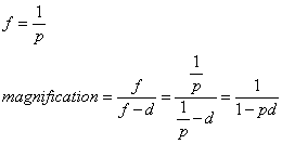
The image magnification induced by this lens is therefore the ratio of the angles ξio. For small angles, the approximation ξ = tan ξ is valid. If ξi and ξo are considered to be sufficiently small, then

However,

So,

Because the power p of a converging lens is the reciprocal of the focal length,

It has further been shown that given a contact lens of power Pc and a spectacle lens of power Ps correcting a given eye, the image magnification of the contact lens relative to that induced by the correcting spectacle is

Thus, as the distance of a convergent correcting optic device from the eye increases, so does the induced image magnifications6 An artificial lens placed in the posterior chamber would theoretically induce no image magnification. As the site of the IOL moves forward to the pupillary plane or the prepupillary space, the induced image magnification increases to the range of 3% to 4%.4,8

OPTIC METHODS OF CORRECTING APHAKIA

Spectacle correlation of aphakia is associated with multiple optic disadvantages, as has been previously noted. Neither contact lenses nor IOLs have these disadvantages, and so in that sense both are optically superior methods of correcting aphakia. Both contact lenses and spectacles can be fitted to provide an essentially exact refractive correction of spherical and regular astigmatic errors. Patients can be evaluated after surgery, and the contact lenses or spectacles can be repeatedly modified as necessary. The power of an IOL, conversely, is determined before cataract extraction, and both the patient and the surgeon must accept the accuracy of the result.

CALCULATION OF INTRAOCULAR LENS POWER

With the advent of easy and accurate ultrasonic determination of the axial length of the globe, multiple methods of preoperative calculation of the necessary IOL power have been introduced, including computer programs and nomograms.9–23 The lens power can be calculated to result in emmetropia or iseikonia (with the necessary accompanying ametropia). Postoperative results have been reported with 1 D of the preoperative calculation in 88% and 91% of eyes.19,20 Generally excellent lens power predictions are found with various formulas.24,25

With a known value for anterior chamber depth (ACD), it is possible to calculate the desired power of the IOL.

  a = axial length of the globe in meters
  k = ACD
  Pc = refractive power of the cornea
  Pl = refractive power of the intraocular lens
  n = index of refraction of the aqueous and vitreous

At the vertex of the cornea in such an eye, light must have a vergence of 1/a to cause the light to reach a focus in distance a, corresponding to the position of the retina. Similarly, at the papillary space, the required focal length is a - k. Because of the refractive index of the media, the reduced focal length of (a - k)/n must be used. Thus, the required vergence at the pupillary space is n/(a - k). However, the vergence contributed by the cornea must be taken into consideration. A cornea of power Pc contributes, at the pupillary plane, a vergence of

Once again it is necessary to use a “reduced” distance of kÞshn owing to the index of refraction of the media.6

The vergence that remains to be supplied at the pupillary space is provided by the IOL.

This formula was used to determine the IOL power of lenses that were inserted into 150 eyes.20 In 136 eyes, the postoperative refraction was within 1 D of the value calculated preoperatively; in 12 eyes, the refraction was within 1.1 to 2 D, andin only two eyes was the difference greater than2 D. Thus, with the use of accurate methods (e.g., ultrasonography) to measure the axial length of the globe, estimates of the required IOL power can be made with some degree of accuracy.21,26–28 Hoffer has tabulated the accuracy of various formulas in predicting emmetropic IOL power.29,30

These so-called first-generation formulas assumed an ACD that was independent of the sizeof the eye. Various theoretically derived first-generation formulas for emmetropic IOL power have been examined graphically and algebraically.31–33 With the exception of Binkhorst's formula, which predicts a value 0.5 D higher than the others, all the theoretically derived formulas are reducible to the same equation.4,31–33

where g(t) is a function of lens thickness and is usually a small value. If lens thickness is not considered (“thin lens assumption”) and g(t) = 0, the previous equation reduces to the earlier derivation.

Back to Top
REGRESSION ANALYSIS

INTRAOCULAR LENS FORMULA

Sanders, Retzlaff, and Kraff Formula

Optic accuracy of IOL implantation depends on the accuracy of the formula used preoperatively to select the lens power. Several investigators have looked retrospectively at the results of larger numbers of IOL implantations in an attempt to approach the problem of predicting IOL power through an empiric approach.34–36 Their data were subjected to multiple regression analyses and mathematical curve-fitting techniques. They found that the power of an IOL could be best predicted by a formula of the form

where A, B, and C are constants for a given lens. Interestingly enough, no significant further accuracy was obtained by adding additional terms representing other factors, such as ACD. (One investigator substituted the actual measured postoperative ACD in the theoretic formula and only improved accuracy 3%.46 This tends to support the conclusion that ACD is only minimally important in IOL power calculation.)

Any additional accuracy was also not obtained by inclusion of multiplicative terms (such as axial length × keratometry) to account for any possible interaction between variables, nor by inclusion of exponential terms (e.g., axial length squared) to account for any nonlinear characteristics in the mathematic model of lens power.34,35 These investigators concluded that the opticophysiologic relationship between IOL power, corneal curvature, and axial length is linear (or rather planar) and that therefore their empirically derived linear formula must be more accurate than the nonlinear theoretical formulas. They point out that many theoretic formulas, for example, tend to overestimate IOL power for axially short eyes, the area in which the theoretic and linear formulas tend to diverge most markedly (Fig. 2). Using a theoretically derived lens formula, Lindstrom and Shammas have noted decreased accuracy with high or low axial length measurements. Their calculated lens was too strong in the short eye and too weak in the long eye.38 Others have also noted this discrepancy between the two types of formulas for long or short eyes.39 Conversely, in one study of 40 myopic eyes that required an IOL of 13 D or less, both the theoretic and Sanders, Retzlaff, and Kraff (SRK) formulas were comparable.40

Fig. 2. The relationship between the IOL predicted by a theoretic formula and a regression analysis-derived linear Formula. Note how the curves separate for extreme values, particularly for shorter axial lengths, at which the theoretic formula tends to predict a significantly stronger IOL power. (Courtesy of Dr. Donald R. Sanders.)

Sanders, and Retzlaff and Kraff, in separate studies, further simplified the regression analysis-derived formula.35,36 It was found that there was a negligible loss of accuracy if the constants B and C in the original formula were universalized for all lenses and manufacturers. B, the coefficient of axial length, was calculated at -2.5, and C, the coefficient of the average keratometry in diopters, was determined to be -0.9. Thus the power of any IOL may be determined by measuring the axial length and keratometry readings only and by knowing the particular A value for the lens in question. Thus, the simplified SRK formula is

This formula has been derived using the measured axial length, and no correction for retinal thickness is necessary. This formula is easier to use than the theoretically derived formulas and can even be calculated by hand with little difficulty. It requires neither a measurement nor an estimate of the preoperative or postoperative ACD or depth at which the lens is to be placed.

The SRK formula was said to be superior to the early theoretic formulas in having a smaller average error per case, a smaller range of error from thehighest minus to the highest plus, and a smaller proportion of cases with greater than 2 D of error.34–36,41Proponents of the theoretic formulas have countered that the difference between immersion versus contact methods of ultrasonography for determining the axial length may be a factor.

Contact methods of A-scan ultrasonography may indent the cornea and result in a falsely lowered axial length measurement.29,31,32,38 This may be one of the reasons for the increased error of the theoretically derived formulas' predicting too-strong lenses for axially short eyes. Nevertheless, in a 1999 survey, immersion A-scan ultrasonography (compared with applanation ultrasonography) was still used by only 5% of responders.42 Another reason for this error in such eyes may be the assumption of a standard ACD for use in the theoretic formulas. Shorter eyes have shallower ACDs.43,44 Use of a standard or average value for ACD in such cases may also contribute to the prediction of too-strong lenses, because the predicted power of the IOL increases with increasing ACD, all other factors remaining constant.

Second- and Third-Generation Intraoperative Lens Formulas

Although the so-called first generation IOL formulas were useful, they were less accurate for long or short eyes. These formulas assume a constant ACD (or position of the IOL) in all eyes, regardless of their axial length. Shorter eyes have shallower ACDs.43,44 Use of a standard or average value for ACD in such cases contributes to the prediction of too-strong lenses, because the predicted power of the IOL increases with increasing ACD, all other factors remaining constant. The measured postoperative ACD was found to be directly proportional to the axial length of the eye; longer eyes had larger ACDs. Several “second generation” theoretic formulas emerged that replaced the constant ACD with one that included a correction for axial length.45

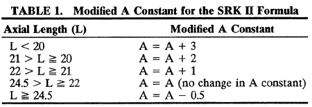
The SRK formula was also modified to improve accuracy for short and long eyes and reemerged as the SRK II formula. This was a simple modification of the original SRK.46 The only change was that the A constant is modified according to the axial length of the eye (Table 1).


Note in Figure 2 that this modification moves the SRK curve closer to the curve of the first-generation theoretic formula. To find the IOL power (1) required to produce a particular postoperative refraction R, I = P - (R × CR), where P is the predicted emmetropic IOL power and CR = 1.25 if P is greater than 14, CR = I otherwise.

Holladay and colleagues have noted that the axial-length versus calculated power graphs for second-generation formulas, both theoretic and regression derived, converge into the same general area.45 Although some have found the newer theoretical formulas to be superior to the SRK47 another group compared the SRK II and four second-generation theoretic formulas.48 They found that the differences in predicted implant powers (in eyes of “normal” size) were smaller than differences due to variations in axial length and keratometer measurements. In addition, significant differences exist in results obtained by different surgeons, thus supporting the idea of modifying the formulas for each individual surgeon, either by modifying the A constant49 or by use of a personalized “surgeon factor” in theoretical formulas.45

The originators of the SRK formula compared regression analysis-derived and theoretic formulas for calculating aphakic spectacle refraction.50 Unlike the situation with pseudophakic formulas, they found the two aphakic formulas approximately of equal accuracy. However, they believed that the SRK-like formula was easier to use.

Thus, with second-generation formulas, ACD was no longer a constant in all eyes but rather varied with axial length. However, IOL power calculations continued to improve. Holladay and associates were the first to consider that the ACD might vary not only with the axial length but also with the corneal curvature.51 Their formula modified the ACD based on the axial length, and also based on the corneal height (distance from the cornea to the IOL's first principal plane). This formula was shown to be significantly more accurate than previous theoretic formulas and the SRK II.52

Hoffer also developed a third-generation IOL formula.53 He speculated on the relationship between ACD and axial length and developed an expression that resulted in an S-shaped curve that fit his impression of what this relationship should be. This formula deepened the ACD with increasing axial length and deepened the ACD with increasing corneal curvature. This modification of ACD, tacked on to his previous Hoffer formula, has become known as the Hoffer Q formula.

The originators of the SRK formulas brought their retrospective analytic approach to third-generation IOL calculations.54 They hoped to combine the theoretic advantages of a physiologic optics approach (which might be more accurate in long or short eyes, out of the range of the database from which the SRK formulas had been developed) with the advantages of retrospective data analysis. They studied a third-generation theoretic approach to IOL calculation and tried to determine which values, correlations, and adjustments gave the most accurate result. They tried to consider all or most of the factors that had been used as modifications in various theoretic formulas. Their formula predicts postoperative ACD based on axial length. It includes a correction for long eyes. They developed a corneal height formula and an expression that predicted the white-to-white corneal width (which had not measured in their data.) They developed a retinal thickness correction to axial length. They studied what the optimum index of refraction was for IOL calculation. They showed how the SRK “A” constant could be correlated with the ACD value. The resulting formula was called the SRK/T (for theoretic) formula. As input, it requires only those same variables as earlier SRK formulas: the corneal curvature, axial length, and IOL A constant. They next compared the SRK/T with the SRK II, the Holladay, Binkhorst II, and Hoffer (note: this was not the Hoffer Q, which had not yet been developed). They found that all formulas had roughly the same accuracy, except for eyes of axial length exceeding 28.4 mm, in which the SRK II became significantly less accurate.

Hoffer compared the accuracy of three third-generation formulas: the Hoffer Q, the SRK/T, and the Holladay. He found all were superior to pre-vious formulas, including the SRK II. There wasno significant difference among the three third-generation formulas, although there was a tendency for the Holladay formula (and in a later study, the SRK/T as well55) to be more accurate in eyes of 24.5 to 26 mm, the SRK/T in eyes of over 26 mm, and the Hoffer Q for eyes under 22 mm. For eyes in the range of 22 to 24.5 mm, Hoffer recommended using the average of all three formulas. One investigator compared formulas for microphthalmic eyes less than 19 mm long.56 He found the SRK/T to be best overall, although all predicted too weak a lens (and postoperative hyperopia) In a study of IOLs in pediatric patients, the three third-generation formulas and the SRK II were compared.57 No difference among the four formulas was statistically significant.

To improve accuracy in short, hyperopic eyes, Holladay further modified his formula by including consideration of white-to-white corneal diameters, and anterior chamber and lens thickness measurements, as well as the patient's age and preoperative refractive error. This Holladay 2 formula was introduced into clinical use in 1996 but has yet to be published though it is available in some software packages. In one study, this Holladay 2 formula, and use of immersion A-scan measurements, resulted in improved accuracy for short eyes.58 Another retrospective analysis of 317 eyes was made and four formulas were compared: the Holladay 1, the Holladay 2, the Hoffer Q, and the SRK/T.55 This study found the Holladay 2 and Hoffer Q were (equivalent and) best in short eyes, the Holladay 1 and Hoffer Q were (equivalent and) best in average eyes, the Holladay 2 and SRK/T were (equivalent and) best in medium long eyes, with the SRK/T producing a trend toward better results in very long eyes. The Holladay 2 formula showed a “trend towards poorer performance in most axial lengths tested (89%).” Especially considering that theHolladay 2 formula requires the additional input of preoperative ACD, lens thickness, corneal diameter, patient's age, and preoperative refraction, this investigator did not recommend its inclusion in the optimal IOL power calculation armamentarium.

The SRK II formula is certainly far easier to use than any of the third-generation formulas; calculations can be made quickly with pencil and paper. Conversely, however, third-generation formulas appear to be more accurate, especially in long and short eyes. Computing technology that calculates IOL power using these formulas is widely available in software packages, handheld devices, and even has been incorporated into many ultrasound A-scan units.

A nonformula approach has been described that might be useful with children or in other patients in whom accurate measurement might be difficult (those who had undergone previous refractive surgery, colobomas, and the like).59 Following lens removal, retinoscopy may be performed, or the patient may be moved and have a subjection refraction performed. The patient is then returned to the operating room for insertion of the IOL.

FACTORS INFLUENCING ACCURACY OF INTRAOCULAR LENS POWER CALCULATION

The accuracy with which IOL power can be calculated depends on several factors.

Measurement of Axial Length

Ultrasonography is the most widely used method to determine the axial length of the eye.21,27,28 Current ultrasonographic techniques permit an accuracy or reproducibility to within 0.1 mm or less in the axial length measurement.4

Binkhorst adds 0.25 mm to the measured axial length to compensate for the thickness of the retina in the area of the sound beam.60 Some studies have found increased accuracy with the addition of retinal thickness to the measured axial length.34 Others, however, have found their calculations more accurate when specifically not including any such retinal thickness correction factor.29,30,61 In a regression analysis of this question, retinal thickness corrections from 0 to 0.5 mm were tested and found to be approximately equivalent in accuracy.54 However, it was noted that in long eyes, smaller correction values were most accurate, whereas in shorter eyes, larger values were better. Optimal values ranged from 0.29 mm for an 18-mm eye to 0 mm for a 32-mm eye. One explanation for this finding is that it may be due to the vagaries of ultrasound measurement; a shorter eye may have, in effect, a faster average speed of sound transmission because a higher percentage of its travel is in the crystalline lens. This would result in a falsely shorter measured axial length, for which the “retinal thickness factor” corrects.

An error of 0.1 mm in the axial length measurement will result in a postoperative refractive error of 0.25 D.4 Axial length measurements may differ depending on the machine used to make the measurement.62

It is also possible that the axial length of the eye may change as a result of the surgical manipulations. This quantity is somewhat variable and unpredictable. One group of investigators found an average postoperative increase in axial length of 0.06 mm,18,27 whereas a second group reported an average decrease in axial length of the same amount. Hoffer found no significant axial length difference between phakic and aphakic eyes.43 Others have found no significant change in axial length in eyes measured after cataract removal and 1OL implantation.37,63

One group noted an average increase of axial length postoperatively of 0.11 mm.64 However, when the measured postoperative axial length value was substituted in the preoperative measurement, they found no increased accuracy and concluded that the measured preoperative axial length was appropriate to use in 1OL formulas.

It is thus impossible at the present time to consider postoperative changes in the axial length of the globe in preoperative calculations of IOL power.

Simplifying assumptions are involved in the use of ultrasound to measure the axial length of a phakic eye. The actual velocity of sound through the crystalline lens may vary with the degree of cataractous change, becoming higher as the lens becomes more cataractous.30,38,65 An unusually high intratissue velocity of ultrasound in a very cataractous lens, for example, may lead to decreased cornea-retina transit time for the ultrasound beam and a falsely low axial length measurement. Hoffer has measured each component of the axial length individually, with the A-scan ultrasound unit reset for the particular speed of ultrasound in each tissue in the path of the ultrasound beam. (An average or estimated velocity of ultrasound in the lens must be used.) When compared with a single measurement with the A-scan unit set for an “average” tissue velocity for the entire eye, however, the more complex method was, if anything, less accurate.30 One investigator compared the accuracy of axial length measurements in phakic versus aphakic patients (for primary versus secondary IOL implants).52 There were no significant differences between the two groups. This implies that any inaccuracy that may be introduced as a result of the variability in the speed of ultrasound in cataractous lenses is not significant.

Others have studied the ultrasound characteristics of cataracts.67 They found that the attenuation of ultrasound was related to the degree of lens hardness; the speed of ultrasound, however, was not significantly different in lenses of different degrees of “hardness.”

The speed of ultrasound used to determine axial length is an approximated average of the speed in the crystalline lens and the speed in the rest of the eye.68 Ultrasound travels at 1532 m/sec though the aqueous and vitreous, and at 1641 m/sec through the cornea and cataractous lens. Adjusting forthe proportion of the eye transmitting at eachspeed yields an average speed through an eye of1555 m/sec. However, this approximation may be less accurate in long or short eyes. In a short eye, for example, a higher proportion of the distance traveled is lens, through which the ultrasound travels faster. This would result in a shorter transit time and a lower measured axial length. Thus, the short eye may be measured as falsely short and the long eye may be measured as falsely long. This error is compounded by another study that revealed that long eyes have thinner lenses (4.5 mm) and very short eyes have thicker lenses (5 mm).69

Corrections to axial length measurement based on the axial length have been presented.68,70 Ultrasound travels through aqueous and vitreous at the same rate, 1532 m/sec. The speed through the corneal and cataract is 1641 m/sec. The “average” speed of sound through the phakic eye is considered to be about 1550 to 1555 m/sec, but obviously this number increases for short eyes (in which a greater proportion of the distance is within the cataract) and decreases for long eyes, which have relatively more aqueous and vitreous. Hoffer and Holladay have suggested that axial length measurement can be made more accurate if these factors are taken into consideration, and this may be especially important in short eyes. Holladay suggests measuring the axial length as if there were no lens present, using an ultrasound speed of 1532 m/sec. (If the A-scan is set at a different ultrasound speed, conversion is simply done. Suppose the A-scan were set at the “average” speed of 1553 m/sec. Multiply the thus measured axial length by 1532/1553 to get the lower axial length that would result if the speed of speed of sound were less because of the absence of the lens.) To this add 0.28 to account for the higher speed of sound in the lens and the resulting shorter-than-actual measurement (Hoffer suggests adding an additional 0.037 for a net add of approximately 0.32, to account for the increased speed of sound in the cornea as well (personal communication 2000) to get the true ultrasonic axial length. (In an aphakic eye, one needs to add only 0.037 for the cornea because there is no lens; such correction may be clinically insignificant in practice.) Using this axial length in a (theoretic) power formula may increase accuracy) The SRK/T formula may already include corrections to maximize IOL accuracy with the A-scan axial length as usually measured and thus may already incorporate this (or a comparable) correction.

Modifications are also available for aphakic or pseudophakic eyes.

When measuring the axial length of a pseudophakic eye, the IOL material must be considered. Because sound travels far more slowly in silicone than in polymethyl methacrylate (PMMA), an apparent axial length increase of 1.045 mm in eyes with silicone IOLs may be noted if this difference in ultrasound speed is not considered. The average speed of sound in an eye with a silicone IOL is 1476-1486 m/sec, compared with 1548 to 1555 m/sec in an eye with a PMMA lens.68,71 Such estimations vary, of course, with the size of the eye, the power of the lens, and perhaps even the body temperature. Similar problems may occur when measuring axial length of a postvitrectomy eye filled with silicone oil. An artifactually longer measurement may be obtained if the speed of sound in silicone oil is not used.72 Some investigators have used 987 m/sec as the speed of sound in 1000 centistoke silicone oil68,73 One approximated the axial length of silicone-filled eyes using the above correction, then remeasured the eye following removal of the silicone oil.73 The error in the axial length was less than 1%. Silicone oils of different viscosities will have different sound speeds and correction factors.

Ultrasound velocities for eyes containing silicone oil or various IOL materials have been suggested.68

Conversely, falsely low axial length measurements have been reported in an eye with asteroid hyalosis as a result of confusingly strong echoes from the midvitreous.74 Others have confirmed falsely short axial length measurements in this situation and have found it more accurate to use the axial length measurement of the non-involved fellow eye for IOL power calculations.75

The technique of ultrasound A-scan measurement that is used may also be a factor.30–32 It has been suggested that applanation methods of axial length determination indent the cornea and falsely shorten the axial length measurement.29,31,32,76 Measurements of axial length made with an applanation technique were, on average, 0.1 mm to 0.32 mm less than measurements made with an immersion technique.77,78 One investigator took multiple axial length measurements. He compared the accuracy of using the average reading compared with the longest value. Although there were similar results in the two groups, he suggested that the longest reading may be the single most accurate, because shorter values may result at least in part from corneal indentation.79

Posterior staphylomas may be present, particularly in longer eyes. One group used B-scan ultrasound to better avoid anomalous A-scan echos from vitreous debris and to avoid staphylomas. They found a nonstatistically significant increase in the accuracy of the resultant refraction.80 Others have also noted decreased IOL accuracy in long eyes, particularly in those requiring negative power IOLs.81 Their eyes over 27 mm were over-minused by 1 to 4 D, leaving the eyes with postoperative hyperopia. They recommend concurrent B-scan of all eyes longer than 27 mm.

A-scan axial length measurements showed less variability on retesting than ACD measurements did.78

It may be prudent to measure both eyes routinely; a malpractice decision in Louisiana faulted the ophthalmologist for not taking comparison axial length readings of the other eye as a check against erroneous measurement.82

Partial coherence interferometry is a new technique for measuring axial length.83 It is a method that does not require direct contact with the tissue being investigated. It measures echo delay and intensity using infrared light reflected back from internal tissue interfaces. Given that the velocity of light is too high to measure echo delay times directly, interferometric methodology must be used instead. This technique provides more precision and reproducibility than ultrasound. The patient must fixate a target; this thus ensures that the optic axis is being measured. The clinical usefulness of this technique is still undetermined. It is likely that a formula such as the SRK/T that already includes corrections for the vagaries of ultrasonic axial length measurement will need reformulation for axial length measurements made in this new way.

Other kinds of eye surgery may affect the axial length of an eye. One study noted a decrease in axial length following trabeculectomy.84 These were more pronounced in cases in which a large decrease in intraocular pressure was achieved (30 mm Hg) and in which antimetabolites were used. An average decrease of 0.46 mm was seen in these cases, at least partly due to an increased thickness of the choroid. Conversely, if these eyes then undergo cataract surgery, there may be an increase in axial length if the intraocular pressure rises. An average increase of 0.275 mm was noted in these cases. They suggest that a pretrabeculectomy axial length be used for IOL calculations rather than posttrabeculectomy axial length.

Scleral buckle surgery may result in an increase in axial length, resulting in a lower IOL power required.85

Measurement of Corneal Curvature

It has been stated that if the average of three readings in each meridian is used, the error is negligible.4 Errors in keratometry have been noted after wearing hard contact lenses.86 Even 2 weeks after use of hard contact lenses, patients had an increase of0.79 mm in corneal measurements, resulting in an average decrease of 0.98 D in calculated IOL power. A malpractice case in Louisiana faulted the ophthalmologist for taking preoperative measurements on a patient who had only just removed his hard contact lenses on arriving at the office.82 The patient was not instructed to remove his contact lenses for a long enough period (speculated to be between 2 hours and 2 weeks) before keratometric measurements were taken.

There may be a postoperative difference in the average keratometric readings (usually a slight flattening of the cornea secondary to the surgical manipulations.87 An average increase in the radius of curvature of approximately 0.08 mm (approximately 0.5 D flatter) has been noted after cataract extraction.87 Binkhorst compensates for this slight flattening by underestimating the index of refraction, using n = 3 rather than 1.336.4 Other studies have revealed a postoperative steepening of thecornea, of 0.16, 0.11, and 0.14 D, respectively.34,38,76Hoffer compared large numbers of phakic, aphakic, and pseudophakic eyes.43 He found a corneal flattening of 0.16 D in aphakic compared with findings in phakic eyes. Interestingly enough, no significant corneal flattening was noted in the pseudophakic eyes. Others have found no significant change in keratometric measurements following cataract removal and IOL implantation.63,88 Following scleral buckle surgery, the corneal curvature may increase.85 An error of 0.1 mm in radius of corneal curvature results in a postoperative refractive error of approximately 0.50 D.4 In otherwise normal patients, computerized videokeratography was, if anything, inferior to standard keratometry (K) in obtaining accurate measurements.89

The problem of selecting which K reading to use is more complex if penetrating keratoplasty is to be combined with IOL implantation. One group found no correlation between preoperative and postoperative K readings.90 Nevertheless, their results were more accurate using preoperative K readings or even using K readings from the other eye, compared with selecting an 1OL power without using a K value from either eye. Another group also noted that penetrating keratoplasty produced marked changes in postoperative corneal curvature and power.91 They found it impossible to develop a single formula for all surgeons to predict IOL power accurately in these cases. Results were quite different from surgeon to surgeon, perhaps owing to differences in surgical technique and suture patterns. In the analysis of results, it was found that the axial length was the single most important factor contributing to the prediction of IOL power. Interestingly enough, the preoperative K reading figured into an SRK-like formula as a positive term; that is, the higher the K reading, the stronger the IOL that was needed. This was explained by the negative correlation, in this series, between preoperative and postoperative keratometry.

Another group suggested using 106% of the K reading of the donor, or an arbitrary value of 45 D (for a 7.5-mm graft placed in a 7-mm recipient bed).92 They also noted no significant change in axial length after surgery and pointed out that an alternative is the deferral of IOL insertion until a second procedure, delayed until after the new K readings have stabilized. Another group obtained best results using the average postkeratoplasty K readings for IOL calculation.93,94 The average K readings 1 year after transplantation were similar, regardless of the differing preoperative K readings.94 The Binkhorst, SRK, and personalized regression formulas all gave similar accuracy.94

Patients who have undergone corneal refractive surgery present unique cataract surgery problems. One group reported four patients undergoing IOL implantation after previous radial keratometry.95 Standard keratometric measurements tended to overestimate the true corneal power, resulting in an underestimation of the required IOL power, possibly because the keratometer may be measuring a more peripheral portion of the cornea that does not fully reflect the flatter central cornea. The Binkhorst and Holladay theoretic formulas were more accurate than the SRK.11 The most accurate corneal curvature to use for IOL calculation was obtained by taking the prerefractive surgery K readings and subtracting the surgical refractive change from that. One investigator found superior IOL accuracy by using computerized videokeratography to measure corneal powers after radial keratotomy.96

After IOL implantation, postoperative corneal flattening was noted, some of which persisted. Severity of this flattening was inversely proportional to the time interval between radial keratometry and cataract surgery, possibly because long-term healing of the radial keratometry incisions may partially blunt the edema-induced hyperopic shift that occurred after IOL surgery.

After photorefractive keratectomy (PRK) for high myopia, the measured postoperative corneal K readings were not most accurate for IOL power calculations96,97 Refraction-derived measurements were obtained by using preoperative K readings and correcting them for the measured change in refraction (at the corneal plane) These numbers proved to be more accurate for IOL calculations.

Hoffer has contrasted four ways of measuring corneal power in eyes that have had refractive surgery.99 The first is standard keratometry. This may measure corneal power at a 3-mm distance from the corneal center and may not fully represent the new more central modified zone. The second is using the refractive history and preoperative K readings. Modify the preoperative K readings by the refractive change postoperatively (at the corneal plane) and use this as the K readings for the IOL power. The third is the contact lens overrefraction method. Take the postoperative refraction, then place a contact lens of known power on the eye and rerefract. Use the base curve of the contact lens for IOL calculation, modified by any change in the manifest refraction and corrected for the power, if any, in the lens. (Another investigator100 points out that this overrefraction method has the advantage of detecting visual loss due to irregular astigmatism rather than cataract, which could conceivable alter the decision to perform cataract surgery in the first place!) The fourth method is to use videokeratography and measure more centrally than is usually possible with standard keratometry.

One study found that direct measurements ofthe cornea after PRK underestimated corneal flat-tening by 24%.101 Another investigator had a de-centered ablation zone after PRK that resulted ina too-steep K reading.102 They recommended usingthe spheric equivalent refractive change (at thecorneal plane) to obtain corneal K readings for IOL calculation. Another investigator had a decentered ablation zone that resulted in a too-steep K reading.

Measurement of Anterior Chamber Depth

The depth of the anterior chamber can be measured by instruments that can be incorporated into some slit lamps (pachometry); alternatively, it can be calculated indirectly by the method of Fyodorov and associates.20 The depth of the anterior chamber has been noted to decrease progressively with age at the rate of approximately 0.1 mm per decade, probably owing to progressive swelling of the lens.4,44 ACD is also related to the degree of ametropia.44 The anterior chamber is 0.06 mm deeper for every diopter of myopia. Thus, an average -6 D myopeis likely to have an ACD 0.6 mm deeper than a+ 4 D hyperope, all other factors being equal. A similar increasing ACD with increasing myopia has also been noted by Hoffer.43

An error of 0.1 mm in estimating the ACD results in a postoperative refractive error of approximately 0.1 D.4 Increasingly, however, the trend has been against measuring ACD values for each patient. It has been emphasized that what the lens formula requires is a predicted postoperative value for ACD (or lens position) rather than the measured preoperative value.103 Hoffer's results for two-loop prepupillary lenses were not as accurate using the measured ACD in his calculations as were those measuring the ACD and using one of three predetermined values.30 Shammas compared preoperative and postoperative ACD in eyes with anterior chamber lenses and concluded that there was no significant difference in postoperative ACD among eyes classified into several different ranges of preoperative ACD measurements.104

Hoffer measured the position of the posterior capsule preoperatively and after extracapsular surgery in 30 patients.30 He found no correlation between the preoperative and postoperative measurements. Realizing that the important measurement is not the ACD in itself but rather the postoperative position of the IOL, Hoffer measured the position of the posterior capsule (supporting a posterior chamber lens) and analyzed factors that could be used to predict it.105 There was no relationship between the position of the posterior capsule and the patient's age or corneal keratometry. There was no relationship between the preoperative position of the posterior capsule and the axial length. However, there was a correlation between the posterior capsule position postoperatively (postoperative ACD) and the axial length L. Hoffer stated that the ACD used to calculate IOL power should reflect the actual postoperative lens position.

Another group measured the distance between the cornea and posterior lens capsule in 60 eyesbefore and 3 months after posterior chamber IOL implantation.106 They found the postoperative posterior capsular position to increase with patient's age, ACD, and axial length.

In contrast to the case of a posterior chamber lens, another investigator applied the techniques of regression analysis to study the postoperative position of an anterior chamber lens.107 For an anterior chamber lens, the postoperative position (postoperative “ACD”) was not correlated with the axial length but was related to the preoperative ACD, corneal curvature and diameter, and thickness of the cataract.

There are other sources of error as well. First, the thickness of the cornea is discounted in all these calculations, and any refraction that occurs at the cornea-aqueous interface is not considered. Second, the IOL is considered as a thin lens, whereas actually the two principal planes of the IOL do not coincide. Light may be thought of as entering the lens at its first principal plane and leaving at the second, with no angular change in the light rays (unit magnification) in the interim.108 Thus, the measured axial length of the eye should be reduced by an amount equal to the distance between the two principal planes. For a convexoplano lens, this distance between principal planes is one tenth of the lens thickness (e.g., 0.05 mm in a 0.5-mm thick lens,0.08 mm in a 0.8-mm thick lens).4

The ACD is usually determined as the distance between the vertex of the cornea and the vertex of the lens. More rigorously, the measurement should reflect the distance between the corneal vertex and the first principal plane of the lens.

Finally, it is possible that the surgical technique itself may have an effect on the postoperative ACD. It was noted that the ACD may be related to the size of the capsulorrhexis. At 90 days after surgery, eyes with a 4-mm capsulorrhexis had a greater anterior chamber than eyes with a 6-mm opening.109 The larger capsulorrhexis may allow more anterior bowing of the IOL and a consequent shallower ACD.

Lens fixation and orientation may have an effect on the ACD. Introduction of capsulorrhexis and more stable fixation in the bag was shown to result in more stable anterior chamber positioning and more accurate IOL power predictions.110 One investigator compared approximately 50 patients with standard in the bag lens fixation, 50 with planned out of the bag IOLs after previous extracapsular surgery, and 50 patients with transscleral suture-fixated IOLs. The IOL was noted to be more anterior with out of the bag and scleral suture-fixated IOLs compared with in the bag fixation.111 This resulted in a myopic shift in the refractive result. In addition, IOL decentration, which occurred with scleral suture fixation, also contributes to a post-operative myopic shift. IOL tilt of greater than 10 degrees was seen in 11.5% of the suture-fixated IOLs; such tilt results in an increase in effective IOL power and contributes to a postoperative myopic shift. These authors recommended dropping0.5 D from the calculated IOL power in cases in which planned sulcus fixation (out of the bag) or scleral suture fixation is planned.

Later YAG capsulotomy may change the effective ACD.112 Capsulotomy caused a backward movement of the IOL, more pronounced with plate haptic IOLs than with one-piece PMMA and three-piece foldables. The resultant hyperopic shift was not clinically significant.

Back to Top
TYPES OF INTRAOCULAR LENSES

PIGGYBACK INTRAOCULAR LENSES

An eye with a shorter axial length requires a stronger IOL. As IOL powers become too high, however, the lens becomes too spheric and image quality decreases. Using two lenses to obtain a needed high-power correction results in an optically superior image compared with that obtainable by a single lens of required power.

A mathematic analysis of image quality of piggyback lenses was performed.113 The axial image quality of piggyback lenses was superior as determined by modulation transfer function analysis, especially at low and middle spatial frequencies, which are those used in daily visual tasks. There is also a greater tolerance for manufacturing and positioning errors with the piggyback technique. Both single lens and piggybacking, however, achieved theoretic Snellen vision of 6/5 (with theoretic optimal lens shapes, which may not necessarily conform to the lenses commercially available.)

Thus, for short or nanophthalmic eyes, so-called piggyback IOLs represent a viable solution.114Holladay implanted six such lens pairs in eyes ranging from 15 to 20 mm in axial length.115 All three third-generation formulas resulted in postoperative hyperopia of about 5 D; the SRK II resulted in an error of about 11 D. This may have occurred for several reasons: First, error of axial length measurement is especially damaging in shorter eye calculations; a given error is higher proportionately in a smaller eye. Furthermore, errors in axial length translate to larger IOL power differences as the IOL powers increase. Second, all third-generation formulas shorten the expected ACD as a function of axial length; for very short eyes, they all predict the ACD to be too shallow and the IOL to be too far from the retina, leading to the calculation of a too-weak lens. Third, the position of the IOLs may be changed. In a piggyback lens system, measurements showed that the anterior IOL was in the normal position, whereas the posterior one was more posterior (thus the more posterior lens was “too weak” for its position). Holladay also points out that we should distinguish the normal short eye, with a normal anterior segment but a short posterior segment, from the nanophthalmic eye with symmetrically short anterior and posterior segments. He proposed modifying IOL calculation in these cases to include determination of the size of the anterior segment, using the white-to-white corneal diameter, ACD, and lens thickness as well ad keratometry and axial length.

Piggyback minus lenses have been used in eyes of long axial lengths alone or combined with high corneal curvature116 to achieve minus powers commercially unavailable in a single lens.

Piggyback IOLs may also be inserted in two stages, with the second IOL inserted (in the ciliary sulcus) to correct an undesirable refractive result following the initial IOL implantation. If the second lens is placed posteriorly (by surgical error), the resulting positions are unstable and unpredictable and the refractive results are less predictable.117

Refractive results in adding a piggybacked IOL as a second procedure should be superior to that of an IOL exchange for several reasons.117 First, the power of the second IOL is based only on the postoperative refraction. Second, the surgeon cannot be certain that an exchanged IOL would be in exactly the same plane as the IOL it replaced. Third, the accuracy of the power labeling of the first IOL is irrelevant, because of the first reason. Fourth, these added IOLs tend to be of lower power, and vergence-distance changes are less important with lower power lenses.

One occasional curious finding in piggyback lenses is an apparent increase in the depth of focus. This may result from contact between the two lenses118 with a slight flattening in the zone of contact. This would result in a decrease in the optic power of the IOL with resultant hyperopia. The power in the peripheral lens area, however, would be unchanged. In three eyes with acrylic IOLs, a difference was seen of 3.0 to 5.5 D between the central contact zone and the peripheral noncontact zone.119 Obviously, spectacle correction of the resulting hyperopia would be needed to allow the patient to take advantage of the increased depth of focus, unless the patient happened to be several diopters overcorrected to begin with. Late hyperopic shifts have similarly been noted in piggyback lenses.120 An alternate reason for this could be a posterior displacement of both IOLs, but to explain the observed hyperopic shift a movement of as much as 1.5 m would be necessary.

TORIC INTRAOCULAR LENSES

It may be possible to correct a part of a preexisting regular astigmatism with an IOL. A cylindric correction of 2 or 3 D was incorporated into IOLs implanted in 47 patients.121 This corresponded to acylindric effect at the spectacle plane of 1.33 or1.98 D. Another investigator implanted lenses with 2, 3.5, or 4 D of toric correction in 37 eyes and found the refractive effect to be 60% to 80% of the power at the IOL plane.122

Some 130 eyes had toric IOLs implanted.123 These eyes had at least 1.5 D of both corneal and refractive cylinder. A 2.00-D toric lens was used for cylinder of 1.5 to 2.25 D, and a 3.5-D cylinder was used for eyes with between 2.25 and 3.5 D of preexisting cylinder. These eyes were compared with 51 similar eyes who were instead treated with spheric IOLs and limbal relaxing incisions. There was no significant difference in postoperative uncorrected vision, although there was a trend to better vision in the toric IOL group. Respectively, 84% and 69% of the toric group had uncorrected vision of at least 20/40 and 20/30, whereas the spheric IOL group had 76% and 51% at these levels. The refractive cylinder was significantly smaller in the toric IOL group. However 12 eyes (9.2%) of the toric group required a second procedure to correct rotational misalignment of the IOL (because of patient dissatisfaction and a rotation of at least 30 degrees). In fact, with the lens design used, 18% of eyes showed IOL rotation of 20 to 40 degrees, with 7% showing rotation of 40 degrees or more. The authors concluded that toric IOLs present a viable method to correct preoperative astigmatism; obviously, improvements in IOL stability will reduce the need for secondary procedures.

The optics may be somewhat more complicated than with spheric IOLs. It has been pointed out that, in deciding on toric IOLs, the clinician should rely on the keratometric cylinder and not the refractive one.124 The refractive cylinder may include toric effects of the natural crystalline lens, which will no longer be operative after cataract removal. The best way to measure the needed toricity at the IOL plane is to calculate the IOL power for each of the two corneal powers; the difference is the amount of toric correction needed at the IOL plane. Obviously, unlike the case of spheric IOLs, the correct orientation of the IOL is critical. The lens must be aligned with the preoperative cylinder. There is still a beneficial effect even if the lens rotates somewhat, as long as the lens is not more than 30 degrees off axis. Although there was some postoperative axial shift, the one-piece plate lens appeared to stabilize by 2 weeks after surgery.122

PHAKIC INTRAOCULAR LENSES

IOLs have been developed for insertion into phakic eyes. Some are designed for anterior chamber placement, angle supported125,126 or clipped onto the iris,127 whereas others are designed to be inserted in the posterior chamber in front of the natural crystalline lens.128 Some have been tried in children.129 To calculate the IOL power for a phakic eye, one needs the preoperative refraction (and vertex distance) and corneal power.130 Because this requires no assumptions of average ultrasound speed or crystalline lens thickness, its accuracy should exceed that of IOL replacement of the natural lens. For a minus power anterior chamber IOL of 10 D or less, there is no improvement in accuracy in calculating the ACD versus using a constant value based on the IOL type.

Posterior chamber phakic IOLs have been used in children for the treatment of anisometropic amblyopia.131

Back to Top
ACCOMMODATION AND INTRAOCULAR LENSES
The IOL, being of constant shape, does not change its focus (accommodate) by modifying its contour, as does the natural crystalline lens. It is possible, however, that especially in young patients, some pseudoaccommodation may occur by movement of the IOL itself.132 The study measured 45 eyes of patients aged 12 to 19. With attempted near vision, anterior movement of the IOL was measured with A-scan ultrasonography. The amount of pseudoaccommodation was greater than could be accounted for by the IOL movement alone. However, this pseudoaccommodation was significantly related to the amount of IOL movement that occurred. There was no relationship to the patient's age (in this group) or the refractive error. As would be expected, pseudoaccommodation is inversely related to pupil size.133

One type of IOL is designed to provide pseudoaccommodation through the mechanism of anteroposterior movement of the optic on near and distance vision. The IOL has a peripheral bulging ring that is supposed to push the optic forward during the effort to accommodate. A study compared a group of patients receiving such a lens with a comparable group receiving a foldable silicone lens.134 ACD was measured after cyclopentolate instillation (ciliary relaxation) and again after pilocarpine installation (ciliary contraction). The accommodating IOL showed a significantly greater axial movement; however, the difference in the measured pseudoaccommodation between the two groups was not significant.

Attempts to refill an evacuated capsular bag with injectable silicone or other deformable material has been studied in animals. This may have the potential to allow some degree of pseudoaccommodation135; however, many questions remain to be answered before it will be known whether this will be a viable technique in humans of presbyopic age.

Back to Top
OPTIC QUALITY OF INTRAOCULAR LENSES
Theoretic considerations of calculation of IOL power are appropriate and important, but they presuppose a minimal quality in the optics and labeling of the IOL. The image quality may be monitored by resolution testing in air, or by modulation transfer function tests.136 Lens powers have been found to be incorrectly labeled, leading some to suggest that a preoperative check of the lens power by the surgeon is warranted.137,138

A method has been proposed to measure the power of an IOL preoperatively, while it is still in its sterile package.139 A standard office keratometer is used. The convex surface of the packaged lens must be visible. The technique is simply to measure the dioptric power of the convex side of the lens with the keratometer. Because of differences in the index of refraction of air, aqueous, and the lens plastic, the keratometer-measured IOL power must be multiplied by 0.46 to give the refractive power of the lens in aqueous. This method assumes a planoconvex lens (with the plano side absolutely plano) and ignores any optic effects due to the thickness of the lens itself (“thin lens” assumption). Comparison of this method with more rigorous laboratory measurements of the power of selected IOLs confirms that this keratometric method is accurate to within 0.50 D. The keratometric method, of course, may be used without disturbing the sterility of the packaged IOL. (Preoperative measurement of the power of a silicone lens is complicated by the fact that its refractive power depends on temperature. A lens that is 20 D at eye temperature would measure 21.35 D at room temperature.140)

It is also possible to measure the power of an IOL after it has been implanted in an eye.141,142 The method is based on the measurement of the horizontal dimension of the corneal reflected image (Purkinje-Sanson I) and the anterior IOL-reflected image (Purkinje-Sanson III). For a biconvex lens, the reflection from the posterior IOL surface (Purkinje-Sanson IV) would also be needed. Alternatively, it is possible to take keratometer readings of the front surface of a convexoplano lens in the eye and, using appropriate tables, to determine the power of the IOL.143

Four hundred lenses from four different manufacturers were examined, their actual dioptric power being compared with their labeled power.137 Almost half were off by greater than 0.25 D, 54 were incorrect by more than 0.5 D, and 26 deviated by 1 D or more from the labeled power. Another series of 300 lenses revealed that 13% of them were 2 D or more different from their labeled power.144 One defective IOL was measured 6.66 D higher than the marked value.145 That same investigator also noted some lenses with poor quality of the fixation loops. Although accuracy of IOL power labeling is very high now, mislabeled IOL powers continue to be a possibility.146

Other types of optic defects may be inherent in some lenses, however, in addition to incorrect power specification. Of 95 lenses from ten manufacturers that were studied for their toricity or “built-in” astigmatism as well as for their maximal resolving power, many lenses had small degrees of toricity, although none had greater than 0.5 D.147 Resolvingpower varied from 313 lines/mm down to 40 lines/mm.Of these patients, 93% were able to resolve 100 lines or more, which is the proposed American National Standards Institute minimum for IOLs.

Two of the 95 lenses produced two distinct images. Lenses like these with “built-in double vision” could cause monocular diplopia in patients who receive them. Better manufacturing quality control should be able to reduce greatly the incidence of lenses with incorrect power designation, doubleimages, exceedingly poor resolution, and large amounts of toricity. These 95 lenses had been manufactured between 1976 to 1978. Another study involved 120 lenses manufactured in mid-1978 and 1979148 and additional lenses studied in 1985.149 Others have also noted better quality control in the more recent IOLs.150,151

In 1989, nine of ten lenses were within 0.5 D of labeled power, although multiple defects still were noted in the optic and haptic portions.152 Subtle surface defects on IOLs may be the result of poor finishing.153

However, even a high-quality, well-centered IOL may produce optic aberrations from factors such as positioning holes seen through a sufficiently dilated pupil.154 That this potential problem may be of clinical significance is suggested by a study of 75 eyes obtained at autopsy. In 50% of cases in which the lens was centered and in 92% of cases in which lens placement was asymmetric, an optic element such as a positioning hole was in the visual axis or within 0.5 mm of the pupillary margin (average pupil diameter 3.45 mm).155 A significantly higher percentage of positioning holes in the pupil was seen in eyes with 6-mm lenses compared with eyes with 7-mm lenses.156 The fact that the positioning holes indeed are responsible for unwanted optic images was clearly demonstrated in one patient in whom a positioning hole was exposed as a result of an iridocapsular adhesion.157 Following YAG laser lysis of the adhesion, the positioning hole was covered by the iris and the disturbing optic images disappeared.

The problem may be compounded by light scattering from positioning holes, as well as from loop insertion sites and lens edges.158 This is more likely to occur when the pupil is relatively dilated, such as at night, even if the lens is perfectly centered.159 Three factors are related to positioning hole complaints: size and reactivity of the pupil, diameter of the clear optic zone of the lens, and distance between the posterior surface of the iris and the anterior surface of the IOL (the less this distance the more of an “iris shield” exists to protect against oblique light passing through the fixation holes).160 Unwanted optic images can be eliminated almost completely by using a 7-mm optic without positioning holes.156,161,162 Partial-depth holes produced their own characteristic images and offered no advantage over full-thickness holes for reducing unwanted optic images.161

Inserting the lens backward, with the optic reversed, could theoretically result in an incorrect power because of the bowing of the lens haptics and the change in ACD. One surgeon was surprised at how little effect one such accidental malpositioning (of a biconvex lens) caused.163 He speculated that the capsular bag tended to flatten and push the IOL into a more uniplanar configuration, in which ACD effects of haptic bowing were minimal. Any ametropic that resulted was too little to necessitate a return to the operating room to correct. However, investigators have noted a decrease in effective IOL power (and consequent hyperopia in the patient) after placing a convexoplano lens with the plano side anteriorly.164,165 Given that the first principal plane of a convexoplano lens coincides with its anterior vertex, there may have been a more drastic change in the position of the principal planes in this case.

One other interesting optic hazard associated with IOLs was reported.166 During vitrectomy, the presence of an IOL was noted to cause a ring scotoma and alter the physician's depth perception during surgery. In one experimental study, adequate visualization was obtained in fluid-filled and air-filled rabbit eyes with both monofocal and multifocal IOLs.167

Back to Top
OTHER OPTIC FACTORS IN INTRAOCULAR LENS DESIGN
The quality of the optic image produced by the IOL depends, at least theoretically, not only on the power of the IOL but also on its shape and optic transmission characteristics.

LENS SHAPE

The dioptric power of a lens depends on its front and back curves (and its thickness). However, other aspects of the quality of the image formed may also be influenced by judicious selection of these parameters in IOL design and production. It has been suggested that spheric aberration may be reduced by implanting a planoconvex lens with the plano side anterior because of decreased lateral spheric aberration.168–170 (Shearing, however, could not distinguish any effective difference in the visual status between having the plano or the convex surface forward.103) Other workers, studying lens design with mathematic calculations, have concluded that a planoconvex IOL (with the plano side an-terior) most closely approximates the theoretic spheric aberration of the Gullstrand eye (although it is not the lens shape that provides the least spheric aberration.)171 Using a different mathematic approach, another group concluded that the spheric aberration of an IOL cannot be made 0, only minimized.172 They believed that the optimal lens shape was almost convexoplano, with the more curved surface anterior. The spheric aberration of an IOL is potentially less than the aberration from the cornea. Taking into account corneal asphericities, on-axis performance of centered IOLs was optimized with shapes near convexoplano, or biconvex, with the more curved surface anterior.173

Investigators have used computer ray tracings to study IOLs of various shapes.174 They concluded that spheric aberration could be almost completely eliminated with an asymmetric biconvex IOL, with the stronger curvature facing the retina. More total refractive power is needed, of course, when the curvature of the IOL is stronger on the side facing the retina, owing to changes in the position of the principal planes.

As the position of the IOL in the eye is moved more posteriorly, the theoretical IOL that produces no spheric aberration changes. As the lens moves posteriorly, the ratio of the curves of the two sides (stronger curvature facing the retina) decreases.174

Others have suggested that the ideal IOL should be located in the posterior chamber with biconvex spheric curves, the posterior curve is approximately three times steeper than the anterior.175 They also point out that a biconvex lens would have fewer reflections from the lens surface than a lens with one plano surface would.

Use of aspheric curves on IOLs has also been suggested to minimize spheric aberration, particularly in patients with a more dilated pupil.171 Conversely, it has been suggested that a biconvex spheric lens might suffer less image degradation from decentration than a lens with aspheric optics.175 A biconvex lens may provide more refractive power, thus allowing for a thinner lens and more lens-iris clearance.176 It has not been demonstrated, however, that there is any clinical significance to spheric aberration in IOL design, nor is there any conclusive clinical evidence that any particular lens design is optically superior.

In eyes filled with silicone oil, the back surface of the IOL has a reduced refractive effect as a result of the different index of refraction of silicone oil.177 These silicone-filled eyes became hyperopic because of the decreased light convergence at the posterior side of the IOL. Meniscus and convexoplano lenses had the least error, biconvex and planoconvex the greatest. The least hyperopic shift is seen with meniscus IOLs.72

Similarly, it has been suggested that convexoplano lenses should be used in patients with diabetes who might need future vitreoretinal surgery.178 During fluid-gas exchange procedures, an eye with a biconvex IOL behaves like a phakic eye and frequently requires a high minus contact lens for visualization of the retina, with consequent minification and reduced stereopsis. With a plano surface posteriorly, minimal changes in the refractive power occur at the lens surface when air or silicone oil fills the vitreous cavity. Therefore, a vitreoretinal surgeon can use standard vitrectomy contact lenses, including prism lenses, for visualization of the fundus in a gas-filled eye with a convexoplano IOL.

Analysis of an optimal IOL design is difficult. One of the problems is determining just which factors are important in obtaining the optimal image. The results depend on what assumptions are made about the corneal curvature, the degree to which the optics of the eye are paraxial, aperture size, and other factors. Some of these alternatives were explored.179 The optimal lens shape and orientation depended on the type of analysis performed.

LENS TRANSMISSION CHARACTERISTICS

Transmission characteristics of the lens material are another factor to be considered in the design of IOLs. The natural crystalline lens absorbs nearly half the incident daylight entering the eye.180 The amount of absorption increases as the lens yellows with age, particularly in the blue and violet regions of the spectrum. Transmission characteristics of an IOL differ markedly. Owing to its thinner shape and the material from which it is made, its absorption in much of the visible spectrum is negligible.174 The only loss of light is that due to reflection at each surface, and the light transmission of the IOL is in the region of 99.4% (Fig. 3). A plastic IOL that allows transmission of wavelengths as short as300 nm produces almost a doubling of chromatic aberration of the image reaching the retina compared with the chromatic aberration in a phakic eye.181,182 This increase in chromatic aberration may tend to produce a haze around the retinal image and may be related to the complaints of increased glare sensitivity noted by patients with implants.183

Fig. 3. Line A: total transmission (T) of direct and scattered light by the natural crystalline lens. Line B: total light transmission of a 0.36-mm thick sample of CQ Perspex immersed in water. (Redrawn from Jalle M: The design of intraocular lenses. Br J Physiol Opt 32:18, 1978.)

One group compared visual thresholds in pseudophakic and aphakic eyes corrected with spectacles.184 They found no significant differences between the two groups in paramacular dark-adapted threshold, foveal luminance threshold, or glare effect on both thresholds. There may be other optic differences between pseudophakic vision and the phakic and aphakic states. In aphakia, the improved transmission characteristics result in changes inthe brightness and color perception that could be matched to the phakic contralateral eye with a combination of yellow and neutral filters.185

Color vision in patients with IOLs was tested.186 The posterior chamber IOL tested transmitted an evenly balanced color spectrum. Conversely, an aphakic patient corrected with spectacles alone experienced significant color distortion, receiving less red and more blue light transmission, possibly owing to the increased chromatic aberration in the spectacle lens compared with the IOL. Also conversely, owing to absorption characteristics of the crystalline lens, the pseudophakic eye (in terms of total color balance) received more red and less blue than the phakic eye.

Contrast attenuation techniques were used to study the optic performance of the pseudophakic eye.187 These investigators point out that Snellen acuity measurements take account only of high-contrast objects. They measured contrast sensitivity in patients with IOLs for object sizes within the resolution limit. In many cases, they detected a contrast loss that was not reflected in the visual acuity measurements. In general, they found a poorer optic performance of IOLs compared with the natural crystalline lens.

The effect of glare (scattering of light in the eye) was also shown to be different in phakic and pseudophakic eyes.188 For a fixed increase in glare luminance, the visual acuity decrease was markedly larger in pseudophakic eyes than in phakic eyes. The IOL scattered about 2.3 times more light than the normal crystalline lens. This probably is because of, first, the larger refractive index of the IOL (1.492 in this study) compared with the mean refractive index of the human lens (1.386) (causing more pronounced internal reflections in the IOL?) and, second, the structure of the human crystalline lens, which is layered so that its refractive index decreases gradually from the center to the periphery of the lens, which also tends to minimize internal reflections.

Evidence suggests that the increased amount of ultraviolet (UV) light reaching the pseudophakic retina may be of potential significance.189 Ordinary PMMA IOLs transmit significant amounts of UV radiation,190 absorbing less UV radiation than the natural crystalline lens.191 This UV radiation has the potential to cause retinal damage.189,192 Aphakic patients or those with a non-UV-filtering IOL have an increased incidence of macular degeneration.193

UV radiation-absorbing IOLs have been shown to reduce retinal damage in animals.194 Such UV radiation-absorbing compounds appear to be chemically safe.195 Two types of these compounds are commonly used in IOLs: hydroxybenzophenones and hydroxyphenylbenzotriazoles. Both absorb UV light energy and transform it into harmless heat energy (only a fraction of a degree) by a process known as photoautomerism.196 In humans, thepresence of a UV radiation-filtering chromophorein the IOL reduced the incidence of fluoresceinangiography-proven cystoid macular edema (although it did not affect visual acuity in the early postoperative period).197 Another group foundno difference in vision or angiographic cystoidmacular edema between patients who received UV-absorbing versus non-UV-absorbing IOLs.198 In another study, the incidence of angiographic and clinical cystoid macular edema was lower if a UV filter was used in the operating microscope; however, the difference was not significant.199

Another possible advantage of UV-filtering IOLs has been reported.200 Several patients were studied who had a non-UV-filtering lens in one eye and were either phakic or had a UV-filtering lens in the other. After excessive exposure to light, these patients reported seeing a red hue in their non-UV-filtered eye, a subtle change that was only noticed by comparison with the other eye. This phenomenon lasted as long as several weeks. It is probably due to excessive bleaching of the blue cones and a relative loss of the blue cone contribution to color vision. This red hue is perceived because of the resulting overabundance of normally functioning red cones.

The long-term clinical importance of UV filtration in IOLs was studied.201 Photoreceptor sensitivities were measured in patients with a UV-filtering lens in one eye and a non-UV-filtering IOL in the other. At 5 years after surgery, the non-UV-filtered eyes showed a selective loss of sensitivity of the short-wave cone photoreceptors compared with the contralateral eyes.

There are wide differences in UV absorption among IOLs made by different manufacturers.202–204 In addition, high-power IOLs have better filtration than weaker IOLs because they are thicker. (In some cases, the weaker IOLs transmitted twice as much radiation as the higher-power IOLs.) A smaller aperture size (i.e., smaller pupil) also results in better filtration; less light reaching the retina restricts the light to the thickest portion of the IOL.

The index of refraction of the IOL may be significant as well. Acrylic IOLs are thinner than others because of the material's high index of refraction, 1.56. Some patients with these lenses have complained of glare, halos around point light sources, and peripheral arcs of light.205 These symptoms were improved following replacement of the IOL with one of a different material. The higher index of refraction results in a correspondingly higher coefficient of reflection. (One manifestation of this is the “twinkle in the eye” phenomenon from front-surface reflections off the IOL.) From the patient's point of view, the problem occurs at the back surface of the IOL. Light entering the IOL obliquely may bounce back from the posterior surface, hit the anterior surface, bounce back again, and enter the eye again in a ray not coincident with the original one. This will create an image slightly displaced from the first image and will be perceived as a ghost image by the patient. This problem may be most acute in patients who are highly observant and slightly younger than the average cataract patient.

LIGHT FROM THE OPERATING MICROSCOPE

Another potential problem related to the optic transmission qualities of IOLs may occur on the operating table. Following the insertion of an IOL, light from the operating microscope is focused on the retina. This intense coaxial light in sharp focus can damage the retina.206–212 Infrared and UV filters did not prevent photic burns, implying that they can be caused by light energy entirely within the visible spectrum.213 This damage may be potentiated by oxygen, thus suggesting a contraindication to supplemental oxygen administration if not otherwise indicated.214

Macular lesions similar to those produced in animals with the light of the operating microscope were noted in six patients after cataract extraction and IOL implantation.215

Choroidal folds emanating from a photic lesion have been reported to result from shrinking or puckering of the underlying choroid,216 as hasdelayed subretinal neovascularization occurring18 months after an operating microscope photic burn.217 Vitrectomy also can result in photic retinal burns in a patient with a clear natural or pseudophakic lens.218 One group of investigators reported ten patients with these light-induced lesions.219 They identified five factors related to the production of retinal lesions from the operating microscope:

  • Immobility of the eye due to anesthesia
  • Focusing of the coaxial microscope light on the retina after the IOL has been placed in the eye
  • Maximal mydriasis, allowing a greater amount of light energy to reach the retina
  • Prolonged duration of the surgical procedure, especially after the IOL has been introduced
  • Use of high illumination levels (particularly in situations in which the procedure is being recorded, for instance)

All lesions were located below the horizontal midline in these cases, probably because of tilting of the globe or displacement of the light source. The site of the microscope light focus is the site of potential damage. With the eye looking straight ahead and the microscope not tilted, the light falls about 5 degrees superior to the fovea.220 (However, if the microscope is moved over the superior limbus, the image of the illuminating element moves inferiorly and may hit the center of the posterior pole.) Tilting the microscope 10 degrees toward the surgeon displaces the image of the illuminating element below the fovea; nevertheless, the fovea may be threatened with a lesser degree of tilt that places the light image centrally.221 Thus the eye should be rotated downward and/or the microscope tilted to produce at least a 10-degree tilt.

Covering the cornea as much as possible reduces the potential for these injuries. It has also been suggested that an air bubble in the anterior chamber defocuses the light and partially protects the retina. Although intuitively this sounds plausible, with an air bubble in the anterior chamber of an eye with a convex-forward convexoplano IOL, the light is still relatively well focused (about 5 mm behind the retina).165 Because of the air bubble in the anterior chamber, the light is greatly defocused by the diverging effect of the posterior cornea-air interface. However, this effect is almost completely neutralized by the augmented converging power of the air-PMMA interface at the anterior surface of the IOL (more power than would be present at an aqueous-PMMA interface, for instance). However, if the IOL is inserted with its plano face anteriorly, this “reconverging” effect of the air-lens interface is lost. In this situation, the light is far more diffused, coming to focus almost 80 mm behind the retina. Indeed, in clinical experience, air in the anterior chamber was no different than hyaluronic acid in protecting the retina from these injuries.222

Back to Top
OPTIC RESULTS OF INTRAOCULAR LENS IMPLANTATION

EFFECTS OF ADDITIONAL SPECTACLE CORRECTION

Despite the fairly close approximation that can be made to IOL power, most patients with IOLs require supplementary spectacle correction to obtain the best possible visual acuity.223,224

After intraocular surgery, many patients are left with a significant residual corneal astigmatism. IOLs, being designed before the time of surgery and being inserted posterior to the cornea, do not correct for this astigmatism; they are spheric lenses. Patients with IOLs may thus also require correction of any residual astigmatic error in their spectacle or contact lens overrefraction.223,224 (Corneal scarring or irregularity may sometimes be effectively compensated for with contact lens correction even in the presence of an IOL.224)

The IOL-spectacle lens combination, however, has an additional effect on the net image magnification, owing to the Galilean telescope principle. For example, if the eye is hyperopic after IOL implantation, it has been calculated that each diopter of spectacle correction required results in a further magnification of the retinal image of about 2%.4 (Obviously it would not take many diopters of residual hyperopia to make patients with IOLs worse off than they would have been with contact lens correction.) Similarly, for every diopter of residual myopia, magnification of the retinal image is decreased by about 2%.4

This principle of telescopic magnification can be used to compensate for the not quite ideal (optic) location of the IOL. For example, a pupillary lens gives an image magnification of 3%.4 If such an eye is made myopic by 1.5 D, the image magnification is eliminated. For this reason, several investigators recommend aiming for a slight residual myopia.4,225 Others have been even more pragmatic. For instance, if IOL power is calculable to within 2 D of error, it has been recommended to aim for 2 D of myopia.225 Thus, the eye will be able to see clearly either at distance or at near without correction. (This may be contrasted to spectacle or contact lens correction of aphakia, in which it certainly is not customary to aim for residual myopia.)

OPTIC IMPORTANCE OF LENS FIXATION

The optic result with IOLs may be somewhat variable and inconstant. If the IOL tilts, a refractive astigmatism results.171 The effect of tilting IOLs was measured in the laboratory using laser beams.149 Tilting of the lens caused a spheric error in addition to any astigmatic error. The spheric error was always in the plus direction and, with higher degrees of tilting, became larger than the astigmatic error. With a 20-D lens, a 10-degree tilt resulted in an increase of spheric power of about 1 D and an astigmatic error of 2 D; a 20-degree tilt increased spheric power about 4 D; and a 30-degree tilt increased spheric power about 12 D, increasing astigmatic error to 6.5 D. Significant IOL-induced astigmatism probably is not often encountered in uncomplicated cases. One group reviewed 127 posterior chamber implants226 and 54 anterior chamber implants.227 In all cases, postoperative astigmatism was correlated with the corneal cylinder; essentially none of the refractive cylinder was thought to be due to the IOL. Another group investigated postmortem eyes containing posterior chamber IOLs.228 The maximum tilt in any of them was 11 degrees; the average tilt was 4.3 degrees. The refractive error that would have been induced was calculated. Although the induced sphere readings often were quite high, the tilted implant did not create much refractive cylinder even in the worst-case situations they considered. Anterior displacement of the IOLs was also noted to induce a plus spheric error. Binkhorst, too, did not believe that tilted IOLs were responsible for significant induced cylindrical errors in patients.229 Nevertheless, tilt of sufficient magnitude may result in a refractive cylinder. A convincing demonstration of this was reported in a case in which a fibrous capsular band was seen to be tilting the posterior chamber IOL.230 After this fibrous band was severed with a Nd:YAG laser, the lens straightened and the refractive cylinder decreased from 6 to 2 D.

In another study, IOL decentration and tilt caused a myopic shift, induced oblique astigmatism, and caused a lateral shift in the focus of the optic system.231 The effect of decentration was larger than that of tilt. Computer modeling of such refractive errors revealed that they were sufficiently “irregular” that they could not be completely corrected by additional toric lenses.232

Tilting and decentration of IOLs may be measured in vivo with image processing233 or Purkinje images,234 which are reflections from different optic surfaces within the eye. A simple new technique to measure lens decentration and tilt has been described using Purkinje images. Lens decentration and tilt may be determined through an undilated pupil merely by noting the relative positions of a handlight's reflections (i.e., its Purkinje images).235

One postmortem study of 222 eyes revealed0.8-mm displacement in 60% of the asymmetrically fixated lenses (resulting in a loss of 17% of the effective optic zone in a 6-mm lens), whereas only 25% of the symmetrically placed lenses were displaced more than 0.6 mm.236 Posterior chamber lenses in which one haptic is in the bag and the other is in the sulcus typically are decentered by 1 to 2 mm.237

A rigid anterior chamber lens may induce a refractive cylinder in another way. One case was reported of corneal flattening of one meridian due to an overly large anterior chamber IOL that stretched and distorted the corneoscleral ring parallel to the axis of the implant.238 (This type of problem may be expected to become less common as rigid anterior chamber lens implants are replaced by more flexible anterior chamber lenses and, of course, by posterior chamber lenses.) If the lens moves anteriorly and posteriorly, its refractive power changes as it does so.171 Finally, if the lens moves parallel to its long axis (up and down or side to side), an induced prismatic displacement of the image occurs.171 Two patients with 2-mm upward displacement of their lenses had vertical diplopia.239

MONOCULAR PATIENTS

For monocular patients, the goal in IOL design should be to leave patients emmetropic for distance (preferring to err on the side of residual myopia rather than residual hyperopia). Such an IOL would eliminate all the optic disadvantages associated with aphakic spectacles. However, an additional spectacle correction is usually necessary to obtain the best possible visual acuity.223,224

Contact lenses also produce sufficiently small image magnification to effectively eliminate the optic disadvantages of aphakic spectacles. Because they are designed and prescribed postoperatively and can be modified or changed as desired, no additional spectacle correction is necessary. Furthermore, an appropriately designed contact lens corrects for the residual astigmatic error as well, and thus no spectacle correction is necessary on that account. In one study of 61 patients undergoing cataract surgery on one eye (the other eye having an unoperated cataract), half were given extended-wear contact lenses and the other half had IOLs implanted. There was no difference in final visual acuity, final visual functioning, and patient contentment between the two groups. (Findings in both groups were superior to those in earlier groups corrected with aphakic spectacles, however.) In a monocular patient, therefore, IOLs do not appear to demonstrate a significant optic superiority to contact lens correction of aphakia.

BINOCULAR PATIENTS

The situation is somewhat more complex in binocular patients, in whom the important consideration is to minimize aniseikonia. It is known that for many monocularly aphakic patients, spectacle correction leads to an aniseikonia so intolerable that it precludes binocular vision. Contact lens correction of such a patient, in contrast, would be expected to yield an aniseikonia of 7% to 12% (if the other eye is emmetropic), which many patients are apparently able to tolerate.4,5 An IOL that makes such an eye emmetropic without additional spectacle correction minimizes aniseikonia for monocularly aphakic patients, providing an aniseikonia of 3% for a pupillary plane lens or 4% for an anterior chamber lens.4

It has been suggested to correct one eye of bilaterally pseudophakic patients for distance and make the other slightly myopic to correct for near work.240,241 One group, implanting bilateral lenses with this idea, found that of patients who had had this kind of bilateral lens implantation, the number who chose to wear bifocals postoperatively was halved.240 However, one case was reported in which a patient developed diplopia after bilateral lens implantation and resultant anisometropia.242 This patient had a final refraction of + 3.00 in her right eye and -1.75 - 3.00 × 25 degrees in her left eye. She demonstrated diplopia (which differed in different positions of gaze) because of varying prismatic effects of her spectacles (when not looking through the center of the lenses) in these gaze positions. A satisfactory solution was obtained by replacing the left lens with plano, leaving the left eye as the “near” eye and the right as the “far” eye.

INTRAOCULAR LENS-SPECTACLE COMBINATIONS

Most patients with IOLs do require additional spectacle correction, and the net image magnification is the product of that induced by the IOL and that of the spectacle.223,224 Because the term aniseikonia refers to a difference in the size of the retinal images of the two eyes, it is also necessary to consider what image magnification or minification is induced by the spectacle correction of the phakic eye. Indeed, it has been pointed out that hyperopic patients are more suitable for contact lens correction of unilateral aphakia than are myopic patients, because the more anterior position of the contact lens may induce an image magnification comparable with that induced by the spectacle lens in the phakic hyperopic eye.5 The same is true, to a lesser extent, of an IOL.4

If a monocularly aphakic patient with an IOL is made iseikonic for distance vision, a problem may arise with near vision. As the patient looks through the near reading add with the aphakic eye, there is a further increase in image magnification in that eye. Little or no magnification occurs, however, in the phakic eye as the patient accommodates for near vision. This results in an increased aniseikonia for near work. This problem can be eliminated by simply prescribing a reading add for the phakiceye as well, regardless of that eye's accommodative capabilities.4,243,244

MACULAR DEGENERATION AND APHAKIC CORRECTION

It has been suggested that for patients with macular degeneration, neither spectacles nor contact lenses provide optimal optic correction for aphakia.245 Patients with bilateral macular degeneration may have difficulty seeing a small contact lens and balancing it on the forefinger for insertion into the eye.245,246 Aphakic spectacles, conversely, constrict the visual fields in these patients as indeed in all patients. However, patients with severe macular degeneration also have a central area of poor vision. Aphakic spectacle correction magnifies the apparent area of the central scotoma. Only a small ring of adequate vision remains, impinged on by the enlarged central scotoma on the one side and the peripheral ring scotoma on the other.247 IOLs, which do not magnify the central scotoma, are optically preferable in these patients.248

Phakic patients with macular degeneration may turn their direction of gaze to allow the object of interest to fall on a paramacular area of functioning retina. This would be difficult or impossible for an aphakic patient, however, owing to the so-called roving ring scotoma. In addition, such patients would have to look obliquely through their lenses, which would thus further increase the distortion.

IOLs have been implanted in patients with dense macular scotomata, with an improvement in their satisfaction and mobility.245 In patients with a less dense central scotoma, others have recommended lens implantation aiming for a final 3.00 D of hyperopia, to achieve a 6% image magnification with the use of spectacles.249,250

Of course, a continuous-wear contact lens would be equally suitable for such patients and, indeed, would provide slightly more image magnification as a result of its more anterior location in the eye.

Back to Top
CLINICAL TRIALS

ANISEIKONIA

In a large series of monocularly pseudophakic patients, aniseikonia was found to vary between 4% minification to 6% magnification in the operated eye.251 Another study reported aniseikonia rang-ing from 0.5% to 6.25% with a pupillary IOL inone eye, with the average aniseikonia at 1.92%.223In contrast, a contact lens-treated group demonstrated aniseikonia ranging from 2% to 12%, on average 6.99%.223 (In this study, eikonometer readings were made to the nearest 0.25%.) In a reported series of 20 patients who had IOLs and in whom the expressed aim was to provide iseikonia, the perceptive aniseikonia ranged from -3.8% to + 3.4%, average -0.6%.18 Another group found an average aniseikonia of 2.8% in 57 pseudophakic patients, compared with 4.6% in 27 contact lens wearers.252

Many patients are able to tolerate contact lens correction of a unilateral aphakic eye. What is the clinical significance, then, of the reduction in aniseikonia afforded by an IOL?

The normal tolerance for aniseikonia as measured by different methods ranges between 5% and 8%.253 However, the ability to fuse images when there is a considerable difference in size ultimately depends on the width of Panum's area.254 If this sensory fusion width falls below physiologic values, lesser degrees of aniseikonia may induce a disturbing effect, possibly even an aniseikonia of 2% or less.253

Apparently, a larger aniseikonia can be tolerated if exposure to it is continuous and unvarying.255 One monocularly aphakic patient had difficulty with his contact lens-induced aniseikonia of 14% with daily-wear contact lenses but had no further problem with spatial disorientation within 24 hours of insertion of a continuous-wear contact lens.255 These investigators concluded that “it would appear to be the rule that patients become readily accustomed to quite high degrees of aniseikonia, given a trouble-free contact lens,” and they did not believe that there was any optic indication for an IOL unless the aniseikonia with contact lenses was greater than 20%. Other groups have concurred with this view and have indicated that IOLs have little optic advantage over contact lenses, even for unilaterally aphakic patients.251,256,257 In one study, 94% of monocularly aphakic hard contact lens wearers were still wearing their lenses after 5.1 years.258

Another group studied the visual evoked response (VER) under conditions of induced aniseikonia.259 Binocular VERs were larger than monocular (binocular summation) with aniseikonia up to 3%, but at 5% no difference was found between binocular and monocular recordings. At higher levels (8% to 10%), binocular inhibition appeared, with binocular VERs smaller than the monocular. This finding implies that the visual system can compensate for an aniseikonia of up to 3% but loses any difference between monocular and binocular VERs at about 5%. At higher degrees of aniseikonia, binocular inhibition results from the inability to fuse different-sized images.

Patients with unilateral two-loop IOL implantations were studied with another instrument for measuring aniseikonia, Aulhorn's phase difference haploscope, which Binkhorst found to be the most reliable method of eikonometric analysis.18,260 When aniseikonia was measured without additional spectacle correction, no relationship was found between refractive error and aniseikonia. When aniseikonia was measured with additional spectacle correction, however, the aniseikonia was directly related to the dioptric difference between the two eyes. Mean aniseikonia with correction was 2.2% in these 23 patients (range, 0 to 6%).

Routine trial of contact lenses has been recommended following cataract extraction, with IOLinsertion performed as a secondary procedure ifnecessary.255,261,262 Other workers, however, strongly disagree with these conclusions.263

STEREOPSIS

A significant correlation has been found between increasing aniseikonia and decreasing stereopsis.254,264 No stereopsis was demonstrated with aniseikonia of greater than 19%. The age of the patient did not appear to be a significant variable, and in adult patients with monocular aphakia corrected with contact lenses, the time that elapsed between the cataract extraction and the fitting of the contact lens did not appear to be important.

Another group compared 30 monocularly aphakic patients whose vision was corrected with contact lenses with 30 patients who had received IOL implants. The average vision of both groups was comparable when the patients with IOLs were wearing supplementary spectacle correction. The aniseikonia of the two groups were 6.99% and 1.92%, respectively; this finding is probably related to the average stereopsis in 46% in the former group and 82% in the latter.

Other investigators have used Wirt's stereotest to compare stereopsis in groups of monocularly aphakic patients: One group was treated with iris clip IOLs, a similar group with contact lenses.265 Of the patients with the IOLs, 17 of 25 (68%) regained 40 to 50 seconds of arc. In comparison, four of ten (40%) of the contact lens-corrected group obtained that degree of stereopsis, with three others (30%) obtaining stereopsis of 100 seconds of arc. Work with Aulhorn's phase difference haploscope has shown that the aniseikonia tolerance to maintain the original stereoacuity was 7% by Titmus's stereotest and 4% with the TNO test in normal study subjects.266 The aniseikonia's tolerance to maintain stereoacuity of 100 seconds of arc (Titmus's circle # 5) was 13%; 40 seconds of arc (Titmus's circle #9) was maintained with 7%.266 In patients with contact lens correction of unilateral aphakia measured with this instrument, stereoacuity of 100 seconds or better was encountered with aniseikonia of 0 to 13% (mean 8%).266

Stereopsis was measured in monocularly and binocularly aphakic patients.267 Binocularly aphakic patients were divided into groups corrected with bilateral spectacles, bilateral contact lenses, and bilateral IOLs. A fourth group had an IOL in one eye and a contact lens in the other. Stereoacuity was measured in the four groups at distance and nearby (Table 2). Monocularly aphakic patients corrected with contact lenses or IOLs were similarly compared (Table 3).


Eikonometric measurements confirmed that image sizes were larger in contact lens wearers, but rarely more than 5%. These eikonometric measurements were not clinically significant. The investigators concluded that contact lenses and IOLs result in visual performance and function superior to that of spectacles. In general, however, they concluded that patients with IOLs did not have stereoacuity superior to that of patients whose vision was corrected with contact lenses. Patients with a contact lens in one eye and an implant in the other may prefer the latter for reasons of convenience ratherthan optic or physiologic superiority of the IOL.267–269Contact lenses were an adequate optic alternative to IOLs.

Aniseikonia and stereoacuity were found to be related when studying 90 unilaterally aphakic patients with Titmus's stereotest.252 Aniseikonia was measured with the New Aniseikonia Test (developed by Awaya in Tokyo). The 57 unilaterally pseudophakic patients had an average aniseikonia of 2.8%, and of these 68.4% had stereoacuity of 100 seconds of arc (Titmus's circle # 5) or better. The 27 contact lens wearers had an average aniseikonia of 4.6%, and of these only 40.7% achieved 100 seconds of stereopsis. The six patients whose vision was corrected with spectacles had average aniseikonia of 17.8%, and only one had stereoacuity of 400 seconds, the other five being unable to perceive even 800 seconds of arc. Interestingly enough, there was no direct linear correlation between the degree of aniseikonia and stereoacuity. This may be because of the visual system's ability to compensate for and overcome small amounts of aniseikonia. A balance in the level of visual acuity of the two eyes was also a factor in good stereoacuity.

OPTIC RESULTS IN CHILDREN

The problem is somewhat more complex in treating unilateral cataracts in children. A child who finds it difficult to fuse the images of both eyes can escape the problem with suppression and amblyopia.224,270 Early reports of results of contact lens correction of monocular aphakia in children were disappointing, with an extremely high incidence of amblyopia and heterotropia.224,243,270–272 Orthoptic and visual results following IOL implantation were said to be superior and led some authorities to conclude that contact lenses should not be used in treatment of childhood unilateral aphakia because they reduced the potential for binocular reeducation.224,270–274 Technological improvements in contact lens design have now allowed successful fitting in pediatric patients.264,275,276

Conversely, others have reported poor results with contact lenses in congenital cataracts, obtaining superior visual results with primary IOL implantation.277 Some have implanted IOLs in children in whom contact lenses had been tried unsuccessfully or in whom it was thought that contact lenses would have been likely to fail.278–280

Pediatric ophthalmologists were initially cautious in introducing the use of IOLs in children, particularly those younger than 2 years of age. As surgical techniques and IOL design have continued to improve, however, results have improved and ophthalmologists have become more aggressive in pursuing this therapy.281 In a 1994 survey, 46% of pediatric ophthalmologists said that they were implanting IOLs in pediatric patients. An editorial in 1996 suggested that IOLs have become the standard of care for pediatric patients older than 2 years of age.282

The importance of early correction of congenital cataracts has been increasingly emphasized in an attempt to prevent the development of irreversible deprivation amblyopia.283 Infants with congenital cataracts operated on later than at 8 weeks of age showed a statistically significant lag in psychological development (which may be reversible).276 Children with cataracts operated on after 10 to 12 weeks of age developed pendular nystagmus.276 In another study, eight patients with monocular cataracts were operated on at age 6 weeks or younger. Contact lens correction was instituted, and patients underwent monthly retinoscopy. Contact lenses were prescribed for a distance of 33 cm (i.e., making the child 3.00-D myopic), and the contact lens was changed whenever retinoscopy revealed a significant change in the eye's refractive status. (Contact lenses in this study were + 26 to + 30 D).

In a full-term neonate, the crystalline lens has a mean power of 34.4 D, compared with a mean value of 18.8 D in an adult's eye.284 An 8-D decrease in corneal curvature was also noted from infancy to adulthood. This obviously raises the question of just what power IOL would be appropriate in a very young infant.

An IOL calculated for a neonate's eye rapidly becomes too strong as the eye grows. If surgery for congenital cataracts is to become more commonly performed in the neonatal period, the usefulness of IOLs in such patients may be limited.285 However, contact lenses were also changed owing to fitting problems of the infant cornea. Although no permanent corneal damage occurred, problems were encountered with contact lens tolerance and reten-tion. Five of the eight patients had visual acuities of 20/30 or better after a mean follow-up of 2.8 years. Interestingly enough, even with the excellent visual acuity results, no patient demonstrated any binocular interaction. It is not clear whether this is because of the aniseikonia induced by the high plus contact lenses used (initially 26 to 30 D) or by other factors, such as the severe deprivation of binocular input during the early period of intensive patching of the phakic eye, which was performed as part of the protocol of this study.

Although the poor results with some of the early contact lens studies may have been related to the increased image magnification and aniseikonia, at least two other factors must be evaluated. First, poorly fitting aphakic contacts may slide around on the cornea, and while they do so they induce a prismatic image displacement that may result in horizontal or vertical diplopia.243 Second, contact lens correction of children is frequently intermittent, because many either do not wear the contact lens consistently or lose it altogether.271 However, other methods of fitting hard or silicone contact lenses in children have been described.264,286

Many studies have reported excellent results with contact lenses in young children.287–289 One group studying 240 children could not identify any who could be selected for primary IOL implantation based on a presumed risk for contact lens failure.290 Another group reported that 85% of 141 infant aphakic eyes tolerated contact lenses well; thus these researchers did not recommend IOLs for primary correction of infantile aphakia.291

The value of continuous and uniform optic correction of unilateral aphakia in restoring binocularity, compared with intermittent correction, has already been discussed in the case of adult patients255 and has been shown in pediatric patients with congenital cataracts as well.276

Some physicians have reported good results with IOLs in young children. One group has used them for unilaterally aphakic children as young as 3 years (in whom contact lenses have failed),292 and others have used them in children as young as 20 months.293 In one study in which social and climactic conditions mitigated against contact lens use, over 300 children as young as age 2 years had generally favorable (short term) results following IOL implantation.294 Secondary IOL placement in the sulcus appeared to be safe as well. Another group reported favorable results in 23 pediatric eyes, none younger than age 2.295 Another study of 39 eyes ages 3 to 12 years found stereopsis in of 43% of traumatic cataracts, 30% of developmental cataracts, and 14% of congenital cataracts.296

Most physicians who have been implanting IOLs in these young patients have chosen a power intermediate between what the formulas would predict for that eye at that time and the much lower power that would be required in the future. Various groups have used 23 to 24 D,284 26 D,277 or simply “astandard adult power.”279 This approach is an ob-vious practical compromise to the refractive power needed by a young infant's eye and the greatly reduced power that would be required as the child matures. However, there may also be some theoretic justification for this maneuver. It has been noted that infants are only able to resolve relatively low spatial frequencies; these low frequencies are degraded by optic blur far less than are high frequencies. Thus, an infant's ability to resolve a target that is near the limit of his or her acuity should not be greatly influenced by optic blur. This, together with the smaller size of an infant's eye, has suggested to some that young infants should have a larger depth of focus than adults.297 One study found that infant acuity, measured with the forced-choice preferential looking technique, was degraded relatively less than adult acuity under identical conditions of optic defocus.298

One group studied the refractive fate of 83 pediatric eyes after IOL implantation.299 The younger the patient, the more pronounced will be the subsequent myopic shift as the patient aged. The myopic shift was greater and more rapid in the operated eye compared with the fellow eye. There was no effect of pre-existing amblyopic, traumatic versus nontraumatic cases, primary versus secondary IOL implantation, and duration of aphakia before IOL implantation in secondary IOL cases. They recommend undercorrecting pediatric patients to aim for hyperopia with the expectation that they will be 1 to 2 D myopic as adults. A different team of pediatric ophthalmologists, however, challenged these recommendations with their own experience of 15 eyes.300 They prefer to aim for emmetropia (in patients greater than 2 years), feeling that it is more important to maximize the image quality during the young formative years. They also point out that there are other methods now available for dealing with the myopia that would develop at maturity.

In the study, 156 pseudophakic eyes of children aged 1 month to 8 years were reviewed.301 All patients were undercorrected because of expected later myopic shift, and spectacles were checked at least twice yearly. Unexpectedly large myopic shifts were treated with IOL exchange. Most eye growth occurred in the first 2 years of life. They found a 10-D change in corneal K readings in the first 6 months of life and do not use them for IOL calcu-lation (using an average adult K reading of 44 D instead). They recommend undercorrecting pediatric IOLs by 20% in infants, 10% in toddlers, even though that would result in a postoperative hyperopia. Their patients did well with spectacle correction of this hyperopia; in fact, most of their amblyopic children were myopic, and their best visual outcomes were in hyperopes.

Eleven infants of mean age 10 weeks received unilateral IOLs.302 They were then observed during a follow-up for a mean of 13 months. Younger age at the time of surgery was correlated with increased risk of complications requiring a reoperation. A mean myopic shift of 5.49 D occurred a year after surgery. No preoperative factors could be identified that correlated with the magnitude of the myopic shift.

Another group reported IOL implantation in 22 eyes of children under age 2 years, including five under age 1 month.303 They found no difference in axial length growth between the operated and fellow eye and no increased complication rate in pseudophakic eyes compared to aphakic eyes. All eyes aimed for a hyperopic postoperative correction.Reliability of IOL calculations in infant eyes are uncertain, with their results showing variability in expected versus actual postoperative refraction. Although the large postoperative hyperopia that resulted from their IOL power choice in infants could be amblyogenic, the authors point out that uncorrected aphakia is even more amblyogenic. They applied patching, spectacle correction, and even contact lenses for the early months (when the hyperopia is most extreme.)

Another group reported 24 pediatric IOLs, six of whom were under 6 months of age, five of whom were under 7 weeks of age.304 When operating on patients younger than 1 year, this team uses an IOL 6 D higher than predicted for emmetropia to compensate for the expected myopic shift as the child grows. They round no residual refractive errors that could not be managed with spectacles.

These investigators continue to be cautious in issuing categoric recommendations for IOLs in infants pending further follow-up and clinical trials.

Another question is whether the presence of an IOL will effect the subsequent growth of a child's eye. In monkeys, the mere presence of an IOL resulted in a (not statistically significant) retardation of axial elongation305 (which would result in a hyperopic shift). As the eye grows, of course, there is a decrease in hyperopia (myopic shift). One group noted a greater decrease in hyperopia as pediatric patients matured in an eye with an IOL compared with a non-IOL aphakic eye. This is probably a purely optic phenomenon. As the eye grows, the distance between the constant-strength IOL and the retina increases. As long as the presence of the IOL does not substantially decrease the rate of growth of the eye, it will cause a greater myopic shift than would have occurred in an aphakic eye.

Phakic IOLs have been tried in children with high myopia.129 Another investigator inserted posterior chamber phakic IOLs in three children ages 9, 14, and 18 years.131 These children had high anisometropia and deep amblyopia. All three showed improvement of visual acuity and fusional abilities, even though they were beyond the age generally considered to be responsive to antiamblyopic therapy.

Back to Top
OPTIC GOALS IN INTRAOCULAR LENS DESIGN
The crucial requirement for comfortable and useful binocular vision following cataract surgery is minimization of aniseikonia. After bilateral cataract extraction, aniseikonia can be minimized with any method of correction, because the image magnification of both eyes is comparable. Any optic method of correcting aphakia is satisfactory as long as both eyes are corrected by the same method.

However, most cataract extractions are not performed simultaneously on both eyes. Spectacles and contact lenses are easily modified as circumstances require; IOLs, however, are permanent and irremediable. What are the optic strategic goals in selecting desired IOL power?

WHEN TO MAKE A PSEUDOPHAKIC EYE EMMETROPIC

In bilateral pseudophakia, of course, there is no reason not to aim for bilateral emmetropia. Actually, some surgeons prefer to aim for residual myopia (as discussed earlier) to compensate for possible error in the final result.4,225 A second possible reason for seeking residual myopia is to mask the lack of accommodation inherent in correction with IOLs partially, as in aphakia in general.

In unilateral pseudophakia, one may justifiably make the pseudophakic eye emmetropic under the following circumstances:

  • If the contralateral eye is 1.5 to 2.5 D hyperopic. (The image magnification of the IOL is then matched by the image magnification of the hyperopic spectacle correction of the contralateral eye.)
  • If there is an impending cataract in the contralateral eye. (One may choose to make this eye ametropic to match the error in the fellow eye and minimize aniseikonia. However, when the second eye is ready for cataract surgery and optic correction in the near future, one will be obligated to make the second eye appropriately ametropic as well, and the opportunity will have been missed to enable a bilaterally aphakic patient to see well with minimal spectacle correction.)
  • In the absence of binocular vision

WHEN TO MAKE THE PSEUDOPHAKIC EYE AMETROPIC

First, it should be emphasized that this consideration should apply only in cases of unilateral aphakia. Functionally, the primary consideration should be to minimize aniseikonia; the anisometropia that may necessarily result is of secondary importance. The image magnification induced by the spectacle lens in the contralateral eye can be calculated.6 The IOL should be selected in such a way that its image magnification and the magnification of the necessary supplementary spectacle lens yield a net image magnification comparable with that of the corrected fellow eye.23 The necessary power may be calculated directly or obtained from computer programs or nomograms.4,21,23 In effect, the IOL should attempt to duplicate the ametropia of the noncataractous eye.252 Unfortunately, if the second eye undergoes cataract surgery and IOL implantation, it is nec-essary to make this eye ametropic as well, and the patient will continue to require significant additional spectacle correction.

WHEN INSERTION OF AN INTRAOPTIC LENS IS CONTRAINDICATED

If a patient is highly myopic before cataract extraction, the residual refractive error may be sufficiently small without additional optic correction that an IOL is not indicated. Whether a small or even zero power intraocular lens is desirable because of some structural integrity it may give the eye is not determined. However, it has been noted that 0/low power IOL implantation is associated with a decreased incidence of posterior capsular opacification and a decreased need for subsequent laser capsulotomy compared with aphakia.306 Another investigator performed cataract removal without IOL insertion in a highly myopic patient. After laser capsulotomy, the vitreous prolapsed and caused pupillary block glaucoma. The authors speculate that 0/low-power IOL implantation in such patients may protect against this possibility.307 Negative-powered IOLs have been implanted in very myopic eyes.308

In patients with unilateral cataracts, IOLs should not be inserted if the second eye has greater than 4 D of axial hyperopia or greater than 7 D of refractive hyperopia; in such cases, contact lens correction of the aphakic eye is better suited to minimization of the aniseikonia.309

We have been using the principle of the Galilean telescope to modify image magnification in IOL-spectacle combinations. This same principle can certainly be applied with equal validity to contact lens-spectacle combinations.4 Such systems have been proposed, using individual eikonometric measurements of aniseikonia in each case.310 Nomograms that are available apply equally well to either contact lens or IOL lens minimization of aniseikonia.23 The residual ametropia is then corrected for in the supplementary spectacle lens. Additionally, if the second eye undergoes cataract surgery, the contact lens prescription can be modified and the patient may be made bilaterally emmetropic.

An interesting recommendation suggested that implant surgeons aim for residual myopia astigmatism.311 When one meridian of the pseudophakic eye is emmetropic and the second meridian myopic, the retina is within the astigmatic conoid of Sturm for all viewing distances from infinity to the near point of the myopic meridian. As long as the retina is within the conoid of Sturm, the shape of the blurred retinal image rather than its size will be the primary change as fixation distance varies. If the visual acuity is related to the size of the blurred image, calculations suggest that the uncorrected visual acuity will change less over distance in myopic astigmatism compared with spherical myopia.

Back to Top
CONCLUSION
The reduced image magnification of an IOL eliminates the optic disadvantages of aphakic spectacles.

Contact lens power can be modified as desired; the power of an intraocular lens usually will remain unchanged for the rest of the patient's life. It is possible that piggyback or iris clip secondary lenses may be developed to permit later modification of the original IOL power or even allow multifocal capabilities to be added to the original prescription.

There is no doubt that IOLs have been the most dramatic advance in clinical optics in our lifetimes. They have become the standard of care for nonphakic rehabilitation. We can look forward to continued innovation in optic correcting devices designed to be placed within the eye. We can expect increasing indications, flexibility, and therapeutic choices to be available to the clinician and his patients for correction of an ever-widening range of refractive disabilities. Our increasing sophistication in our understanding of the optics of IOLs helps us to select the optimum ophthalmic option for our patients.

Back to Top
REFERENCES

1. Casanova de Seingalt G: Memoirs (translated by Machen A). Vol 8. New York: Limited Editions Club, 1940:47

2. Ridley H: Intraocular acrylic lenses. Trans Ophthalmol Soc UK 71:617, 1951

3. Ridley H: Intra-ocular acrylic lenses after cataract extraction. Lancet 1:118, 1952

4. Binkhorst RD: The optical design of intraocular lens implants. Ophthalmic Surg 6:17, 1975

5. Ogle KN, Burian HM, Bannon RE: On the correction of unilateral aphakia with contact lenses. Arch Ophthalmol 59:639, 1958

6. Schechter RJ: Image magnification, contact lenses, and visual acuity. Ann Ophthalmol 10:1665, 1978

7. Ogle KN: Optics, p 192. 2nd ed. Springfield, IL: Charles C Thomas, 1968

8. Ridley H: Intraocular acrylic lenses: Past, present, and future. Trans Ophthalmol Soc UK 84:5, 1964

9. Etienne CE: A micro-computer program package for the computation of intraocular lens powers. Ophthal Surg 15:386, 1984

10. Paviin CJ: Use of electronic spread-sheet programs for intraocular lens power calculation. Ophthalmic Surg 15:58, 1984

11. Thompson JT, Maumenee AK, Baker CC: A new posterior chamber intraocular lens formula for axial myopes. Ophthalmology 91:484, 1984

12. Murphy CG, Murphy GE: A BASIC program for deriving linear regression formulas for intraocular lens power prediction. J Cataract Refract Surg 12:188, 1986

13. Lugo M: A simple BASIC computer program to individualize the SRK formula. Arch Ophthalmol 104:687, 1986

14. Sharvelle DJ: A BASIC language computer program for intraocular lens power calculations. Am Intraocular Implant Soc J 11:400, 1985

15. McEwan JR, Cinotti DJ, Maltzman BA: Estimates of primary implant power using an intraocular lens table. J Cataract Refract Surg 12:401, 1986

16. Want G, Pomerantzeff O, Miao T: A slide rule for calculating the power of an intraocular lens. Am Intraocular Implant Soc J 9:335, 1983

17. McDonald JE II: A menu-driven Lotus 1-2-3 template for intraocular lens calculation and automatic generation of an SRK formula. Ann Intraocular Implant Soc J 11:75, 1985

18. Binkhorst CD: Intraocular lens power. Trans Am Acad Ophthalmol Otolaryngol 81:70, 1976

19. Ogachi Y. van Balen AThM: Determination of the expected power of the implant lens by ultrasound. Ophthalmologica 171:281, 1975

20. Fyodorov SN, Galin MA, Linksz A: Calculation of the optical power of intraocular lenses. Invest Ophthalmol Vis Sci 14:625, 1975

21. Binkhorst RD: Pitfalls in the determination of intraocular lens power without ultrasound. Ophthalmic Surg 7:69, 1976

22. van der Heijde GL: A nomogram for calculating the power of the pre-pupillary lens in the aphakic eye. Bibl Ophthalmol 83:273, 1975

23. van der Heijde GL: The optical correction of unilateral aphakia. Trans Am Acad Ophthalmol Otolaryngol 81:80, 1976

24. Olsen T: Theoretical vs empirical prediction of aphakic refraction. Arch Ophthalmol 105:1042, 1987

25. Olsen T: Theoretical, computer-assisted prediction versus SRK prediction of postoperative refraction after intraocular lens implantation. J Cataract Refract Surg 13:146, 1987

26. Hillman JS: Intraocular lens power calculation for emmetropia: A clinical study. Br J Ophthalmol 66:53, 1982

27. Leonard PAM: Ultrasonography and lens implantation. Ophthalmologica 171:726, 1975

28. Weinstein GW, Baum G. Binkhorst RD et al:A comparison of ultrasonographic and optical methods for determining the axial length of the aphakic eye. Am J Ophthalmol 62:1194, 1966

29. Hoffer KJ: Accuracy of ultrasound intraocular lens calculation. Arch Ophthalmol 99:1819, 1981

30. Hoffer KJ: Preoperative cataract evaluation: Intraocular lens power calculation. Ophthalmol Clin 22:37, 1982

31. Hoffer KJ: Intraocular lens calculation: The problem of the short eye. Ophthalmol Surg 12:269, 1981

33. Fritz KJ: Letter: Intraocular lens power formulas. Am J Ophthalmol 91:414, 1981

34. Retzlaff J: A new intraocular lens calculation formula. Am Intraocular Implant Soc J 6:148, 1980

35. Sanders DR, Kraff MC: Improvement of intraocular lens power calculation using empirical data. Am Intraocular Implant Soc J 6:263, 1980

36. Retzlaff J: Posterior chamber implant power calculation: Regression formulas. Am Intraocular Implant Soc J 6:268, 1980

37. Freudiger H, Artaria L, Niesel P: Influence of intraocular lenses on ultrasound axial length measurement: In vitro and in vivo studies. Am Intraocular Implant Soc J 10:29, 1984

38. Lindstrom RL: Accuracy of lens implant power determination using A-scan. Contact Lens 5:61, 1979

39. Hillman JS: Intraocular lens power calculation: The selection of formula. Trans Ophthalmol Soc UK 104:693, 1985

40. Armstrong TA, Lichtenstein SB: Intraocular lenses in myopes. Ophthalmic Surg 15:653, 1984

41. Sanders D, Retzlaff J. Kraff M et al:Comparison of the accuracy of the Binkhorst, Colenbrander, and SRK implant power prediction formulas. Am Intraocular Implant Soc J 7:337, 1981

42. Leaming DV: Practice styles and preferences of ASCRS members: 1999 Survey. J Cataract Refract Surg 26:913, 2000

43. Hoffer KJ: Biometry of 7,500 cataractous eyes. Am J Ophthalmol 90:360, 1980

44. Fontana ST, Brubaker RF: Volume and depth of the anterior chamber in the normal aging human eye. Arch Ophthalmol 98:1803, 1980

45. Holladay JT, Musgrove KH, Prager TC et al: A three-part system for refining intraocular lens power calculations.J Cataract Refract Surg 14:17, 1988

46. Sanders DR, Retzlaff J, Kraff MC: Comparison of the SRK II formula and other second generation formulas. J Cataract Refract Surg 14:136, 1988

47. Olsen T, Thim K, Corydon L: Theoretical versus SRK I and SRK II calculation of intraocular lens power. J Cataract Refract Surg 16:217, 1990

48. McEwan JR, Massengill RK, Friedel SD: Effect of keratometer and axial length measurement errors on primary implant power calculations. J Cataract Refract Surg 16:61, 1990

49. Retzlaff J, Sanders D, Kraff M: A Manual of Implant Power Calculation. Copyright Retzlaff J, Sanders D, Kraff M. Distributed by Cilco Inc, 1982:17a–17b

50. Sanders DR, Retzlaff J, Kraff MC: Comparison of empirically derived and theoretical aphakic refraction formulas. Arch Ophthalmol 101:965, 1983

51. Holladay JT, Prager TC, Chandler TY et al: A three-part system for refining intraocular lens power calculations.J Cataract Refract Surg 14:17, 1988

52. Hoffer KJ: Lens power calculation for multifocal IOLs. In Maxwell WA, Nordan LT (eds): Current Concepts of Multifocal Intraocular Lenses. Thorofare, NJ: Slack, 1991:193–208

53. Hoffer KJ: The Hoffer Q formula: A comparison of theoretic and regression formulas. J Cataract Refract Surg 19:700, 1993

54. Retzlaff JA, Sanders DR, Kraff MC: Development of the SRK/T intraocular lens implant power calculation formula. J Cataract Refract Surg 16:333, 1990 [Errata: J Cataract Refract Surg 16:528, 1990; J Cataract Refract Surg 19:442, 1993]

55. Hoffer KJ: Clinical Results using the Holladay 2 intraocular lens power formula. J Cataract Refract Surg 26:1233, 2000

56. Inatomi M, Ishii K, Koide R et al: Intraocular lens power calculation for microphthalmos. J Cataract Refract Surg 23:1208, 1997

57. Andreo LK, Wilson ME, Saunders RA: Predictive value of regression and theoretical IOL formulas in pediatric intraocular lens implantation. J Pediatr Ophthalmol Strabismus 34:240,1997

58. Fenzl RE, Gills JP, Cherchio M: Refractive and visual outcome of hyperopic cataract cases operated on before and after implementation of the Holladay II formula. Ophthalmology 105:1759, 1998

59. MacKool RJ: The cataract extraction-refraction-implantation technique for IOL power calculation in difficult cases. J Cataract Refract Surg 24:434, 1998 (Letter)

60. Binkhorst RD: Biometric A scan ultrasonography and intraocular lens power calculation. In Emery JM (ed): Current Concepts in Cataract Surgery. St Louis: CV Mosby, 1978:175–182

61. Olsen T: Calculating axial length in the aphakic and pseudophakic eye. J Cataract Refract Surg 14:413, 1988

62. Sanders DR, Kraff MC: A comparison of the Digital Biometric Ruler-300 and Echo-oculometer-3000: A report of two hundred cases.Am Intraocular Implant Soc J 8:365, 1982

63. Richards SC, Olson RJ, Richards WL: Factors associated with poor predictability by intraocular lens calculation formulas. Arch Ophthalmol 103:515, 1985

64. Naeser K, Naeser A, Boberg-Ans J et al: Axial length following implantation of posterior chamber lenses. J Cataract Refract Surg 15:673, 1989

65. Coleman DJ, Lizzi FL, Jack RL: Ultrasonography of the Eye and Orbit. Philadelphia: Lea & Febiger, 1977:91–129

66. Shammas HJF: Axial length measurement and its relation to intraocular lens power calculations. Am Intraocular Implant Soc J 8:346, 1982

67. Tabandeh H, Wilkins M, Thompson G et al: Hardness and ultrasonic characteristics of the human crystalline lens.J Cataract Refract Surg 26:838, 2000

68. Hoffer KJ: Ultrasound velocities for axial eye length measurement. J Cataract Refract Surg 20:554, 1994

69. Hoffer KJ: Axial dimension of the human cataractous lens. Arch Ophthalmol 111:914, 1993

70. Holladay JT: Standardizing constants for ultrasonic biometry, keratometry, and intraocular lens power calculations. J Cataract Refract Surg 23:1356, 1997

71. Milauskas AT: Pseudo axial length increase after silicone lens implantation as determined by ultrasonic scans. J Cataract Refract Surg 14:400, 1988

72. McCartney DL, Miller KM, Stark WJ et al: Intraocular lens style and refraction in eyes treated with silicone oil. Arch Ophthalmol 105:1385, 1987

73. Murray DC, Potamitis T, Good P et al: Biometry of the Silicone filled eye. Eye 13:319, 1999

74. Margin RG, Safir A: Asteroid hyalosis affecting the choice of intraocular lens implant. J Cataract Refract Surg 13:62, 1987

75. Erkin EF, Tarhan S, Ozturk F: Axial Length measurement and asteroid hyalosis. J Cataract Refract Surg 25:1400, 1999

76. Oguchi Y, van Balen ATM: Ultrasonic study of the refraction of patients with pseudophakos. Ultrasound Med Biol 3:267, 1974

77. Shelenz J, Kammann J: Comparison of contact and immersion techniques for axial length measurement and implant power calculation. J Cataract Refract Surg 15:425, 1989

78. Giers U, Epple C: Comparison of A-scan device accuracy. J Cataract Refract Surg 16:235, 1990

79. Murphy GE, Murphy CG: Comparison of efficacy of longest-average, and shortest axial length measurements with a solid-tip ultrasound probe in predicting intraocular lens power. J Cataract Refract Surg 19:644, 1993

80. Berges O, Puech M, Assouline M et al: B-mode-guided vector-A-mode versus A-mode biometry to determine axial length and intraocular lens power. J Cataract Refract Surg 24:529, 1998

81. Zaldivar R, Shultz MC, Davidorf JM et al: Intraocular lens power calculations in patients with extreme myopia. J Comput Assist Tomogr 26:668, 2000

82. Insler MS: Liability for intraocular lens calculations. Am J Ophthalmol 110:578, 1990 (Letter)

83. Drexler W, Findl O, Menapace R et al: Partial coherence interferometry: A novel approach to biometry in cataract surgery. Am J Ophthalmol 126:524, 1998

84. Cashwell FL, Martin CA: Axial length decrease accompanying successful glaucoma filtration surgery. Ophthalmology 106:2307, 1999

85. Malukiewicz-Wisniewska G, Stafiej J: Changes in axial length after retinal detachment surgery. Eur J Ophthalmol 9:115, 1999

86. Cleasby GW, Dadson AA: The effects of hard contact lenses on intraocular lens power calculations. Am Intraocular Implant Soc J 11:603, 1985

87. Floyd G: Changes in the corneal curvature following cataract extraction. Am J Ophthalmol 34:1525, 1951

88. Shammas HJF: The fudged formula for intraocular lens power calculations. Am Intraocular Implant Soc J 8:350, 1982

89. Husain SE, Kohnen T, Maturi R et al: Computerized videokeratography and keratometry in determining intraocular lens calculations. J Cataract Refract Surg 22:362, 1996

90. Katz HR, Forster RK: Intraocular lens calculation in combined penetrating keratoplasty, cataract extraction and intraocular lens implantation. Ophthalmology 92:1203, 1985

91. Crawford GJ, Stulting RD, Waring GO et al: The triple procedure: Analysis of outcome, refraction, and intraocular lens power calculation. Ophthalmology 93:817, 1986

92. Abdel-Hakin AS, Khalil A: Intraocular lens power calculations in the triple procedure. Br J Ophthalmol 73:709, 1989

93. Mattax JB, McCulley JP: The effect of standardized keratoplasty technique on IOL power calculation for the triple procedure. Acta Ophthalmol 67(Suppl):24, 1989

94. Musch DC, Meyer RF: Prospective evaluation of a regression-determined formula for use in triple procedure surgery. Ophthalmology 95:79, 1988

95. Koch DD, Liu IF, Hyde LL et al: Refractive complications of cataract surgery after radial keratotomy. Am J Ophthalmol 108:676, 1989

96. Celikkol L, Pavolpoulos G, Weinstein B et al: Calculation of intraocular lens power after radial keratotomy with computerized videokeratography. Am J Ophthalmol 120:739, 1995

97. Kalski RS, Danjoux JP, Fraenkel GE et al: Intraocular lens power calculation for cataract surgery after photorefractive keratectomy for high myopia. J Refract Surg 13:362, 1997

98. Gimbel HV, Sun R, Furlong MT et al: Accuracy and predictability of intraocular lens power calculation after photorefractive keratectomy. J Cataract Refract Surg 26:1147, 2000

99. Hoffer KJ: Intraocular lens power calculation for eyes after refractive keratotomy. J Refract Surg 11:490, 1995

100. Zeh WG, Kock DD: Comparison of contact lens overrefraction and standard keratometry for measuring corneal curvature in eyes with lenticular opacity. J Cataract Refract Surg 25:898, 1999

101. Seitz B, Langenbucher A, Nguyen NX et al: Underestimation of intraocular lens power for cataract surgery after myopic photorefractive keratectomy. Ophthalmology 106:693, 1999

102. Speicher L, Gottinger W: Intraocular lens power calculation after decentered photorefractive keratectomy. J. Cataract Refract Surg 25:140, 1999

103. Panel discussion: Determination of intraocular lens power. In Emery JM, Jacobson AC (eds): Current Concepts in Cataract Surgery: Selected Proceedings of the Sixth Biennial Cataract Surgical Congress. St Louis: CV Mosby, 1980:104–109

104. Shammas HJF: Postoperative anterior chamber depth for anterior chamber lenses. Am Intraocular Implant Soc J 6:153, 1980

105. Hoffer KJ: Biometry of the posterior capsule: A new formula for anterior chamber depth of posterior chamber lenses. In Emery JM, Jacobson AC (eds): Current Con-cepts in Cataract Surgery: Selected Proceedings of the Eighth Biennial Cataract Surgical Congress. Norwalk, CT: Appleton-Century-Crofts, 1984:56

106. Naeser K, Boberg-Ans J, Bargum R: Biometry of the posterior lens capsule: A new method to predict pseudophakic anterior chamber depth. J Cataract Refract Surg 16:202, 1990

107. Olsen T: Prediction of intraocular lens position after cataract extraction. J Cataract Refract Surg 12:376, 1986

108. Binkhorst RD, Weinstein GW, Troutman RC: A weightless iseikonic intraocular lens. Am J Ophthalmol 58:73, 1964

109. Cekic O, Batman C: The relationship between capsulor-rhexis size and anterior chamber depth relation. Ophthal Surg Lasers 30:185, 1999

110. Olsen T, Gimbel H: Phacoemulsification, capsulorrhexis, and intraocular lens power prediction accuracy. J Cataract Refract Surg 19:695, 1993

111. Hayashi K, Hayashi H, Nakao R et al: Intraocular lens tilt and decentration, anterior chamber depth, and refractive error after transscleral suture fixation surgery. Ophthalmology 106:878, 1999

112. Findl O, Drexler W, Menapace R et al: Changes in intra-ocular lens position after neodymium:YAG capsulotomy. J Cataract Refract Surg 25:659, 1999

113. Hull CC, Liu CSC, Sciscio A: Image quality in polypseudophakia for extremely short eyes. Br J Ophthalmol 83:656, 1999

114. Gayton JL, Sanders VN: Implanting two posterior chamber intraocular lenses in a case of microphthalmos. J Cataract Refract Surg 19:776, 1993

115. Holladay JT, Gills JP, Leidlein J et al: Achieving emmetropia in extremely short eyes with two piggyback posterior chamber intraocular lenses. Ophthalmology 103:1118, 1996

116. Gills JP: Piggyback minus-power lens implantation in keratoconus. J Cataract Refract Surg 24:566, 1998

117. Gayton JL, Sanders V, Van Der Kar M et al: Piggybacking intraocular implants to correct pseuodphakic refractive error. Ophthalmology 106:56, 1999

118. Findl O, Menapace R, Rainer G et al: Contact zone of piggyback acrylic intraocular lenses. J Cataract Refract Surg 25:860, 1999

119. Findl O, Menapace R: Piggyback intraocular lenses. J Cataract Refract Surg 26:308, 2000 (Letter)

120. Shugar JK, Schwartz T: Interpseudophakos Elschnig pearls associated with late hyperopic shift: A complication of piggyback posterior chamber intraocular lens implantation.J Cataract Refract Surg 25:863, 1999.

121. Shimizu K, Misawa A, Suzuki Y: Toric intraocular lenses: Correcting astigmatism while controlling axis shift. J Cataract Refract Surg 20:523, 1994

122. Ruhswurm I, Scholz U, Zehermayer M et al: Astigmatism correction with a foldable toric intraocular lens in cataract patients. J Cataract Refract Surg 26:1022, 2000

123. Xiao-Yi S, Vicary D, Montgomery P et al: toric intraocular lenses for correcting astigmatism in 130 eyes. Ophthalmology 107:1776, 2000

124. Novis C: Astigmatism and the toric intraocular lines and other vertex distance effects. Surv Ophthalmol 42:268, 1997

125. Alio JL, de la Hoz F, Ruiz-Moreno JM et al: Cataract surgery in highly myopic eyes corrected by phakic anterior chamber angle-supported lenses. J Cataract Refract Surg 26:1303, 2000

126. Perez-Santonja JJ, Alio JL, Jimenez-Alfaro I: Surgical correction of severe myopia with an angle-supported phakic intraocular lens. J Cataract Refract Surg 26:1288, 2000

127. Budo C, Hessloehl JC, Izak M: Multicenter study of the artisan phakic intraocular lens. J Cataract Refract Surg 26:1163, 2000

128. Fechner PU, Haigis W, Wichmann W: Posterior chamber myopia lenses in phakic eyes. J Cataract Refract Surg 22:178, 1996

129. Lesueur LC, Arne JL: Phakic posterior chamber lens implantation in children with high myopia. J Cataract Refract Surg 25:1571, 1999

130. Holladay JT: Refractive power calculations for intraocular lenses in the phakic eye. Am J Ophthalmol 116:63, 1993

131. Ben-Ezra D, Cohen E, Karshai I: Phakic posterior chamber intraocular lens for the correction of anisometropia and treatment of amblyopia. Am J Ophthalmol. 130:292, 2000

132. Lesiewska-Junk H, Kaluzny J: Intraocular lens movement and accommodation in eyes of young patients. J Cataract Refract Surg 26:562, 2000

133. Nakazawa M, Ohtsuki K: Apparent accommodation in pseudophakic eyes after implantation of posterior chamber intraocular lenses: optical analysis. Invest Ophthalmol Vis Sci 25:1458, 1984

134. Legeais JM, Werner L, Werner L et al: Pseudoaccommodation: BioComFold versus a foldable silicone intraocular lens. J Cataract Refract Surg 25:262, 1999

135. Nishi O, Nishi K: Accommodation amplitude after lens refilling with injectable silicone by sealing the capsule with a plug in primates. Arch Ophthalmol 116:1358, 1998

136. Norrby NES, Grossman LW, Geraghty EP et al: Determining the imaging quality of intraocular lenses. J Cataract Refract Surg 24:703, 1998

137. Gills JP: Letter to the editor. Ann Intraocular ImplantSoc J 4:163, 1978

138. Lloyd T, Montgomery D, Gills JP: Deviation from labeled dioptic power for 400 lenses. Am Intraocular Soc J 5:229, 1979

139. Miller D. Manning, Miller R et al: Intraocular lens power check. Am J Ophthalmol 91:462, 1981

140. Holladay JT, Van Gent S, Ting AC et al: Silicone intraocular lens power vs. temperature. Am J Ophthalmol 107:428, 1989 (Letter)

141. Holladay JT, Prager TC, Long SA et al: Determining intra-ocular lens power within the eye. Am Intraocular Implant Soc J 11:353, 1985

142. Olsen R: Measuring the power of an in situ intraocular lens with the keratometer. J Cataract Refract Surg 14:64, 1988

143. Binkhorst CD, Colenbrander MC, Loones LH: Determination of the power of a convex-piano intraocular lens in situ from the dioptric keratometer readings of its front surface: Extension table for the Javal-Schiotz ophthalmometer. Br J Ophthalmol 71:473, 1987

144. McReynolds WU, Snider NL: The quick, simple measurement of intraocular lens power and lens resolution at surgery. Am Intraocular Implant Soc J 4:15, 1978

145. Olson RJ, Drandall AS, Welch RC: Intraocular lens quality control. Am Intraocular Implant Soc J 8:361, 1982

146. Kohnen S: Postoperative refractive error resulting from incorrectly labeled intraocular lens power. J Cataract Refract Surg 26:777, 2000

147. Olson RJ, Kolodner H, Kaufman HE: The optical quality of currently manufactured intraocular. Am J Ophthalmol 88:548, 1979

148. Olson RJ: Intraocular lens optical quality: Update 1979. Am Intraocular Implant Soc J 6:16, 1980

149. Sivak JG, Kreuzer RO, Hildebrand T: Intraocular lenses, tilt, and astigmatism. Ophthalmic Res 17:54, 1985

150. Drews RC: Quality control and changing indications for lens implantation. Ophthalmology 90:301, 1983

151. Olson RJ, Waters SW: The clinical use, accuracy, and reliability of the Veri-Vu Lensometer. Arch Ophthalmol 98:2060, 1980

152. Cameron JO, Lane SS, Lindstrom RL: The importance of intraocular lens inspection prior to implantation. Ophthalmic Surg 20:250, 1989

153. Ohara K, Okada K, Akahoshi T: Surface quality of intraocular lenses. J Cataract Refract Surg 15:105, 1989

154. Rosner M, Sharir M, Blumenthal M: Letter: Optical aberrations from a well-centered intraocular lens implant. Am J Ophthalmol 101:117, 1986

155. Brems RN, Apple OF, Pfeffer BR et al: Posterior chamber intraocular lenses in a series of 75 autopsy eyes: Part III. Correlation of positioning holes and optic edges with the papillary aperture and visual axis.J Cataract Refract Surg 12:367, 1986

156. McDonnell PJ, Spalton DJ, Falcon MG: Decentration of the posterior chamber lens implant: The effect of optic size on the incidence of visual aberrations. Eye 4:132, 1990

157. Schechter RJ: Pupillary peaking with exposure of an intra-ocular lens positioning hole corrected by Nd:YAG laser treatment. J Cataract Refract Surg 14:86, 1988

158. Ohara K, Abe Kuniomi A: Role of positioning holes in intraocular lens glare. J Cataract Refract Surg 15:647, 1989

159. Apple DJ, Lichtenstein SB, Heerlein K et al: Visual aberrations caused by optic components of posterior chamber intraocular lenses. J Cataract Refract Surg 13:431, 1987

160. Landry RA: Unwanted optical effects caused by intraocular lens positioning holes. J Cataract Refract Surg 13:421, 1987

161. Friedberg HL, Kline OR, Friedberg AH: Comparison of the unwanted optical images produced by 6 mm and 7 mm intraocular lenses. J Cataract Refract Surg 15:541, 1989

162. Sharir M, Rosner M, Blumenthal M: Choosing an intraocular lens for patients with large pupils. J Cataract Refract Surg 14:88, 1988

163. Halpern BL, Gallagher SP: Refractive error consequence of reversed-optic AMO SI-40NB lens. Ophthalmology 106:901, 1999

164. Downing JE, Sayano RR: Change in effective power of posterior chamber lenses placed with the piano surface anterior. Am Intraocular Implant Soc J 9:297, 1983

165. Fechner PU, Barth R: Effect on the retina of an air cushion in the anterior chamber and coaxial illumination. Am J Ophthalmol 96:600, 1983

166. DeJuan E Jr, McCuen BW, Tiedeman J: Optical hazards of intraocular lenses during vitreous surgery. Am J Ophthalmol 97:386, 1984 (Letter)

167. Lim JI, Kupperman BD, Gwon A et al: Vitreoretinal surgery through multifocal intraocular lenses compared with monofocal intraocular lenses in fluid-filled and air-filled rabbit eyes. Ophthalmology 107:1083, 2000

168. Simcoe CW: Ridley revisited: Anatomic and lens design considerations in posterior chamber pseudophakia. InEmery JM, Jacobson AC (eds): Current Concepts in Cataract Surgery: Selected Proceedings of the Sixth Biennial Cataract Surgical Congress. St Louis: CV Mosby, 1980:133–143

169. Lindstrom RL, Harris WS: Management of the posterior capsule following posterior chamber lens implantation. Am Intraocular Implant Soc J 6:255, 1980

170. Choyce DP: The theoretical ideal for an artificial lens implant to correct aphakia. Trans Ophthalmol Soc UK 97:94, 1977

171. Jalie M: The design of intraocular lenses. Br J Physiol Optics 32:1, 1978

172. Smith G, Cheng-Wan L: The spherical aberration of intraocular lenses. Ophthalmic Physiol Opt 8:287, 1988

173. Atchison DA: Optical design of poly(methyl methacrylate) intraocular lenses. J Cataract Refract Surg 16:178, 1990

174. Wang G, Pomerantzeff O: Obtaining a high-quality retinal image with a biconvex intraocular lens. Am J Ophthalmol 94:87, 1982

175. Holladay JT, Bishop JE, Prager TC et al: The ideal intraocular lens. CLAO J 9:15, 1983

176. Pearlstein CS, Lane SS, Lindstrom RL: The incidence of secondary posterior capsulotomy in convex-posterior vs. convex-anterior posterior chamber intraocular lenses.J Cataract Refract Surg 14:578, 1988

177. McCartney DL, Miller KM, Stark WJ et al: Intraocular lens style and refraction in eyes treated with silicone oil. Arch Ophthalmol 105:1385, 1987

178. McCuen BW, Klombers L: The choice of posterior chamber intraocular lens style in patients with diabetic retinopathy. Arch Ophthalmol 108:1376, 1990

179. Pomerantzeff O. Pankratov MM, Want G: Calculation of an IOL from the wide-angle optical model of the eye. Am Intraocular Implant Soc J 11:37, 1985

180. Duke-Elder S: System of Ophthalmology. Vol 4: Physiology of the Eye and of Vision. St Louis: CV Mosby, 1968:461

181. Miller D: Intraocular lenses. Ann Ophthalmol 13:541, 1981

182. Zigman S: Tinting of intraocular lens implants. Arch Ophthalmol 100:998, 1982

183. Miller D, Lazenby GW: Glare sensitivity in corrected aphakes. Ophthalmol Surg 8:54, 1977

184. Abraham FA, Levartovsky S, Blumenthal M: Visual thresholds in aphakia and pseudophakia. J Cataract Refract Surg 15:432, 1989

185. Aarnisaio E: Unilateral intraocular lens: Matching brightness and colour perception against the phakic fellow eye. Acta Ophthalmol 66:104, 1988

186. Jay JL, Gautam VB, Allan D: Colour perception in pseudophakia. Br J Ophthalmol 66:658, 1982

187. Hess RF, Woo GO, White PD: Contrast attenuation characteristics of iris clipped intraocular lens implants in situ. Br J Ophthalmol 69:129,1985

188. Van der Heijde GL, Weber J, Boukes R: Effects of stray light on visual acuity in pseudophakia. Doc Ophthalmol 59:81, 1985

189. Kirkness CM, Weale RA: Does light pose a hazard to the macula in aphakia? Trans Ophthalmol Soc UK 104:699, 1985

190. Thomas M, Fishman GA, Vander Meulan D: Spectral transmission characteristics of intraocular and aphakic contact lenses. Arch Ophthalmol 101:92, 1983

191. Werner JS, Hardenbergh FE: Spectral sensitivity of the pseudophakic eye. Arch Ophthalmol 101:758, 1983

192. Mainster MA: Spectral transmittance of intraocular lenses and retinal damage from intense light sources. Am J Ophthalmol 85:167, 1978

193. Mainster MA: Light and macular degeneration: A biophysical and clinical perspective. Eye 1:304, 1987

194. Peyman GA, Zak R, Sloane H: Ultraviolet-absorbingpseudophakos: An effcacy study. Am Intraocular ImplantSoc J 9:161, 1983

195. Peyman GA, Sloan HD, Lim J: Ultraviolet light-absorbing pseudophakos. Am Intraocular Implant Soc J 8:357, 1982

196. Lindstrom RL, Doddi N: Ultraviolet light absorption in intraocular lenses. J Cataract Refract Surg 12:285, 1986

197. Kraff MC, Sanders DR, Jampol LM et al: Effect of an ultraviolet-filtering intraocular lens on cystoid macular edema. Ophthalmology 92:366, 1985

198. Komatsu M, Kanagami S, Shimizu K: Ultraviolet-absorbing intraocular lens versus non-UV-absorbing intraocular lens: Comparison of angiographic cystoid macular edema. J Cataract Refract Surg 15:654, 1989

199. Jampol LM, Kraff MC, Sanders DR et al: Near-UV radiation from the operating microscope and pseudophakic cystoid macular edema. Arch Ophthalmol 103:28, 1985

200. Lawrence HM, Reynolds TR: Erythropsial phototoxicity associated with nonultraviolet-filtering intraocular lenses. J Cataract Refract Surg 15:569, 1989

201. Werner JS, Steele VG, Pfoff DS: Loss of human photoreceptor sensitivity associated with chronic exposure to ultraviolet radiation. Ophthalmology 96:1552, 1989

202. Mainster MA: The spectra, classification, and rationale of ultraviolet-protective intraocular lenses. Am J Ophthalmol 102:727, 1986

203. Clayman HM: Ultraviolet-absorbing chromophores: Chemical and ultraviolet transmission characteristics. J Cataract Refract Surg 12:529, 1986

204. Miller SA, James RH: Variables associated with ultraviolet transmittance measurements of intraocular lenses. Am J Ophthalmol 106:256, 1988

205. Farbowitz MA, Zabriskie NA, Crandall AS et al: Visual complaints associated with the AcrySof acrylic intraocular lens. J Cataract Refract Surg 26:1339, 2000

206. Stamler JF, Blodi CR, Verdier D et al: Microscope light induced maculopathy in combined penetrating keratoplasty, extracapsular cataract extraction, and intraocular lens implantation. Ophthalmology 95:1142, 1988

207. Jaffe GJ, Wood IS: Retinal phototoxicity from the operating microscope: A protective effect by the fovea. Arch Ophthalmol 106:445, 1988 (Letter)

208. Donzis PB, DeBartolo DF, Lewen RM et al: Light-induced maculopathy and cystoid macular edema. J Cataract Refract Surg 14:84, 1988

209. Hupp SL: Delayed, incomplete recovery of macular function after photic retinal damage associated with extracapsular cataract extraction and posterior lens insertion. Arch Ophthalmol 105:1022, 1987 (Letter)

210. Cech JM, Choromokos EA, Sanitato JA: Light-induced maculopathy following penetrating keratoplasty and lens implantation. Arch Ophthalmol 105:751, 1987 (Letter)

211. Irvine AR, Wood I, Morris BW: Retinal damage from the illumination of the operating microscope. Arch Ophthalmol 102:1358, 1984

212. Colvard DM: Operating microscope light-induced retinal injury: Mechanisms, clinical manifestations, and preventive measures. Am Intraocular Implant Soc J 10:438, 1984

213. Robertson DM, McLaren JW: Photic retinopathy from the operating room microscope: Study with filters. Arch Ophthalmol 107:373, 1989

214. Jaffe GJ, Irvine AR, Wood IS et al: Retinal phototoxicity from the operating microscope: The role of inspired oxygen. Ophthalmology 95:1130, 1988

215. McDonald HR, Irvine AR: Light-induced maculopathy from the operating microscope in extracapsular cataract extraction and intraocular lens implantation. Ophthalmology 90:945, 1983

216. Johnson R, Schatz H, McDonald HR: Photic maculopathy: Early angiographic and ophthalmoscopic findings and late development of choroidal folds. Arch Ophthalmol 105:1633, 1987 (Letter)

217. Leonardy NJ, Dabbs CK, Sternberg P Jr: Subretinal neovascularization after operating microscope burn. Am J Ophthalmol 109:224, 1990 (Letter)

218. McDonald HR, Harris MJ: Operating microscope-induced retinal phototoxicity during pars plane vitrectomy. Arch Ophthalmol 106:521, 1988

219. DeLaey JJ, De Wachter A, Van Oye R et al: Retinal phototrauma during intra-ocular lens-implantation. Int Ophthalmol 7:109, 1984

220. Brod RD, Ball SF, Packer AJ: A model for predicting the site of paraxial retinal lesions secondary to “coaxial” operating microscope illumination systems. Am J Ophthalmol 104:516, 1987

221. Brod RD, Olsen KR, Ball SF et al: The site of operating microscope light-induced injury on the human retina. Am J Ophthalmol 107:390, 1989

222. Khwarg SO, Linstone FA, Daniels SA et al: Incidence, risk factors, and morphology in operating microscope light retinopathy. Am J Ophthalmol 103:255, 1987

223. Girard LJ, Friedman B, Moore CD et al: Intraocular implants and contact lenses: A comparison of the visual functions of monocularly aphakic patients treated by pupillary intraocular lens implants and corneal contact lenses. Arch Ophthalmol 68:762, 1962

224. Binkhorst CD, Gobin MH, Leonard PAM: Posttraumatic artificial lens implants (pseudophakoi) in children. Br J Ophthalmol 53:518, 1969

225. Drews RC: A practical approach to lens implant power. Am Intraocular Implant Soc Newslett 1:50, 1975

226. Maltzman BA, Cinotti DJ, Horan CA et al: Posterior chamber implants and postoperative refractive astigmatism. CLAO J 9:229, 1983

227. Baltzman BA, Haupt EJ, Capiello L et al: Anterior chamber implants and postoperative astigmatism. CLAO J 12:32, 1986

228. Lakshminarayanan V, Enoch JM, Raasch T et al: Refractive changes induced by intraocular lens tilt and longitudinal displacement. Arch Ophthalmol 104:90, 1986

229. Binkhorst RD: The cause of excessive astigmatism with intraocular lens implants. Ophthalmology 86:672, 1979

230. Jolson AS, Seidl FJ: Postoperative astigmatism inducedby intraocular lens tilt. Am Intraocular Implant Soc J 10:213, 1984

231. Korynta J, Bok J, Cendelin J: Changes in refraction induced by changes in intraocular lens position. J Refract Corneal Surg 10:556, 1994

232. Korynta J, Bok J, Cendelin J et al: Computer modeling of visual impairment caused by intraocular lens misalignment. J Cataract Refract Surg 25:100, 1999

233. Sasaki K, Sakamoto Y, Shibata T et al: Measurement of postoperative intraocular lens tilting and decentration using Scheimpflug images. J Cataract Refract Surg 15:454, 1989

234. Phillips P. Rosskothen HD, Perez-Emmanuelli J et al: Measurement of intraocular lens decentration and tilt in vivo. J Cataract Refract Surg 14:129, 1988

235. Guyton DL, Uozato H, Wisnicki HJ: Rapid determination of intraocular lens tilt and decentration through the undilated pupil. Ophthalmology 97:1259, 1990

236. Hansen SO, Tetz MR, Solomon KD et al: Decentration of flexible loop posterior chamber intraocular lenses in a series of 222 postmortem eyes. Ophthalmology 95:344, 1988

237. McDonnell PJ, Champion R, Green WR: Location and composition of haptics of posterior chamber intraocular lenses. Ophthalmology 94:136, 1987

238. Abdel-Hakim AS: Corneal astigmatism induced by oversized rigid anterior chamber implants. Am Intraocular Implant Soc J 11:474, 1985

239. Hamed LM, Helveston EM, Ellis FD: Persistent binocular diplopia after cataract surgery. Am J Ophthalmol 103:741, 1987

240. Boerner CF, Thrasher BH: Results of monovision correction in bilateral pseudophakes. Am Intraocular Implant Soc J 10:49, 1984

241. Wilson DG: Achieving a predictable refraction following IOL implantation. Cataract 1:7, 1984

242. Schipper I: Anisophoria after implantation of an intraocular lens. Am Intraocular Implant Soc J 11:290, 1985

243. Binkhorst CD, Gobin MH: Injuries to the eye with lens opacities in young children. Ophthalmologica 148:169, 1964

244. Wang G. Pomerantzeff O: Iseikonia and accommodation in the fellow eye in monocular IOL implantation. Am Intraocular Implant Soc J 9:441, 1983

245. Shoch D: Cataracts and macular degeneration. Am J Ophthalmol 88:499, 1979

246. Stark WJ, Kracher GP, Cowan CL et al: Extended-wear contact lenses and intraocular lenses for aphakic correction. Am J Ophthalmol 88:535, 1979

247. Faye EE: Clinical Low Vision. Boston: Little, Brown, 1976:228

248. Newsome DA, Stark WJ, Maumenee IH: Cataract extraction and intraocular lens implantation in patients with retinitis pigmentosa or Usher's syndrome. Arch Ophthalmol 104:852, 1986

249. Moore CR: A scan accuracy. In Emery JM (ed): Current Concepts in Cataract Surgery. St Louis: CV Mosby, 1978:184–185

250. Tennant JL: Calculation of the power of the Peter Choyce anterior chamber intraocular lens. In Emery JM (ed): Current Concepts in Cataract Surgery. St Louis: CV Mosby, 1978:194

251. Troutman RC: Artiphakia and aniseikonia. Am J Ophthalmol 56:602, 1963

252. Katsumi O, Miyanaga Y, Hirose T et al: Binocular function in unilateral aphakia: Correlation with aniseikonia andstereoacuity. Ophthalmology 95:1088, 1988

253. Crone RA, Leuridan OMA: Unilateral aphakia and tolerance of aniseikonia. Ophthalmologica 171:258, 1975

254. Burian HM: Optics: Fusion in unilateral aphakia. Trans Am Acad Ophthalmol Otolaryngol 66:285, 1962

255. Highman VN: Stereopsis and aniseikonia in uniocular aphakia. Br J Ophthalmol 61:30, 1977

256. Troutman RC: Artiphakia and aniseikonia. Trans Am Acad Ophthalmol Otolaryngol 60:590, 1962

257. Troutman RC: Correction of unilateral aphakia: The use of intraocular lens implants. Arch Ophthalmol 68:861, 1962

258. Weis DR: Long-term results wearing hard contact lenses in monocular aphakia. Ophthalmology 89:1003, 1982

259. Katsumi O, Tanino T, Hirose T: Effects of aniseikonia on binocular fusion. Invest Ophthalmol Vis Sci 27:601, 1986

260. Miyake S, Awaya S, Miyake K: Aniseikonia in patients with a unilateral artificial lens, measured with Aulhorn's phase difference haploscope. Am Intraocular ImplantSoc J 7:36, 1981

261. Nolan J, Hawkswell A, Becket S: Fusion in aphakia. Proceedings of the Third International Orthoptic Congress, Boston, 1975

262. Girard LJ: Ultrasonic Fragmentation for Intraocular Surgery. St Louis: CV Mosby, 1979:194

263. Choyce P: Intraocular Lenses and Implants. London: Lewis, 1964

264. Saunders RA, Ellis FD: Empirical fitting of hard contact lenses in infants and young children. Ophthalmology 88:127, 1981

265. Percival SPB, Yousef KM: Treatment of uniocular aphakia: A comparison of iris clip lenses with hard corneal contact lenses. Br J Ophthalmol 60:642, 1976

266. Isomura Y, Awaya S: Studies on aniseikonia and binocular fusion with special reference to stereoacuity [English abstract and tables]. Nippon Ganka Gakkai Zasshi (Acta Soc Ophthalmol Jpn) 84:1619, 1980

267. Galin MA, Baras I: Stereoscopic acuity measurement in aphakia. Am J Ophthalmol 86:825, 1978

268. Hales RH: Silicone extended-wear contact lenses in aphakic patients: A comparison with intraocular lenses over four years of continuous use. Contact Lens 7:219, 1981

269. Stein HA: Aphakia: Selection of patients for contact lenses, intraocular lenses or spectacles—review of 1,000 cataract operations. Contact Lens 7:210, 1981

270. Binkhorst CD, Gobin MH: Pseudophakia after lens injury in children. Ophthalmologica 154:81, 1967

271. van Balen AThM: Binkhorst's method of implication of Pseudophakia in unilateral traumatic cataract. Ophthalmologica 165:490, 1972

272. Daniel R: An evaluation of contact lenses in unilateral post-traumatic aphakic children. Contact Lens 4:19, 1974

273. Bierlaagh JJM, van der Wee A, Kats A et al: Techniques and perspectives of lens implants (pseudophakoi) in children. Proceedings of Second International Orthoptics Congress, Amsterdam (International Congress Series No. 245). Amsterdam: Excerpta Medica, 1971

274. Dulaney DD, Freeman LW: Shearing-style vs. Platina-style IOLs. Am Intraocular Implant Soc J 6:369, 1980 (Letter)

275. Harris M: Correction of pediatric aphakia with silicone contact lenses. CLAO J 11:343, 1985

276. Rogers GL, Tishler CL, Tsou BH et al: Visual acuities in infants with congenital cataracts operated on prior to 6 months of age. Arch Ophthalmol 99:999, 1981

277. Ben-Ezra D, Paez JH: Congenital cataract and intraocular lenses: Am J Ophthalmol 96:311, 1983

278. Hiles DA: Intraocular lenses: Visual rehabilitation of aphakic children. Surv Ophthalmol 34:371, 1990

279. Hiles DA: Intraocular lens implantation in children with monocular cataracts 1974-1983. Ophthalmology 91:1231, 1984

280. Aron JJ, Aron-Rosa D: Intraocular lens implantation in unilateral congenital cataract: A preliminary report. Am Intraocular Implant Soc J 9:306, 1983

281. Wilson ME, Bluestein EC, Want XH: Current trends in the use of intraocular lenses in children. J Cataract Refract Surg 20:579, 1994

282. Wilson ME: Intraocular lens implantation: Has it become the standard of care for children? Ophthalmology 103:1719, 1966 (Editorial)

283. Beller R. Hoyt C, Marg E et al: Good visual function after neonatal surgery for congenital monocular cataracts. Am J Ophthalmol 91:559, 1981

284. Gordon RA, Donzis PB: Refractive development of the human eye. Arch Ophthalmol 103:785, 1985

285. Stark WJ, Taylor HR, Michels RG et al: Management of congenital cataracts. Trans Am Acad Ophthalmol Otolaryngol 86:1571, 1979

286. Rogers GL: Extended wear silicone contact lenses in children with cataracts. Ophthalmology 87:867, 1980

287. Del Monte MA: Diagnosis and management of congenital and developmental cataracts. Ophthalmol Clin North Am 3:205, 1990

288. Vila-Coro AA, Mazow ML: Initiation of amblyopia treatment in monocular congenital cataracts. Arch Ophthalmol 107:1113, 1989 (Letter)

289. Drummond GR, Scott WE, Keech RV: Management of monocular congenital cataracts. Arch Ophthalmol 107:45, 1989

290. Levin AV, Edmonds SA, Nelson LB et al: Extended-wear contact lenses for the treatment of pediatric aphakia. Ophthalmology 95:1107, 1988

291. Amaya LG, Speedwell L, Taylor D: Contact lenses for infant aphakia. Br J Ophthalmol 74:150, 1990

292. Hiles DA, Hered RW: Modern intraocular lens implants in children with new age limitations. J Cataract Refract Surg 13:493, 1987

293. Sinsky RM, Karel F, Ri ED: Management of cataracts in children. J Cataract Refract Surg 15:196, 1989

294. Zwaan J, Mullaney PB, Awad A et al: Pediatric intraocular lens implantation. Ophthalmology 105:112, 1998

295. Cavallaro BE, Madigan WP, O'Hara MA et al: Posterior chamber intraocular lens use in children. J Pediatr Ophthalmol Strabismus 35:254, 1998

296. Zubcov AA, Stahl E, Rossillion B et al: Stereopsis after primary in-the-bag posterior chamber implantation in children. JAAPOS 3:227, 1999

297. Green DG, Powers MK, Banks MS: Depth of focus, eye size, and visual acuity. Vis Res 20:827, 1980

298. Powers MK, Dobson V: Effect of focus on visual acuity of human infants. Vis Res 22:521, 1982

299. Enyedi LB, Peterseim MW, Freedman SF et al: Refractive changes after pediatric intraocular lens implantation. Am J Ophthalmol 126:772, 1998

300. Basti S, Jensen A, Greenwald MJ: Refractive changes after pediatric intraocular lens implantation. Am J Ophthalmol 128:394, 1999 (Letter)

301. Dahan E, Drusedau MUH: Choice of lens and dioptric power in pediatric pseudophakia. J Cataract Refract Surg 23(Suppl 1):618, 1997

302. Lambert SR, Buckley EG, Plager DA et al: Unilateral intraocular lens implantation during the first six months of life. JAAPOS 3:344, 1999

303. Hutchinson AK, Wilson ME, Saunders RA: Outcomes and ocular growth rates after intraocular lens implantation in the first 2 years of life. J Cataract Refract Surg 24:846, 1998

304. Knight-Nanan D, O'Keefe M, Bowell R: Outcomes and complications of intraocular lenses in children with cataract. J Cataract Surg 22:730, 1996

305. Lambert SR, Fernandes A, Drews-Botsch C et al: Pseudophakia retards axial elongation in neonatal monkey eyes. Invest Ophthalmol Vis Sci 37:451, 1996

306. Badr IA, Hussain HM, Jabak M et al: Extracapsular cataract extraction with or without posterior chamber intraocular lens in eyes with cataract and high myopia. Ophthalmology 102:1139, 1995

307. Ball JL, Brittain GPH: Zero or low-power IOL implantation rather than planned aphakia. J Cataract Refract Surg 26:1437, 2000 (Letter)

308. Kohnen S, Brauweiler P: First results of cataract surgery and implantation of negative power intraocular lenses in highly myopic eyes. J Cataract Refract Surg 22:416, 1996

309. Duke-Elder S, Abrams D: System of Ophthalmology, Vol 5. Ophthalmic Optics and Refraction. St. Louis: CV Mosby, 1970:71

310. Christman EH: Correction of aniseikonia in monocular aphakia. Arch Ophthalmol 85:148, 1971

311. Huber C: Planned myopic astigmatism as a substitute for accommodation in pseudophakia. Am Intraocular Implant Soc J 7:244, 1981

Back to Top