Chapter 72C
Nutritional and Environmental Influences on Risk for Cataract
ALLEN TAYLOR
Main Menu   Table Of Contents

Search

CATARACT AS A PUBLIC HEALTH ISSUE
AGE-RELATED CHANGES IN LENS FUNCTION
CLINICAL FEATURES IN CATARACT
HIGH-ENERGY RADIATION, OXIDATION, SMOKING, AND FAILURE OF PRIMARY AND SECONDARY DEFENSE SYSTEMS
EPIDEMIOLOGIC AND CLINICAL STUDIES REGARDING ASSOCIATIONS BETWEEN ANTIOXIDANTS AND CATARACT
CONCLUSION
ACKNOWLEDGMENTS
REFERENCES

The number of associations between nutriture and eye lens cataract has burgeoned in the past decade, inspired in part by early studies regarding antioxidant properties of nutrients.1 Such studies include laboratory, clinical, and epidemiologic investigations, as well as human intervention trials. Because this volume has as its focus relationships between nutritional and environmental influences on risk for age-related eye diseases, data regarding associations between nutriture and eye health are given the most thorough treatment. For a review of data regarding animal or cell-free studies, readers can refer to other recent summaries and the rich body of pioneering work which is, of necessity, given limited coverage here.2–11
Back to Top
CATARACT AS A PUBLIC HEALTH ISSUE
Cataract is one of the major causes of blindness throughout the world.12–14 In the United States, the prevalence of visually significant cataract increases from approximately 5% at age 65 to around 50% for persons older than 75 years of age.15–17 In the United States and much of the developed world, cataract surgery, albeit costly, is readily available and routinely successful in restoring sight. In less-developed countries, such as India,18 China,19 and Kenya,20 cataracts are more common and develop earlier in life than in more-developed countries. For example, for persons age 60 and older, cataract with low vision or aphakia (i.e., absence of the lens, which usually is the result of cataract extraction) is approximately five times more common in India than in the United States.17,18 The impact of cataract on impaired vision is much greater in less-developed countries, where more than 90% of the cases of blindness and visual impairment are found14,21–25 and where there is a dearth of ophthalmologists to perform lens extractions.

Given both the extent of disability caused by age-related cataract and its costs, $5 to $6 billion per year26 (Congressional Testimony of S.J. Ryan, May 5, 1993) in the United States, it is urgent that we elucidate causes of cataract and identify strategies to slow the development of this disorder. It is estimated that a delay in cataract formation of approximately 10 years would reduce the prevalence of visually disabling cataract by approximately 45%.12 Such a delay would enhance the quality of life for much of the world's older population and substantially reduce the economic burden due to cataract-related disability and cataract surgery. It is such data that provide the impetus for this research.

Back to Top
AGE-RELATED CHANGES IN LENS FUNCTION
The primary function of the eye lens is to collect and focus light on the retina (Figs. 1 and 2a). To do so, it must remain clear throughout life. The lens is located posterior to the cornea and iris and receives nutriture from the aqueous humor. Although the clarity of the lens frequently is inter-preted as indicative of an absence of structure, the lens is exquisitely organized. A single layer of epithelial cells is found directly under the anterior surface of the collagenous membrane in which it is encapsulated (see Fig. 2b). The epithelial cells at the germinative region divide, migrate posteriorly, and differentiate into lens fibers. As their primary gene products, the fibers elaborate the predominant proteins of the lens, called crystallins. They also lose their organelles. New cells are formed throughout life, but older cells usually are not lost. Instead, they are compressed into the center or nucleus of the lens. There is a coincident dehydration of the proteins and of the lens itself. Consequently, protein concentrations rise to hundreds of milligrams per milliliter.27 Together with other age-related modifications of the protein (noted below) and other constituents, these changes result in a less-flexible lens with limited accommodative capability.

Fig. 1. Cross-section of the eye. (Taylor A, DoreyCK, Nowell T Jr. Oxidative stress and ascorbate in relation to risk for cataract and age-related maculopathy. In Packer L, Fuchs J [eds]: Vitamin C in Health and Disease. New York: Marcel Dekker, 1997.)

Fig. 2. Clear and cataractous lens. a. Clear lens allows an unobstructed view of the wire grid placed behind it. b. Cartoon of the structure of the lens. The anterior surface of the lens has a unicellular layer of epithelial cells (youngest tissue). Cells at the anterior equatorial region divide and migrate to the cortex as they are overlaid by less-mature cells. These cells produce most the crystallins. As develop-ment and maturation proceed, the cells denucleate and elongate. Tissue originally found in the embry-onic lens is found in the core or nucleus (oldest tissue). c. The cataractous lens prohibits viewing the wire grid behind it. d. Artist's view through a clear uncolored young lens. The image is clear and crisp. e. Artist's view through a lens with developing cataract. The image is partially obscured,and the field is darkened due to browning of the lens that accompanies aging. (Taylor A, Dorey CK, Nowell T Jr. Oxidative stress and ascorbate in relation to risk for cataract and age-related maculopathy. In Packer L, Fuchs J [eds]: Vitamin C in Health and Disease. New York: Marcel Dekker,1997.)

In addition, as the lens ages, the proteins are photo-oxidatively damaged, aggregate, and accumulate in lens opacities. Dysfunction of the lens due to opacification is called cataract. The term age-related cataract is used to distinguish lens opacification associated with old age from opacification associated with other causes, such as congenital and metabolic disorders or trauma.28

Back to Top
CLINICAL FEATURES IN CATARACT
There are several systems for evaluating and grading cataracts. Most of these use an assessment of extent, or density, and location of the opacity.29 Usually evaluated are opacities in the posterior subcapsular, nuclear, cortical, and multiple (mixed) locations (see Fig. 2b). However, it is not established that cataract at each location has completely different etiology. Coloration or brunescence also is quantified, since these diminish visual function (see Figs. 2c-e).30,31
Back to Top
HIGH-ENERGY RADIATION, OXIDATION, SMOKING, AND FAILURE OF PRIMARY AND SECONDARY DEFENSE SYSTEMS
Only a brief introduction to some of these topics is offered here. The solid mass of the lens is about 98% protein. These proteins undergo minimal turnover as the lens ages. Accordingly, on aging, they are subject to the chronic stresses of exposure to light or other high-energy radiation and oxygen. Several, if not all, of these insults cause oxidative damage to lens constituents, and this damage is thought to be causally related to cataractogenesis. A schematic summary of insults and protective species, along with a proposal of their interactions, is indicated in Figure 3.

Fig. 3. Proposed interaction between lens proteins, oxidants, light, smoking, antioxidants, antioxidant enzymes, and proteases. Lens proteins are extremely long lived. Lens proteins are subject to alteration by light and various forms of oxygen. They are protected indirectly by antioxidant enzymes: superoxide dismutase, catalase, and glutathione reductase/peroxidase. These enzymes convert active oxygen to less-damaging species. Direct protection is offered by antioxidants: glutathione (GSH), ascorbate (vita-min C), tocopherol (vitamin E), and carotinoids. Levels of reduced and oxidized forms of some, but perhaps not all (?), of these molecules are determined by interaction among the three and with the environment.166–171 Proteins that are damaged may accumulate and precipitate in cataract if there is insufficient proteolytic capability. When the proteolytic capability is sufficient, obsolete and damaged proteins may be reduced to their constituent amino acids. On aging, some of the eye antioxidant supplies are diminished, antioxidant enzymes inactivated, and proteases less active. This appears to be related to the accumulation, aggregation, and eventual precipitation in cataractous opacities of damaged proteins. (Taylor A, Dorey CK, Nowell T Jr. Oxidative stress and ascorbate in relation to risk for cataract and age-related maculopathy. In Packer L, Fuchs J [eds]: Vitamin C in Health and Disease. New York: Marcel Dekker, 1997.)

LIGHT EXPOSURE AS A RISK FACTOR FOR CATARACT

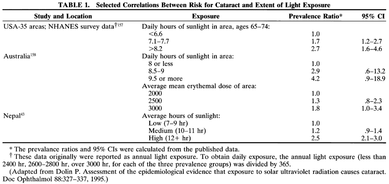
Various epidemiologic studies show associations between elevated risk of various forms of cataract and exposure to higher intensities of incident or reflected ultraviolet light or both (Table 1).15,32–40 Greater light exposure was (weakly) associated with an increased risk for cortical opacity in Chesapeake Bay watermen32 and in men (but not women) in Wisconsin.36 Light-related risk for cataract alsowas increased among Italians but not among resi-dents of Massachusetts.37 Risk for posterior subcapsular cataract was weakly related to light exposure in Chesapeake Bay watermen and (nonsignificantly) in residents of Wisconsin. Other studies (Massachusetts and Italy) did not find associations between posterior subcapsular cataract risk and light exposure. Nuclear cataract appears unrelated to risk for cataract in most studies.


Geographic data provide some support for purported relationships between light exposure and cataract risk.41 Persons living closer to the equator42 and living at higher elevations appear to have anelevated risk of various forms of cataract.35,38–41,43 Indeed, one of the strongest predictors of cataract surgery likelihood in a Medicare beneficiary is a person's latitude of residence.40 Although not a uniform observation,2,44,45 these epidemiologic data have been corroborated or anticipated by exposure of squirrels to ultraviolet light in vivo46 and in many experiments in vitro6–8,42,47–52 and references cited within. As an aggregate, the latter references indicate that exposure of lens constituents to various wavelengths of light results in alterations that are quite similar to those found in cataract.

HIGH-ENERGY RADIATION AS A RISK FACTOR FOR CATARACT

Cataractogenesis is also clearly related to exposure to high-energy radiation. Taylor and associates showed a dose-response relationship betweenx-irradiation and risk for cataract in rats.53 In a study with 99 patients, the 89 who received whole-body irradiation (10 g) had cataract develop in less than 4 years.54 The 10 patients who were treated for aplastic anemia and did not receive radiation treatment did not show evidence of cataractogenesis.

EXPOSURE TO HIGH LEVELS OF OXYGEN AS A RISK FACTOR FOR CATARACT

Perhaps the clearest causal association between oxidative stress and cataract comes from experiences involving elevated levels of oxygen. Nuclear cataract was observed in patients treated with hyperbaric oxygen therapy,55 and markedly elevated levels of mature cataract were observed in mice that survived exposure to 100% oxygen twice weekly for 3 hours.56 A decline in glutathione (GSH) and an increase in glutathione disulfide (oxidative changes normally related to aging or cataract) also were noted. A higher incidence of cataract was noted in lenses exposed to hyperbaric oxygen in vitro,57 and Giblin also noted very early stages of cataract in guinea pigs exposed to hyperbaric oxygen.58 However, there was difficulty in repeating these results (Taylor and coworkers, unpublished data). Oxidative damage to membrane lipids in fiber cells also is associated with lens opacities.59

SMOKING AS A RISK FACTOR FOR CATARACT

Smoking and tobacco chewing appear to induce oxidative stress and have been associated with both diminished levels of antioxidants, ascorbate, and carotenoids60–65 and with enhanced cataract at a younger age.66–69 Of interest are the following recent observations:

  1. For male smokers, there appears to be an inverse relationship between serum levels of α-carotene, 13-cryptoxanthin, lutein, and severity of nuclear sclerosis70 (but the reverse may be true for women).
  2. There is diminished risk for cataract in smokers who use multivitamins.25

CELLULAR ANTIOXIDANTS AS PRIMARY DEFENSES AGAINST LENS DAMAGE

Protection against photo-oxidative insult can be conceived as due to two interrelated processes. Primary defenses offer protection of proteins and other constituents by lens antioxidants and antioxidant enzymes (see Fig. 3). Secondary defenses include proteolytic and repair processes, which degrade and eliminate damaged proteins and other biomolecules in a timely fashion.71

The major aqueous antioxidants in the lens are ascorbate72 and GSH.3,73–75 Both are present in the lens at millimolar concentrations.76–78 Ascorbate is probably the most effective, least toxic antioxidant identified in mammalian systems.79,80 Interest in the function of ascorbate in the lens was prompted by teleologic arguments, which suggested age-related compromises in ascorbate and compromises in lens function might be related. Thus, the following observations were noted:

  1. The lens and aqueous concentrate ascorbate greater than 10-fold the level was found in guinea pig and human plasma72,73,81,82 (Fig. 4).
  2. In the lens core (see Fig. 2b), the oldest part of the lens, and the region involved in much senile cataract, the concentration of ascorbate is only 25% of the surrounding cortex.83
  3. Lens ascorbate concentrations are lower in cataract than in the normal lens.84
  4. Ascorbate levels in the lens are significantly lower in old guinea pigs than in young animals with the same dietary intake of ascorbate.72,81 The same pertains in Emory mice.78

Fig. 4. Tissue or plasma ascorbate versus ascorbate intake. A. Guinea pig. B. Plasma ascorbate versus ascorbate intake in men (light line and square symbols) and women (bold line and filled circles).C. aqueous ascorbate versus ascorbate intake in men (open squares) and women (filled circles). D. Lens ascorbate versus ascorbate intake in men (dashed lines and open squares) and women (solid line and filled circles). (A adapted from Berger J, Shepard D, Morrow F et al: Reduced and total ascorbate in guinea pig eye tissues in response to dietary intake. Curr Eye Res 7:681-86, 1988; B-D adapted from Taylor A, Jacques P, Nowell T Jr et al: Vitamin C in human and guinea pig aqueous, lens, and plasma in relation to intake. Curr Eye Res 16:857–864, 1997.)

These data suggest either that there is age-related depletion of ascorbate in the lens or that the bioavailability of this compound changes with age. Enthusiasm for nutrient antioxidants has been fueled by observations that ocular levels of ascorbate are related to dietary intake in humans and animals that require exogenous ascorbate (see Fig. 4).72,81,82 Thus, the concentration of vitamin C in the lens was increased with dietary supplements beyond levels achieved in persons who already consumed more than two times the recommended daily allowance (60 mg/day) for vitamin C.72,73

Feeding elevated ascorbate delayed progress of, or prevented galactose cataract in guinea pigs85 and rats,86 selenite-induced cataracts in rats,87 lens opacification in GSH-depleted chick embryos,88 and delayed UV-induced protein and protease damage in guinea pig lenses.7–9,89 Increasing lens ascorbate concentrations by only twofold is associated with protection against cataract-like damage.8

Because ascorbate is a carbohydrate, it is biochemically plausible that vitamin C induces damage in the lens in vivo.90,91 However, there currently are no data to support this as a medical concern. Mice fed 8% of the weight of their diet as ascorbate did not develop cataract.92 If glycation induces pathologic lens damage, antiglycating agents such as phenacylthiazoliums may be useful.93 It is interesting that comparable compounds have been tried as anticataractogens and were assumed to act as reducing cysteine prodrug agents.94

GSH levels are several-fold the levels found in whole blood and orders of magnitude greater than the concentration observed in the plasma. GSHlevels also diminish in the older and cataractouslens.74 There have been several attempts to exploitthe reducing capabilities of GSH. Injection ofGSH-OMe was associated with delayed buthioninesulfoxamine-induced95 and naphthalene cataract.74,96,97Preliminary evidence from studies with galactose-induced cataract also indicates some advantage of maintaining elevated GSH status in rats.76 However, it is not clear that feeding GSH is associated with higher ocular levels of this antioxidant.76 Other compounds, such as pantetheine, which also includes sulfhydryls, are under investigation as anticataractogenic agents.98 However, the efficacy of this compound in later-life cataract remains to be established.99

Pharmacologic opportunities are suggested by observations that incorporating the industrial antioxidant 0.4% butylated hydroxytoluene in diets of galactose-fed (50% of diet) rats diminished prevalence of cataract.100

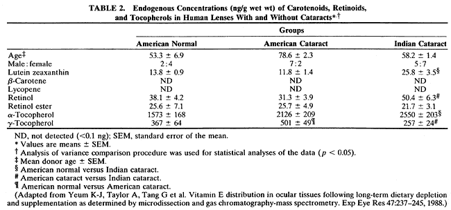
Tocopherols and carotenoids are lipid-soluble antioxidants101,102 with probable roles in maintaining membrane integrity103 and GSH recycling.104 Concentrations of tocopherol in the whole lens are in the micrometer range105 (Table 2), but it appears that lens and dietary levels of tocopherol are unrelated.106 Because most of the compound is found in the membranes, particularly in the younger tissues (Taylor and coworkers, unpublished data), the concentrations can be orders of magnitude higher. Age-related changes in levels of tocopherol and carotenoids have not been documented. Tocopherol is reported to be effective in delaying a variety of induced cataracts in animals, including galactose28,107,108 and aminotriazole-induced cataracts in rabbits.109


Elevated carotenoid intake frequently is associated with health benefits. However, little experimental work has been done regarding lens changes in response to variations in levels of this nutrient. It is intriguing that β-carotene levels in the human lenses are limited105 (see Table 2). Instead, major lens carotenoids are lutein/zeaxanthin. These also are the major carotenoids in the macula.110 Also present are retinol and retinol ester, and tocopherols. In beef, β-carotene occasionally was observed in lenses. This apparent quixotic appearance of the β-carotene appears to be caused by seasonal and dietary availability.

The lens also contains the following antioxidant enzymes: glutathione peroxidase/reductase, catalase, and superoxidase dismutase and enzymes of the glutathione redox cycle.47,48,96,111,112 These interact via the forms of oxygen, as well as with the antioxidants (i.e., GSH is a substrate for glutathione peroxidase). The activities of many antioxidant enzymes are compromised on development, aging, and cataract formation.59

PROTEASES AS SECONDARY DEFENSES

Proteolytic systems can be considered secondary defense capabilities that remove cytotoxic damaged or obsolete proteins from lenses and other eye tissues.2,71,85,113–124 Such proteolytic systems exist in young lens tissue, and damaged proteins usually are maintained at harmless levels by primary and secondary defense systems in younger lenses and in younger lens tissues within older lenses.

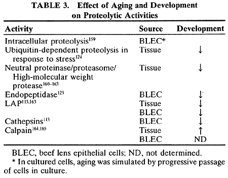
Two studies indicate interactions between primary and secondary defense systems. A direct sparing effect of ascorbate on a photo-oxidatively induced compromise of proteolytic function has been shown.7 GSH also spares activity of enzymes involved in the conjugation of ubiquitin to substrates.115,122 Ubiquitin conjugation is required for selective targeting of substrates for degradation. However, on aging or oxidative stress, most of these enzymatic capabilities are found in a state of reduced activity71 (Table 3). The observed accumulation of oxidized (and/or otherwise modified) proteins in older lenses is consistent with the failure of these protective systems to keep pace with the insults that damage lens proteins. This occurs in part because, like bulk proteins, enzymes that comprise some of the protective systems are damaged by photo-oxidation.2,7,115,122,125 From these data, it is clear that the young lens has significant primary and secondary protection. However, age-related compromises in the activity of antioxidant enzymes, con-centrations of the antioxidants, and activities ofsecondary defenses may lead to diminished protection against oxidative insults.123 This diminished protection leaves the long-lived proteins and other constituents vulnerable. Lens opacities develop as the damaged proteins aggregate and precipitate.2 Current data predict that elevated antioxidant intake can be exploited to extend the function of some of these proteolytic capabilities.


Back to Top
EPIDEMIOLOGIC AND CLINICAL STUDIES REGARDING ASSOCIATIONS BETWEEN ANTIOXIDANTS AND CATARACT
Approximately a dozen epidemiologic studiesexamined the associations between cataract andantioxidants.25,37,126–135 Comparisons are not always straightforward since each of the studies varied in design. Nevertheless, comparisons of the data appear to indicate some agreement with respect to use of nutriture to diminish risk for cataract.

Nine of the studies were retrospective case-controlor cross-sectional studies in which levels of cataract patients were compared with levels of individuals with clear lenses.37,101,126–128,130,131,133,136 Our ability to interpret data from retrospective studies, such as these, is limited by the concurrent assessment of lens status and levels. Prior diagnosis of cataract might influence behavior of cases including diet, and it also might bias reporting of usual diet.

Seven studies25,132,135,137–140 assessed levels or supplement use or both and then followed individuals with intact lenses for up to 8 years. Prospective studies, such as these, are less prone to bias because assessment of exposure is performed before the outcome is present. Some of these studies25,132,135 did not directly assess lens status, but used cataract extraction or reported diagnosis of patients with cataract as a measure of cataract risk. Extraction may not be a good measure of cataract incidence (i.e., development of new cataract), because it incorporates components of both incidence and progression in severity of existing cataract. However, extraction is the result of visually disabling cataract and is the endpoint that we wish to prevent.

The length of time that dietary intake of nutrients is measured also may affect the accuracy of these analyses since cataract develops over many years; one measure may not provide as accurate an assessment of usual intake. Instead, multiple measures over time may offer a better nutritional correlate of cataract.

Hankinson and coworkers132 measured intake several times over a 4-year period, whereas other studies used only one measure of serum antioxidant status, dietary intake, or supplement use. Jacques and associates141 measured supplement intake for more than 10 years and found different risk ratios (RRs) for cataract in persons who took vitamin C supplements for different periods of time (see section A below).

In addition to the different study designs noted above, various studies used different lens classification schemes, different definitions of high and low levels of nutrients, and different age groups of subjects. A study (n = 367) that monitored cataract in vivo and cataract extraction but did not find associations between nutriture and cataract is not described further because the cataract classifications do not match those used on other work.34

ASCORBATE

As noted above, dietary ascorbate intake is related to eye tissue ascorbate levels. Given potential anti-cataractogenic and putative procataractogenic roles for ascorbate, the available epidemiologic data regarding ascorbate intake and risk for cataract are particularly intriguing.

Vitamin C was considered in 9 published studies37,127,128,130–133,139,141 and observed to be inversely associated with at least one type of cataract in 8 of these studies (Fig. 5).

Fig. 5. Cataract risk ratio, high versus low intake (with or without supplements) for plasma levels of vitamin C. Types of cataract are any, moderate/advanced, nuclear, cortical, posterior subcapsular, mixed, or cataract extraction. Data for retrospective and prospective studies are presented independently. (Adapted from Taylor A, Dorey CK, Nowell T Jr. Oxidative stress and ascorbate in relation to risk for cataract and age-related maculopathy. In Packer L, Fuchs J [eds]. Vitamin C in Health and Disease. New York: Marcel Dekker, 1997.)

Several studies found correlations between vitamin C supplement use and risk of cataract. In our nutrition and vision project,141 age-adjusted analyses based on 165 women with high vitamin C intake (mean, 294 mg/day) and 136 women with low vitamin C intake (mean, 77 mg/day) indicated that the women who took vitamin C supplements for 10 years or longer had over 70% lower prevalence of early opacities (RR, 0.23; CI, 0.09–0.60; see Fig. 5A) and greater than 80% lower risk of moderate opacities (RR, 0.17; CI, 0.03–0.87) at any site compared with women who did not use vitamin C supplements.141 Recent reexamination of 600 of the members of the same cohort indicates that comparable data can be anticipated. This corroborated work by Hankinson and associates,132 who noted that women who consumed vitamin C supplements for more than 10 years had a 45% reduction in rate of cataract surgery (RR, 0.55; CI, 0.32–0.96; see Fig. 5G). However, after controlling for nine potential confounders, including age, diabetes, smoking, and energy intake, they did not observe an association between total vitamin C intake and rate of cataract surgery (see below).

In comparison to the data noted above, Mares-Perlman and coworkers126 report that past use of supplements containing vitamin C was associated with a reduced prevalence of nuclear cataract (RR, 0.7; CI, 0.5–1.0; see Fig. 5C) but an increased prevalence of cortical cataract (adjusted RR, 1.8; CI, 1.2–2.9) after controlling for age, sex, smoking, and history of heavy alcohol consumption (see Fig. 5D).

The inverse relationship is corroborated by data from other studies. Robertson and coworkers127 compared cases (with cataracts that impaired vision) to age and sex-matched control subjects who were either free of cataract or had minimal opacities that did not impair vision. The prevalence of cataract in consumers of daily vitamin C supplements of greater than 300 mg/day was approximately one third the prevalence in persons who did not consume vitamin C supplements (RR, 0.30; CI, 0.24–0.77; see Fig. 5B).

Elevated dietary ascorbate also was related to benefit with respect to cataract in some studies. Leske and coworkers128 observed that persons with vitamin C intake in the highest 20% of their population group had a 52% lower prevalence for nuclear cataract (RR, 0.48; CI, 0.24–0.99) compared with persons who had intakes among the lowest 20% after controlling for age and sex (see Fig. 5C). Weaker inverse associations were noted for other types of cataract (see Fig. 5F). Jacques andChylack130 observed that among persons with higher vitamin C intakes (over 490 mg/day), the prevalence of cataract was 25% of the prevalence among persons with lower intakes (less than 125 mg/day; RR, 0.25; CI, 0.06–1.09; see Fig. 5A).

However, Vitale and coworkers133 observed no differences in cataract prevalence between per-sons with high (over 261 mg/day) and low (lessthan 115 mg/day) vitamin C intakes. The Italian-American Studies group37 also failed to observe any association between prevalence of cataract and vitamin C intake. In addition, in a large prospective study, comparison of women with high intakes (median, 705 mg/day) to women with low intakes (median, 70 mg/day) failed to show any significant correlation with risk for cataract extraction (RR, 0.98; CI, 0.72–1.32).132

Attempts to corroborate the above inverse associations between cataract risk and intake using plasma vitamin C levels generally were frustrating. Jacques and Chylack130 observed that persons with high plasma vitamin C levels (greater than 90 μmol/L) had less than one-third the prevalence of early cataract as did persons with low plasma vitamin C (less than 40 μmol/L), although this difference was not statistically significant (RR, 0.29; 95% CI, 0.06–1.32) after adjustment for age, sex, race, and history of diabetes (see Fig. 5A). Mohan and associates131 noted an 87% (RR, 1.87; CI, 1.29–2.69) increased prevalence of mixed cataract (posterior subcapsular and nuclear involvement) for each standard deviation increase in plasma vitamin C levels. Vitale and coworkers133 observed that persons with plasma levels greater than 80 μmol/L and less than 60 μmol/L had similar prevalences of both nuclear (RR, 1.31; CI, 0.61–2.39) and cortical (RR, 1.01; CI, 0.45–2.26) cataract after controlling for age, sex, and diabetes. Results from one intervention trial have been published and are described below.142

VITAMIN E

Vitamin E, a natural lipid-soluble antioxidant, can inhibit lipid peroxidation103 and appears to stabilize lens cell membranes.143 The efficacy of vitamin E as an antioxidant may be affected by ascorbate (see legend to Fig. 3) and also enhances glutathione recycling, perhaps helping to maintain reduced glutathione levels in the lens and aqueous humor.104

Consumption of vitamin E supplements was inversely correlated with cataract risk in two studies (Fig. 6). Robertson and coworkers127 found among age- and sex-matched case and control subjects that the prevalence of advanced cataract was 56% lower (RR, 0.44; CI, 0.24–0.77; see Fig. 6B) in persons who consumed vitamin E supplements (greater than 400 IU/day) than in persons not consuming supplements. Jacques and Chylack (unpublished data) observed a 67% (RR, 0.33; CI, 0.12–0.96) reduction in prevalence of cataract for vitamin E supplement users after adjusting for age, sex, race, and diabetes. Mares-Perlman and coworkers126 observed only weak, nonsignificant associations between vitamin E supplement use and nuclear (RR, 0.9; CI, 0.6–1.5; see Fig. 6C) and cortical (RR, 1.2; CI, 0.6–2.3; see Fig. 6D) cataract.

Fig. 6. Cataract risk ratio, high versus low intake (with or without supplements) for plasma levels of vitamin E (α-tocopherol). (Adapted from Taylor A, Dorey CK, Nowell T Jr. Oxidative stress and ascorbate in relation to risk for cataract and age-related maculopathy. In Packer L, Fuchs J [eds]: Vitamin C in Health and Disease. New York: Marcel Dekker, 1997.)

The inverse association between vitamin E intake and risk for cataract was corroborated by Leske and associates.128 They observed that after controlling for age and sex, persons with vitamin E intakes among the highest 20% had an approximately 40% lower prevalence of cortical (RR, 0.59; CI, 0.36–0.97; see Fig. 6D) and mixed (RR, 0.58; CI, 0.37–0.93; see Fig. 6F) cataract relative to personswith intakes among the lowest 20%. Jacques andChylack130 observed a nonsignificant inverse association when they related total vitamin E intake (i.e., combined dietary and supplemental intake) to cataract prevalence. Persons with vitamin E intake over 35.7 mg/day had a 55% lower prevalence of early cataract (RR, 0.45; CI, 0.12–1.79) than did per-sons with intakes less than 8.4 mg/day.130 However,Hankinson and coworkers132 found no association between vitamin E intake and cataract surgery. Women with high vitamin E intakes (median,210 mg/day) had a similar rate of cataract surgery (RR, 0.96; CI, 0.72–1.29) as did women with low intakes (median, 3.3 mg/day). In partial contrast with their positive correlations between serumα-tocopherol levels and cataract, Mares-Perlman and coworkers129 found that dietary vitamin E was associated (nonsignificantly) with diminished risk of nuclear cataract in men but not in women (see Fig. 6C).

Four studies assessing plasma vitamin E levels also reported significant inverse associations with cataract. Knekt and coworkers135 followed a cohort of 1419 Finns for 15 years and identified 47 patients admitted to ophthalmologic wards for mature cataract. They selected two controls per patient matched for age, sex, and municipality. These investigators reported that persons with serum vitamin E concentrations above approximately 20 μmol/L had approximately half the rate of subsequent cataract surgery (RR, 0.53; CI, 0.24–1.1; see Fig. 6G) compared with persons with vitamin E concentrations below this concentration. Vitale and coworkers133 observed the age-, sex-, and diabetes-adjusted prevalence of nuclear cataract to be approximately 50% less (RR, 0.52; CI, 0.27–0.99; see Fig. 6C) among persons with plasma vitamin E concentrations greater than 29.7 μmol/L compared with persons with levels less than 18.6 μmol/L. A similar comparison showed that the prevalence of cortical cataract did not differ between those with high and low plasma vitamin E levels (RR, 0.96; CI, 0.52–0.1.78; see Fig. 6D). Jacques and Chylack130 also observed the prevalence of posterior subcapsular cataract to be 67% (RR, 0.33; CI, 0.03–4.13; see Fig. 6E) lower among persons with plasma vitamin E levels above 35 μmol/L relative to persons with levels below21 μmol/L after adjustment for age, sex, race, anddiabetes; however, the effect was not statistically significant. Prevalence of any early cataract (RR, 0.83; CI, 0.20–3.40; see Fig. 6A) or cortical cataract (RR, 0.84; CI, 0.20–3.60; see Fig. 6D) did not differ between those with high and low plasma levels. Plasma vitamin E also was inversely associated with prevalence of cataract in a large Italian study after adjusting for age and sex, but the relationship was no longer statistically significant after adjusting for other factors such as education, sunlight exposure, and family history of cataract.37 Leske and coworkers101 also showed that individuals with high plasma vitamin E levels had significantly lower prevalence of nuclear cataract (RR, 0.44; CI, 0.21–0.90), but vitamin E was not associated with cataracts at other lens sites.

Mares-Perlman and coworkers noted a significant elevated prevalence of nuclear cataract in men with high serum vitamin E (RR, 3.74; CI, 1.25–11.2) but not in women (RR, 1.47; CI, 0.57–3.82; see Fig. 6C).70 One other study failed to observe any association between cataract and plasma vitamin E levels.131

Mares-Perlman and coworkers70 observed an inverse (nonsignificant) relationship (RR, 0.61; CI, 0.32–1.19) between serum γ-tocopherol, which has lower biologic vitamin E activity compared toα-tocopherol (Fig. 7B), and severity of nuclear sclerosis, but a positive, significant relationship between elevated serum α-tocopherol levels and severity of nuclear cataract (RR, 2.13; CI, 1.05–4.34; see Fig. 6C).

Fig. 7. Cataract risk ratio, high versus low intake (with or without supplements) for plasma levels ofγ-tocopherol. (Adapted from Taylor A, Dorey CK, Nowell T Jr. Oxidative stress and ascorbate in relation to risk for cataract and age-related maculopathy. In Packer L, Fuchs J [eds]: Vitamin C in Health and Disease. New York: Marcel Dekker, 1997.)

Whereas serum α-tocopherol appeared to be associated with nonsignificant increases in risk for cortical or any cataract, serum γ-tocopherol was not significantly associated with cortical or any cataract in these studies.

Two prospective studies showed a reduced risk for cataract progress among individuals with higher plasma vitamin E. Rouhiainen and coworkers138 found a 73% reduction in risk for cortical cataract progression (RR, 0.27; CI, 0.08–0.83; see Fig. 6D), whereas Leske and coworkers139 reported a 42% reduction in risk for nuclear cataract progression (RR, 0.58; CI, 0.36–0.94; see Fig. 6C). Vitamin E supplementation was related to a lower risk for progress of nuclear opacity (RR, 0.43; CI, 0.19–0.99).139

CAROTENOIDS

The carotenoids, like vitamin E, also are natural lipid-soluble antioxidants.103 β-carotene is the best-known carotenoid because of its importance as a vitamin A precursor. It exhibits particularly strong antioxidant activity at low partial pressures of oxygen (15 torr).144 This is similar to the -20 torr partial pressure of oxygen in the core of the lens.145 However, it is only one of approximately 400 naturally occurring carotenoids,146 and other carotenoidsmay have similar or greater antioxidant potential.102,103,147,148 In addition to β-carotene, α-carotene, lutein, and lycopene are important carotenoid components of the human diet.149 Carotenoids have been identified in the lens in 10 ng/g or greater net weight concentrations (see Table 1).105,150 There are a dearth of laboratory data that relate carotenoids to cataract formation, and there also are no data that relate carotenoid supplement use to risk for cataracts.

Jacques and Chylack130 were the first to ob-serve that persons with carotene intakes above18,700 IU/day had the same prevalence of cataract as those with intakes below 5677 IU/day (RR, 0.91; CI, 0.23–3.78; Fig. 8A). Hankinson and associates132 followed this report with a study that specified that the multivariate-adjusted rate of cataract surgery was approximately 30% lower (RR, 0.73; CI, 0.55–0.97) for women with high carotene intakes (median, 14,558 IU/day) compared with women with low intakes of this nutrient (median, 2935 IU/day; see Fig. 8E). However, although cataract surgery was inversely associated with total carotene intake, it was not strongly associated with consumption of carotene-rich foods, such as carrots. Rather, cataract surgery was associated with lower intakes of foods, such as spinach, that are rich in lutein and xanthin carotenoids rather than β-carotene. This would appear to be consistent with our observation that the human lens contains lutein and zeaxanthin but not β-carotene. Unfortunately, cataract surgery was not an endpoint in other studies that considered xanthaphylls.70,126

Fig. 8. Cataract risk ratio, high versus low intake (with or without supplements) for plasma levels of carotenoids (generally measured as β-carotene). (Adapted from Taylor A, Dorey CK, Nowell T Jr. Oxidative stress and ascorbate in relation to risk for cataract and age-related maculopathy. In Packer L, Fuchs J [eds]: Vitamin C in Health and Disease. New York: Marcel Dekker, 1997.)

Jacques and Chylack130 also noted that persons with high plasma total carotenoid concentrations (over 3.3 μmol/L) had less than one fifth the prevalence of cataract compared with persons with low plasma carotenoid levels (less than 1.7 μmol/L; RR, 0.18; CI, 0.03–1.03) after adjustment for age, sex, race, and diabetes (see Fig. 8A). However, they were unable to observe an association between carotene intake and cataract prevalence. Knekt and coworkers135 reported that among age- and sex-matched case and control subjects, persons with serum β-carotene concentrations above approximately 0.1 AM had a 40% reduction in the rate of cataract surgery compared with persons with concentrations below this level (RR, 0.59; CI, 0.261.25).

The most recent study that correlated serum carotenoids and severity of nuclear and conical opaci-ties70 indicates that higher levels of individual or total carotenoids in the serum were not associated with less-severe nuclear or cortical cataract overall, although same sex-related differences in risk were noted. Associations between risk for some forms of cataract and nutriture differed between men and women (e.g., nuclear cataract and α-carotene intake; Fig. 9B).129 Other nutrients for which cataract risk in women versus men showed opposing relationships to include serum β-carotene (Fig. 10) and serum lycopene (Fig. 11). A marginally significant trend for lower RR for cortical opacity with increasing serum levels of β-carotene was observed in men but not women. Higher serum levels of α-carotene, β-cryptoxanthin, and lutein were significantly related to lower risk for nuclear sclerosis only in men who smoked. In contrast, higher levels of some carotenoids often were directly associated with elevated risk for nuclear sclerosis and cortical cataract (see Figs. 10 and 12), particularly in women.

Fig. 9. Cataract risk ratio, high versus low intake (with or without supplements) for plasma levels of α-carotene. (Adapted from Taylor A, Dorey CK, Nowell T Jr. Oxidative stress and ascorbate in relation to risk for cataract and age-related maculopathy. In Packer L, Fuchs J [eds]: Vitamin C in Health and Disease. New York: Marcel Dekker, 1997.)

Fig. 10. Cataract risk ratio, high versus low intake (with or without supplements) for plasma levels of β-carotene (also see data in Fig. 8). (Adapted from Taylor A, Dorey CK, Nowell T Jr. Oxidative stress and ascorbate in relation to risk for cataract and age-related maculopathy. In Packer L, Fuchs J [eds]: Vitamin C in Health and Disease. New York: Marcel Dekker, 1997.)

Fig. 11. Cataract risk ratio, high versus low intake (with or without supplements) for plasma levels of lycopene. (Adapted from Taylor A, Dorey CK, Nowell T Jr. Oxidative stress and ascorbate in relation to risk for cataract and age-related maculopathy. In Packer L, Fuchs J [eds]: Vitamin C in Health and Disease. New York: Marcel Dekker, 1997.)

Fig. 12. Cataract risk ratio, high versus low intake (with or without supplements) for plasma levels of β-cryptoxanthin. (Adapted from Taylor A, Dorey CK, Nowell T Jr. Oxidative stress and ascorbate in relation to risk for cataract and age-related maculopathy. In Packer L, Fuchs J [eds]: Vitamin C in Health and Disease. New York: Marcel Dekker, 1997.)

Vitale and colleagues133 also examined the relationships between plasma β-carotene levels and age-, sex-, and diabetes-adjusted prevalence of cortical and nuclear cataract (see Fig. 10B and C). Although the data suggested a weak inverse association between plasma β-carotene and cortical cataract and a weak positive association between this and nuclear cataract, neither association was statistically significant. Persons with plasma β-carotene concentrations above 0.88 μmol/L had a 28% lower prevalence of cortical cataract (RR, 0.72; CI, 0.37–1.42) and a 57% (RR, 1.57; CI, 0.84–2.93) higher prevalence of nuclear cataract compared with persons with levels below 0.33 μmol/L (Fig. 13).

Fig. 13. Cataract risk ratio, high versus low intake (with or without supplements) for plasma levels of lutein. (Adapted from Taylor A, Dorey CK, Nowell T Jr. Oxidative stress and ascorbate in relation to risk for cataract and age-related maculopathy. In Packer L, Fuchs J [eds]: Vitamin C in Health and Disease. New York: Marcel Dekker, 1997.)

Hankinson and coworkers132 observed correlations between β-carotene intake but not intake of β-carotene-containing foods and risk for cataract extraction. Instead, they saw an inverse relation be-tween risk for cataract extraction and intake of lutein and zeaxanthin-containing foods such as spinach (Fig. 14). This observation would appear to be consistent with observations that lutein and zeaxanthin are the most prevalent carotenoids in lens (see Table 2). However, Mares-Perlman126,129 did not detect significantly altered risk for cataract among consumers of these nutrients.

Fig. 14. Cataract risk ratio, high versus low intake (with or without supplements) for plasma levels of antioxidant nutrient index using multiple antioxidants. (Adapted from Taylor A, Dorey CK, Nowell T Jr. Oxidative stress and ascorbate in relation to risk for cataract and age-related maculopathy. In Packer L, Fuchs J [eds]: Vitamin C in Health and Disease. New York: Marcel Dekker, 1997.)

ANTIOXIDANT COMBINATIONS

To more closely approximate combined effects on cataract risk of the multiple antioxidants that are contained in food, this group was the first to adopt “antioxidant indices.” However, it is possible that single nutrients appear to have strong influences on the indices, and we now question the utility of theindices. Nevertheless, in an attempt to offer a complete summary, data regarding relationships between antioxidant indices and cataract risk are presented below (see Fig. 14). To be consistent with the earlier sections, we first describe effects of supplement use. Then the effects of intake, including foods, are summarized.

Robertson and coworkers127 found no enhanced benefit to persons taking both vitamin E and vitamin C supplements compared with persons who only took either vitamin C or vitamin E. Leske and coworkers128 found that the use of multivitamin supplements was associated with decreased prevalence for each type of cataract: 60%, 48%, 45%, and 30%, respectively, for posterior subcapsular (RR, 0.40; CI, 0.21–0.77), cortical (RR, 0.52; CI, 0.36–0.72), nuclear (RR, 0.55; CI, 0.33–0.92), and mixed (RR, 0.70; CI, 0.51–0.97) cataracts (see Figs. 14B-E). Luthra and coworkers136 and Leske and coworkers151 reported that supplement consumption was associated with a slight reduction in risk of cortical cataract in a black population (RR, 0.77; CI, 0.61–0.98) in those younger than 70 years of age (see Fig. 14C).

Multivitamins also were reported to reduce the risk of incident cataracts, as well as progression of existing cataracts in several studies (see Fig. 14). Seddon and coworkers25 observed a reduced risk for incident cataract for users of multivitamins (RR, 0.73; CI, 0.54–0.99). Mares-Perlman and coworkers137 reported that multivitamin users had significant 20% (RR, 0.8; CI, 0.6–1.0) and 30% (RR, 0.7; CI, 0.5–1.0) reduction of cortical cataract progression and incidence, respectively (see Fig. 14C). Leske and coworkers139 reported a 31% reduced risk for progression of nuclear cataract among users of multivitamins (RR, 0.69; CI, 0.48–0.99). Hankinson and coworkers132 found no relationship between multivitamin use and risk of cataract extraction. In these studies, it is not clear whether synergy between nutrients, with respect to conferring diminished risk for cataract, is indicated.

The first, and perhaps most important, study in terms of showing the utility of diet indicates that persons who consumed at least 1.5 servings of fruits or vegetables or both had only a 20% risk of having cataract develop as did those who did not (see Fig. 14A).130 Hankinson and coworkers132 calculated an antioxidant score based on intakes of carotene, vitamin C, vitamin E, and riboflavin and observed a 24% reduction in the adjusted rate of cataract surgery among women with high antioxidant scores relative to women with low scores (RR, 0.76; CI, 0.57–1.03; see Fig. 14F).

Using a similar index based on combined antioxidant intakes (vitamin C, vitamin E, and carotene, as well as riboflavin), Leske and coworkers128 found that persons with high scores had 60% lower adjusted prevalence of cortical (RR, 0.42; CI, 0.18–0.97; see Fig. 14C) and mixed (RR, 0.39; CI, 0.19–0.80; see Fig 14E) cataract compared with those who had low scores.

Jacques and Chylack130 also found that the adjusted prevalence of all types of cataract was 40% (RR, 0.62; CI, 0.12–1.77) and 80% (RR, 0.16; CI, 0.04–0.82) lower for persons with moderate and high-antioxidant index scores (based on combined plasma vitamin C, vitamin E, and carotenoid lev-els) compared with persons with low scores (seeFig. 14A). Mohan and coworkers131 constructed asomewhat more complex antioxidant scale that in-cluded erythrocyte levels of glutathione peroxidase,glucose-6-phosphate dehydrogenase, and plasma levels of vitamin C and vitamin E. Even though Mohan and coworkers failed to see any protective associations with any of these individual factors and even reported a positive association between plasma vitamin C and prevalence of cataract, they found that persons with high antioxidant index scores had a substantially lower prevalence of cataracts involving the posterior subcapsular region (RR, 0.23; CI, 0.06–0.88; see Fig. 14D) or mixed cataract with posterior subcapsular and nuclear components (RR, 0.12; CI, 0.03–0.56) after multivariate adjustment (see Fig. 14E). Knekt and coworkers135 observed that the rate of cataract surgery for persons with high levels of both serum vitamin E and β-carotene concentrations appeared lower than the rate for persons with either high vitamin E or high β-carotene levels (see Fig. 14F). Persons with high serum levels of either had a rate of cataract surgery that was 40% less than did persons with low levels of both nutrients (RR, 0.38; CI, 0.15–1.0).

Vitale and coworkers133 also examined the relationship between antioxidant scores (based on plasma concentrations of vitamin C, vitamin E, and β-carotene) and prevalence of cataract, but did not see evidence of any association. The age-, sex-, and diabetes-adjusted RRs were close to one for both nuclear (RR, 0.96; CI, 0.54–1.70) and cortical (RR, 1.17; CI, 0.62–2.20) cataract.

INTERVENTION STUDIES

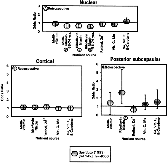
Only one intervention trial designed to assess the effect of vitamin supplements on cataract risk has been completed. Sperduto and coworkers142 took advantage of two ongoing, randomized, double-masked vitamin and cancer trials to assess the impact of vitamin supplements on cataract prevalence. The trials were conducted among almost 4000 participants aged 45 to 74 years from rural communes in Linxian, China. Participants in the first trial received either a multisupplement or placebo. In the second trial, a more complex factorial design was usedto evaluate the effects of four different vitamin/mineral combinations: retinol (5000 IU) and zinc(22 mg); riboflavin (3 mg) and niacin (40 mg); vitamin C (120 mg) and molybdenum (30 μg); and vitamin E (30 mg), β-carotene (15 mg), and selenium (50 μg). At the end of the 5- to 6-year follow-up, the investigators conducted eye examinations to determine the prevalence of cataract (Fig. 15).

Fig. 15. Cataract risk ratio, high versus low intake (with or without supplements) for intervention trials. (Adapted from Taylor A, Dorey CK, Nowell T Jr. Oxidative stress and ascorbate in relation to risk for cataract and age-related maculopathy. In Packer L, Fuchs J [eds]: Vitamin C in Health and Disease. New York: Marcel Dekker, 1997.)

In the first trial, there was a significant 43% reduction in the prevalence of nuclear cataract for persons aged 65 to 74 years receiving the multisupplement (RR, 0.57; CI, 0.36–0.90; see Fig. 15A). The second trial showed a significantly reduced prevalence of nuclear cataract in persons receiving the riboflavin/niacin supplement relative to those persons not receiving this supplement (RR, 0.59; CI, 0.45–0.79). The effect was strongest in those aged 65 to 74 years (RR, 0.45; CI, 0.31–0.64). However, the riboflavin/niacin supplement appeared to increase the riskof posterior subcapsular cataract (RR, 2.64; CI,1.31–5.35; see Fig. 15C). The results further sug-gested a protective effect of the retinol/zinc supple-ment (RR, 0.77; CI, 0.58–1.02) and the vitaminC/molybdenum supplement (RR, 0.78; CI, 0.59–1.04) on prevalence of nuclear cataract.

CALORIE RESTRICTION AND CONTROL OF BODY MASS INDEX AS A MEANS TO DELAY CATARACT

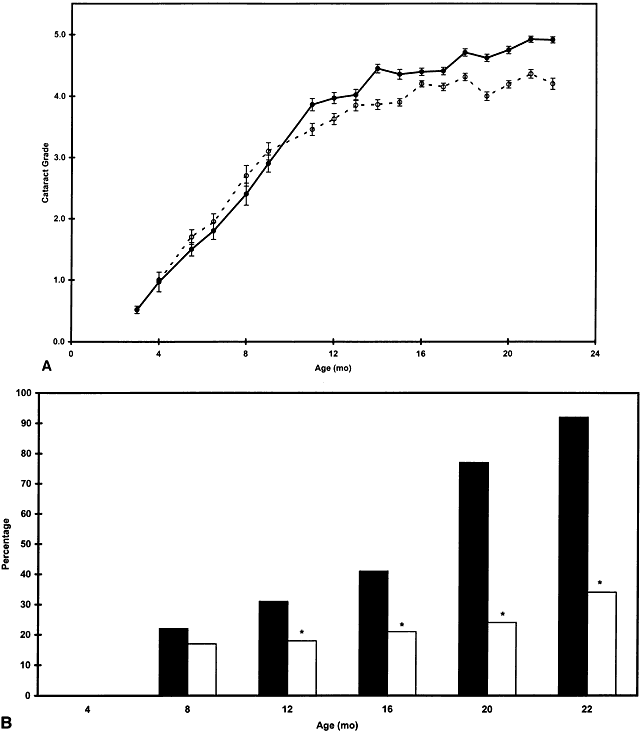
Restriction of caloric intake extends youth anddelays age-related cataract (as well as many other late-life diseases) in these animals (Fig. 16). The decrease in risk for cataract in calorie-restricted Emory mice has some parallels in a recent study, which indicates that well-fed male physicians with body mass index below 22 enjoyed less risk for cataract compared with physicians with body mass indices above 25.152 Because cataract is associated with oxidative stress, it might be anticipated that the delay in cataract would be accompanied by elevated ascorbate levels in the protected animals. Nevertheless, in young and old animals, plasma-ascorbate concentrations were lower than in the nonrestricted mice.77,78,153

Fig. 16. A. Mean cataract grade in calorie-restricted and control Emory mice at 3 to 22 months of age (filled circles, control mice; open circles, restricted mice; values are means ± standard error of the mean. At older ages, error bars are approximately equal in size to the symbol for control animals. B. Percentage of lenses with grade 5 cataract (*p = 0.05). (Taylor A, Jahngen-Hodge J, Smith D et al: Dietary restriction delays cataract and reduces ascorbate levels in Emory mice. Exp Eye Res 61:55–62, 1995.)

Back to Top
CONCLUSION
Light and oxygen appear to be both a boon and a bane. Although necessary for physiologic function, when present in excess or in uncontrolled circumstances, they appear to be causally related to cataractogenesis. On aging, compromised function of the lens is exacerbated by depleted or diminished primary antioxidant reserves, antioxidant enzyme capabilities, and diminished secondary defenses such as proteases. Smoking appears to provide an oxidative challenge and also is associated with an elevated risk of cataract.

The impression created by the literature is that there is some benefit to enhanced antioxidant intake with respect to diminished risk for cataract. Optimal levels of ascorbate appear to be 250 mg/day; however, more information is essential before describing optimal nutriture vis à vis cataract. It is difficult to compare the various studies. That the correlations were not always with the same form of cataract may indicate, in addition to the conclusions reached, that the cataracts were graded differently or that there are common etiologic features of each of the forms of cataract described or both. Most of the studies noted above used case-control designs, and most assessed status only once. Because intake or status measures are highly variable and the effects of diet are likely to be cumulative, studies should be performed on populations for which long-term dietary records are available. It appears that intake studies are preferable to use for plasma measures if a single measure of status must be chosen. Longitudinal studies and more intervention studies are certainly essential to truly establish the value of antioxidants and to determine the extent to which cataract progress is affected by nutriture. More uniform methods of lens evaluation, diet recording, and blood testing, for example, would facilitate conclusions regarding the merits of antioxidants. Optimization of nutriture can be achieved through better diets and supplement use once appropriate levels of specifically beneficial nutrients are defined. In addition to quantifying optimal intake, it is essential to know for how long or when intake of the nutrients would be useful with respect to delaying cataract. It is possible to adjust normal dietary practice to obtain close-to-saturating levels of plasma ascorbate (less than 250 mg/day).72,132,141,154 Because the bioavailability of ascorbate may decrease with age, slightly higher intakes may be required in the elderly. Thus, the overall impression created by these data suggests that further research in this field will bring significant health benefits.

Poor education and lower socioeconomic status also markedly increase risk for these debili-ties.128,131,155,156 These are related to poor nutrition.Because cost-benefit analysis regarding remediation clearly indicates that cataract prevention is preferable (and essential where there is a dearth of surgeons) to surgery, it is not premature to contemplate the value of intervention for populations at risk. The work available, albeit preliminary, indicates that nutrition may provide the least costly and most practicable means to attempt the objectives of delaying cataract.

Back to Top
ACKNOWLEDGMENTS
We thank Tom Nowell in the preparation of figures and Paul Jacques for invaluable assistance in evaluating the epidemiologic data.
Back to Top
REFERENCES

1. Muller HK, Buschke W: Vitamin C in Linse, Kammerwasser and Blut normalem and pathologischem Linsentstoffwech. Arch F Augenh 108:368–390, 1934

2. Taylor A, Jacques PF, Dorey CK: Oxidation and aging: Impact on vision. J Toxicol Indust Health 9:349-71, 1993

3. Bunce GE, Kinoshita J, Horwitz J: Nutritional factors in cataract. Annu Rev Nutr 10:233–254, 1990

4. Jacques PF, Chylack LT Jr, Taylor A: Relationships between natural antioxidants and cataract formation. In Frei B (ed): Natural Antioxidants Human Health and Disease. Orlando, FL: Academic Press

5. Taylor A: Vitamin C. In Hartz SC, Russell RM, Rosenberg IH (eds): Nutrition in the Elderly: The Boston Nutritional Status Survey. London: Smith Gordon Limited, 1992:147–150

6. Taylor A: Cataract: Relationships between nutrition and oxidation. J Am Coll Nutr 12:138–146, 1993

7. Blondin J, Taylor A: Measures of leucine aminopeptidase can be used to anticipate UV-induced age-related damage to lens proteins: Ascorbate can delay this damage. Mech Ageing Dev 41:39–46, 1987

8. Blondin J, Baragi VJ, Schwartz E et al: Delay of UV-induced eye lens protein damage in guinea pigs by dietary ascorbate. Free Radiol Biol Med 2:275–281, 1986

9. Taylor A, Jacques PF: Relationships between aging, antioxidant status, and cataract. Am J Clinical Nutr 62:1439S–1447S, 1995

10. Taylor A: Oxidative stress and antioxidant function in relation to risk for cataract. In Sies H (ed): Antioxidants in Disease Mechanisms and Therapeutic Strategies (A Volume of Advances in Pharmacology Series). San Diego: Academic Press, 1997:515–536

11. Taylor A, Jacques P: Antioxidant status and risk for cataract. In Bendich A, Deckelbaum RJ (eds): Preventive Nutrition: The Guide for Health Professionals. Totawa, NJ: Humana Press, 1997:267–283

12. Kupfer C: The conquest of cataract: A global challenge. Trans Ophthal Soc UK 104:1–10, 1984

13. Schwab L: Cataract blindness in developing nations. Internat Ophthalmol Clin 30:16–18, 1990

14. World Health Organization: Use of intraocular lenses in cataract surgery in developing countries. Bull WHO 69:657–666, 1991

15. Klein BEK, Klein R, Linton KLP: Prevalence of age-related lens opacities in a population: The Beaver Dam Eye Study. Ophthalmology 99:546–552, 1992

16. Klein R, Klein BE, Linton KL et al: The Beaver Dam Eye Study: The relation of age-related maculopathy to smoking. Am J Epidemiol 37:190–200, 1993

17. Leibowitz H, Krueger D, Maunder C et al: The Framingham Eye Study Monograph. Surv Ophthalmol (Suppl) 24:335–610, 1980

18. Chatterjee A, Milton RC, Thyle S: Prevalence and etiology of cataract in Punjab. Br J Ophthalmol 66:35–42, 1982

19. Wang G-M, Spector A, Luo C-Q et al: Prevalence of age-related cataract in Ganzi and Shanghai: The Epidemiological Study Group. Chinese Med J 103:945–951, 1990

20. Whitfield R, Schwab L, Ross-Degnan D et al: Blindness and eye disease in Kenya: Ocular status survey results from the Kenya Rural Blindness Prevention Project. Br J Ophthalmol 74:333–340, 1990

21. Chan CW, Billson FA: Visual disability and major causes of blindness in NSW: A study of people aged 50 and over attending the Royal Blind Society 1984 to 1989. Aust N Zealand J Ophthalmol 19:321–325, 1991

22. Dana MR, Tielsch JM, Enger C et al: Visual impairment in a rural Appalachian community: Prevalence and causes. JAMA 264:2400–2405, 1990

23. Salive ME, Guralnik J, Christian W et al: Functional blindness and visual impairment in older adults from three communities. Ophthalmology 99:1840–1847, 1992

24. Wormald RPL, Wright LA, Courtney P et al: Visual problems in the elderly population and implications for services. Br Med J 304:1226–1229, 1992

25. Seddon JM, Christen WG, Manson JE et al: The use of vitamin supplements and the risk of cataract among US male physicians. Am J Public Health 84:788–792, 1994

26. Young RW: Optometry and the preservation of visual health. Optom Vis Sci 70:255–262, 1992

27. Taylor A, Tisdell FE, Carpenter FH: Leucine aminopeptidase (bovine lens): Synthesis and kinetic properties of ortho, meta, and para substituted leucyl-anilides. Arch Biochem Biophys 210:90–97, 1981

28. Jacques PF, Taylor A: Micronutrients and age-related cataracts. In Bendich A, Butterworth CE (eds): Micronutrients in Health and in Disease Prevention. New York: Marcel Dekker, 1991:359–379

29. Chylack LT Jr, Wolfe JK, Singer DM et al: The lens opaci-ties classification system III. Arch Ophthalmol 111:831–836, 1993

30. Chylack LT Jr, Wolfe JK, Friend J et al: Nuclear cataract: Relative contributions to vision loss of opalescence and brunescence. Invest Ophthalmol Vis Sci 35:42632, 1994 (abstract)

31. Wolfe JK, Chylack LT Jr, Leske MC et al: Lens nuclear color and visual function. Invest Ophthalmol Vis Sci 34:2550, 1993 (abstract)

32. Taylor HR, West SK, Rosenthal FS et al: Effect of ultraviolet radiation on cataract formation. N Engl J Med 319:1429–1433, 1988

33. Zigman S, Datiles M, Torczynski E: Sunlight and human cataract. Invest Ophthalmol Vis Sci 18:462–467, 1979

34. Wong L, Ho SC, Coggon D et al: Sunlight exposure, antioxidant status, and cataract in Hong Kong fishermen. J Epidemiol Community Health 47:46–49, 1993

35. Hirvela H, Luukinen H, Laatikainen L: Prevalence and risk factors of lens opacities in the elderly in Finland: A population-based study. Ophthalmology 102:108–117, 1995

36. Cruickshanks KJ, Klein BE, Klein R: Ultraviolet light exposure and lens opacities: The Beaver Dam Eye Study. Am J Public Health 82:1658–1662, 1992

37. The Italian-American Cataract Study Group: Risk factors for age related cortical, nuclear, and posterior subcapsular cataracts. Am J Epidemiol 133:541–553, 1991

38. Wang G-M, Spector A, Luo C-Q et al: Prevalence of age-related cataract in Ganzi and Shanghai. Chinese Med J 103:945–951, 1990

39. Klein BE, Cruickshanks KJ, Klein R: Leisure time, sunlight exposure and cataracts. Doc Ophthalmol 88:295–305, 1994-1995

40. Javitt JC, Taylor HR: Cataract and latitude. Doc Ophthalmol 88:307–325, 1995

41. Dolin P: Assessment of the epidemiological evidence that exposure to solar ultraviolet radiation causes cataract. Doc Ophthalmol 88:327–337, 1995

42. Zigman S: Effects of near ultraviolet radiation on the lens and retina. Doc Ophthalmol 55:375–391, 1983

43. Brilliant LB, Grasset NC, Pokhrel RP et al: Associations among cataract prevalence, sunlight hours, and altitude in the Himalayas. Am J Epidemiol 118:250–264, 1983

44. Minassian DC, Baasanhu J, Johnson GJ et al: The relationship between cataract and climatic droplet keratopathy in Mongolia. Acta Ophthalmol 72:490–495, 1994

45. Wolff SP: Cataract and UV radiation. Documenta Ophthalmol 88:201–204, 1995

46. Zigman S, Paxhia T, McDaniel T et al: Effect of chronic near-ultraviolet radiation on the gray squirrel lens in vivo. Invest Ophthalmol Vis Sci 32:1723–1732, 1991

47. Zigler JS, Goosey JD: Singlet oxygen as a possible factor in human senile nuclear cataract development. Curr Eye Res 3:59–65, 1984

48. Varma SD, Chand O, Sharma YR et al: Oxidative stress on lens and cataract formation. Role of light and oxygen. Curr Eye Res 3:35–57, 1984

49. Taylor A, Jahngen-Hodge J, Huang LL et al: Aging in the eye lens: Roles for proteolysis and nutrition in formation of cataract. AGE 14:65–71, 1991

50. Rao CM, Qin C, Robison WG Jr et al: Effect of smoke condensate on the physiological integrity and morphology of organ cultured rat lenses. Curr Eye Res 14:295–301, 1995

51. Shalini VK, Luthra M, Srinivas L et al: Oxidative damage to the eye lens caused by cigarette smoke and fuel smoke condensates. Ind J Biochem Biophys 31:261–266, 1994

52. Zigman S, McDaniel T, Schultz JB et al: Damage to cultured lens epithelial cells of squirrels and rabbits by UV-A (99.9%) plus UV-B (0.1%) radiation and alpha tocopherol protection. Mol Cell Biochem 143:35–46, 1995

53. Smith D, Palmer V, Kehyias J et al: Induction of cataracts by X-ray exposure of guinea pig eyes. Lab Animal 22:34–39, 1993

54. Calissendorff BM, Lonnqvist B, el Azazi M: Cataract development in adult bone marrow transplant recipients. Acta Ophthalmol Scand 73:52–154, 1995

55. Palmquist BM, Phillipson B, Barr PO: Nuclear cataract and myopia during hyperbaric oxygen therapy. Br J Ophthalmol 60:113–117, 1984

56. Schocket SS, Esterson J, Bradford B et al: Induction of cataracts in mice by exposure to oxygen. Israel J Med 8:1596–1601, 1972

57. Giblin FJ, Schrimscher L, Chakrapani B et al: Exposure of rabbit lens to hyperbaric oxygen in vitro: Regional effects on GSH level. Invest Ophthalmol Vis Sci 29:1312–1319, 1988

58. Giblin FJ, Padgaonkar VA, Leverenz VR et al: Nuclear light scattering, disulfide formation and membrane damage in lenses of older guinea pigs treated with hyperbaric oxygen. Exp Eye Res 60:219–235, 1995

59. Berman ER: Biochemistry of the Eye. New York: Plenum Press, 1991:210–308

60. Schectman G, Byrd JC, Gruchow HW: The influence of smoking on vitamin C status in adults. Am J Health 79:158–162, 1989

61. Russell-Briefel R, Bates MW, Kuller LH: The relationship of plasma carotenoids to health and biochemical factors in middle-aged men. Am J Epidemiol 22:741–749, 1985

62. Giraud DW, Martin HD, Driskell JA: Plasma and dietary vitamin C and E levels of tobacco chewers, smokers, and nonusers. J Am Diet Assoc 95:798–800, 1995

63. Chow CK, Thacker RR, Changchit C et al: Lower levels of vitamin C and carotenes in plasma of cigarette smokers. J Am Coll Nutr 5:305–312, 1986

64. Mezzetti A, Lapenna D, Pierdomenico SD et al: Vitamins E, C, and lipid peroxidation in plasma and arterial tissue of smokers and non-smokers. Atherosclerosis 112:91–99, 1995

65. Bolton-Smith C, Casey CE, Gey KF et al: Antioxidant intakes assessed using a food-frequency questionnaire: Correlation with biochemical status in smokers and non-smokers. Br J Nutr 65:337–346, 1991

66. Flaye DE, Sullivan KN, Cullinan TR et al: Cataracts and cigarette smoking: The City Eye Study. Eye 3:379–384, 1989

67. West SK, Munoz B, Emmett EA et al: Cigarette smoking and risk of nuclear cataracts. Arch Ophthalmol 107:1166–1169, 1989

68. West S: Does smoke get in your eyes? JAMA 268:1025–1026, 1992

69. Hankinson SE, Willett WC, Colditz GA et al: A prospective study of cigarette smoking and risk of cataract surgery in women. JAMA 268:994–998, 1992

70. Mares-Perlman JA, Brady WE, Klein BEK et al: Serum carotenoids and tocopherols and severity of nuclear and cortical opacities. Invest Ophthalmol Vis Sci 36:276–288, 1995

71. Taylor A, Davies KJA: Protein oxidation and loss of protease activity may lead to cataract formation in the aged lens. Free Radic Biol Med 3:371–377, 1987

72. Taylor A, Jacques PF, Nadler D et al: Relationship in humans between ascorbic acid consumption and levels of total and reduced ascorbic acid in lens, aqueous humor, and plasma. Curr Eye Res 10:751–759, 1991

73. Taylor A, Jacques P, Nowell T Jr et al: Vitamin C in human and guinea pig aqueous, lens, and plasma in relation to intake. Curr Eye Res 16:857–864, 1997

74. Reddy VN: Glutathione and its function in the lens—anoverview. Exp Eye Res 150:771–778, 1990

75. Mune M, Meydani M, Jahngen-Hodge J et al: Effect of calorie restriction on liver and kidney glutathione in aging Emory mice. Age 18:49, 1995

76. Sastre J, Meydani M, Martin A et al: Effect of glutathione monoethyl ester administration on galactose-induced cataract in the rat. Life Chem Rep 12:89–95, 1994

77. Taylor A, Jahngen-Hodge J, Smith D et al: Dietary restriction delays cataract and reduces ascorbate levels in Emory mice. Exp Eye Res 61:55–62, 1995

78. Taylor A, Lipman RD, Jahngen-Hodge J et al: Dietary calorie restriction in the Emory mouse: Effects on lifespan, eye lens cataract prevalence and progression, levels of ascorbate, glutathione, glucose, and glycohemoglobin, tail collagen breaktime, DNA and RNA oxidation, skin integrity, fecundity and cancer. Mech Ageing Dev 79:33–57, 1995

79. Levine M: New concepts in the biology and biochemistry of ascorbic acid. N Engl J Med 314:892–902, 1986

80. Frei B, Stocker R, Ames BN: Antioxidant defenses and lipid peroxidation in human blood plasma. Proc Nat Acad Sci USA 85:9748–9752, 1988

81. Berger J, Shepard D, Morrow F et al: Relationship between dietary intake and tissue levels of reduced and total vitamin C in the guinea pig. J Nutr 119:1–7, 1989

82. Berger J, Shepard D, Morrow F et al: Reduced and total ascorbate in guinea pig eye tissues in response to dietary intake. Curr Eye Res 7:681–686, 1988

83. Nakamura B, Nakamura O: Ufer das vitamin C in der Linse and dem Kammerwasser der menschlichen Katarakte. Graefes Arch Clin Exp Ophthalmol 134:197–200, 1935

84. Wilczek M, Zygulska-Machowa H: Zawartosc Witaminy C W: Roznych typackzaem. J Klin Oczna 38:477–480, 1968

85. Kosegarten DC, Mayer TJ: Use of guinea pigs as model to study galactose-induced cataract formation. J Pharm Sci 67:1478–1479, 1978

86. Vinson JA, Possanza CJ, Drack AV: The effect of ascorbic acid on galactose-induced cataracts. Nutr Rep Int 33:665–668, 1986

87. Devamanoharan PS, Henein M, Morris S et al: Prevention of selenite cataract by vitamin C. Exp Eye Res 52:563–568, 1991

88. Nishigori H, Lee JW, Yamauchi Y et al: The alteration of lipid peroxide in glucocorticoid-induced cataract of developing chick embryos and the effect of ascorbic acid. Curr Eye Res 5:37–40, 1986

89. Blondin J, Baragi VJ, Schwartz E et al: Dietary vitamin C delays UV-induced age-related eye lens protein damage. Ann NY Acad Sci 498:460–463, 1987

90. Garland DD: Ascorbic acid and the eye. Am J Clin Nutr 54:1198S–1202S, 1991

91. Naraj RM, Monnier VM: Isolation and characterization of a blue fluorophore from human eye lens crystallins: In vitro formation from Maillard action with ascorbate and ribose. Biochim Biophys Acta 1116:34–42, 1992

92. Bensch KG, Fleming EE, Lohmann W: The role of ascorbic acid in senile cataract. Proc Nat Acad Sci USA 82:7193–7196, 1985

93. Vasan S, Zhang X, Zhang X et al: An agent cleaving glucose-derived protein crosslinks in vitro and in vivo. Nature 382:275–278, 1996

94. Rathbun WB, Holleschau AM, Cohen JF et al: Prevention of acetaminophen- and naphthalene-induced cataract and glutathione loss by CySSME. Invest Ophthalmol Vis Sci 37:923–929, 1996

95. Martenssen J, Steinhertz R, Jain A et al: Glutathione ester prevents buthionine sulfoximine-induced cataracts and lens epithelial cell damage. Biochemistry 86:8727–8731, 1989

96. Rathbun WB, Killen CE, Holleschau AM et al: Maintenance of hepatic glutathione homeostasis and prevention of acetaminophen induced cataract in mice by L-cysteine prodrugs. Biochem Pharmacol 51:1111–1116, 1996

97. Vina J, Perez C, Furukawa T et al: Effect of oral glutathione on hepatic glutathione levels on rats and mice. Br J Nutr 62:683–691, 1989

98. Clark JI, Livesey JC, Steele JE: Delay or inhibition of rat lens opacification using pantethine and WR-77913. Exp Eye Res 62:75–84, 1996

99. Congdon NG, Duncan DD, Fisher D et al: UV light and lenticular opacities in the Emory mouse. Invest Ophthalmol Vis Sci 38:S10–20, 1997

100. Srivastava S, Ansari NH: Prevention of sugar induced cataractogenesis in rats by butylated hydroxytoluene. Diabetes 37:1505–1508, 1988

101. Leske MC, Wu SY, Hyman L et al: Biochemical factors in the lens opacities. Case-control study. The Lens Opacities Case-Control Study Group. Arch Ophthalmol 113:1113–1119, 1995

102. Schalch W, Weber P: Vitamins and carotenoids: A promising approach to reducing the risk of coronary heart disease, cancer and eye diseases. Adv Exp Med Biol 366:335–350, 1994

103. Machlin LJ, Bendich A: Free radical tissue damage: Protective role of antioxidants. FASEB J 1:441–445, 1987

104. Costagliola C, Iuliano G, Menzione M et al: Effect of vitamin E on glutathione content in red blood cells, aqueous humor and lens of humans and other species. Exp Eye Res 43:905–914, 1986

105. Yeum K-J, Taylor A, Tang G et al: Measurement of carotenoids, retinoids, and tocopherols in human lenses. Invest Ophthalmol Vis Sci 36:2756–2761, 1995

106. Stevens RJ, Negi DS, Short SM et al: Vitamin E distribution in ocular tissues following long-term dietary depletion and supplementation as determined by microdissection and gas chromatography-mass spectrometry. Exp Eye Res 47:237–245, 1988

107. Creighton MO, Ross WM, Stewart-DeHaan PJ et al: Modeling cortical cataractogenesis. VII: Effects of vitamin E treatment on galactose induced cataracts. Exp Eye Res 40:213–222, 1985

108. Bhuyan DK, Podos SM, Machlin LT et al: Antioxidant in therapy of cataract. II: Effect of all roc-alpha-tocopherol (vitamin E) in sugar-induced cataract in rabbits. Invest Ophthalmol Vis Sci 24:74, 1983

109. Bhuyan KC, Bhuyan DK: Molecular mechanism of cataractogenesis. III: Toxic metabolites of oxygen as initiators of lipid peroxidation and cataract. Curr Eye Res 3:67–81, 1984

110. Hammond BR, Wooten BR, Snodderly DM: The density of the human crystalline lens is related to the macular pigment carotenoids, lutein and zeaxanthin. Optom Vis Sci 74:499–504, 1997

111. Fridovich I: Oxygen: Aspects of its toxicity and elements of defense. Curr Eye Res 3:1–2, 1984

112. Giblin FJ, McReady JP, Reddy VN: The role of glutathione metabolism in detoxification of H2O2 in rabbit lens. Invest Ophthalmol Vis Sci 22:330–335, 1992

113. Eisenhauer DA, Berger JJ, Peltier CZ et al: Protease activities in cultured beef lens epithelial cells peak and then decline upon progressive passage. Exp Eye Res 46:579–590, 1988

114. Jahngen-Hodge J, Laxman E, Zuliani A et al: Evidence for ATP ubiquitin-dependent degradation of proteins in cultured bovine lens epithelial cells. Exp Eye Res 52:341–347, 1991

115. Shang F, Taylor A: Oxidative stress and recovery from oxidative stress are associated with altered ubiquitin conjugating and proteolytic activities in bovine lens epithelial cells. Biochem J 307:297–303, 1995

116. Huang LL, Jahngen-Hodge J, Taylor A: Bovine lens epithelial cells have a ubiquitin-dependent proteolysis system. Biochim Biophys Acta 1175:181–187,1993

117. Jahngen JH, Lipman RD, Eisenhauer DA et al: Aging and cellular maturation cause changes in ubiquitin-eye lens protein conjugates. Arch Biochem Biophys 276:32–37, 1990

118. Jahngen-Hodge J, Cyr D, Laxman E et al: Ubiquitin and ubiquitin conjugates in human lens. Exp Eye Res 55:897–902, 1992

119. Obin MS, Nowell T, Taylor A: The photoreceptor G-protein transducin (G1) is a substrate for ubiquitin-dependent proteolysis. Biochem Biophys Res Comm 200:1169–1176, 1994

120. Jahngen JH, Haas AL, Ciechanover A et al: The eye lens has an active ubiquitin protein conjugation system. J Biol Chem 261:13760–13767, 1986

121. Obin MS, Jahngen-Hodge J, Nowell T et al: Ubiquitinylation and ubiquitin-dependent proteolysis invertebratephotoreceptors (rod outer segments): Evidence for ubiquitinylation of G, and rhodopsin. J Biol Chem 271:14473–14484, 1996

122. Jahngen-Hodge J, Obin MS, Nowell TR Jr et al: Regulation of ubiquitin conjugating enzymes by glutathione following oxidative stress. J Biol Chem 272:28218–28226, 1997

123. Shang F, Gong X, Palmer H et al: Age-related decline in ubiquitin conjugation in responses to oxidative stresses in the lens. Exp Eye Res 64:21–30, 1997

124. Shang F, Gong X, Taylor A: Activity of ubiquitin-dependent pathway in response to oxidative stress: Ubiquitin-activating enzyme is transiently unregulated.J Biol Chem 272:23086–23093, 1997

125. Shang F, Gong X, Taylor A: Changes in ubiquitin conjugation activities in young and old lenses in response to oxidative stress. Invest Ophthalmol Vis Sci 36:S528, 1995

126. Mares-Perlman JA, Klein BEK, Klein R et al: Relationship between lens opacities and vitamin and mineral supplement use. Ophthalmology 101:315–355, 1994

127. Robertson J McD, Donner AP, Trevithick JR: Vitamin E intake and risk for cataracts in humans. Ann N Y Acad Sci 570:372–382, 1989

128. Leske MC, Chylack LT Jr, Wu S: The lens opacities case-control study risk factors for cataract. Arch Ophthalmol 109:244–251, 1991

129. Mares-Perlman JA, Brady WE, Klein BEK et al: Diet and nuclear lens opacities. Am J Epidemiol 141:322–334, 1995b

130. Jacques PF, Chylack LT Jr: Epidemiologic evidence of a role for the antioxidant vitamins and carotenoids in cataract prevention. Am J Clin Nutr 53:3525-355S, 1991

131. Mohan M, Sperduto RD, Angra SK et al: India-US case-control study of age-related cataract. Arch Ophthalmol 107:670–676, 1989

132. Hankinson SE, Stampfer MJ, Seddon JM et al: Intake and cataract extraction in women: A prospective study. Br Med J 305:335–339, 1992

133. Vitale S, West S, Hallfrisch J et al: Plasma antioxidants and risk of cortical and nuclear cataract. Epidemiology 4:195–203, 1994

134. Jacques PF, Lahav M, Willett WC et al: Relationship between long-term vitamin C intake and prevalence of cataract and macular degeneration. Exp Eye Res 55(Suppl 1):5152, 1992 (abstract)

135. Knekt P, Heliovaara M, Rissanen A et al: Serum antioxidant vitamins and risk of cataract. Br Med J 305:1392–1394, 1992

136. Luthra R, Wa S-Y, Leske MC et al: Lens opacities and use of nutritional supplements: The Barbados study. Invest Ophthalmol Vis Sci 8:S450, 1997

137. Mares-Perlman JA, Brady WE, Klein BEK et al: Supplement use and 5-year progression of cortical opacities. Invest Ophthalmol Vis Sci 37:137, 1996

138. Rouhiainen P, Rouhiainen H, Salonen TJ: Association between low plasma vitamin E concentration and progression of early cortical lens opacities. Am J Epidemiol 144:496–500, 1996

139. Leske MC, Chylack LT Jr, He Q et al: Antioxidant vitamins and nuclear opacities: The longitudinal study of cataract. Ophthalmology 105:831–836, 1998

140. Leske MC, Chylack LT Jr, He Q et al: Risk factors for a nuclear opalescence in a longitudinal study. Am J Epidemiol Jan 1, 1998

141. Jacques PF, Taylor A, Hankinson SE et al: Long-term vitamin C supplement use and prevalence of early age-related lens opacities. Am J Clin Nutr 66:911–916, 1997

142. Sperduto RD, Hu T-S, Milton RC et al: The Linxian Cataract Studies: Two nutrition intervention trials. Arch Ophthalmol 111:1246–1253, 1993

143. Libondi T, Menzione M, Auricchio G: In vitro effect of alpha-tocopherol on lysophosatiphatidylcholine-induced lens damage. Exp Eye Res 40:661–666, 1985

144. Burton W, Ingold KU: Beta-carotene: An unusual type of lipid antioxidant. Science 224:569–573, 1984

145. Kwan M, Niinikoski J, Hunt TK: In vivo measurement of oxygen tension in the cornea, aqueous humor, and the anterior lens of the open eye. Invest Ophthalmol Vis Sci 11:108–114, 1972

146. Erdman J: The physiologic chemistry of carotenes in man. Am J Clin Nutr 7:101–106, 1988

147. Di Mascio P, Murphy ME, Sies H: Antioxidant defense systems: The role of carotenoids, tocopherols and thiols. Am J Clin Nutr 53:194S–200S, 1991

148. Krinsky NI, Deneke SS: Interaction of oxygen and oxy-radicals with carotenoids. J Natl Cancer Inst 69:205–210, 1982

149. Micozzi MS, Beecher GR, Taylor HR et al: Carotenoid analyses of selected raw and cooked foods associated with a lower risk for cancer. J Natl Cancer Inst 82:282–285, 1990

150. Daicker B, Schiedt K, Adnet JJ et al: Canthaxamin retinopathy. An investigation by light and electron microscopy and physiochemical analyses. Graefes Arch Clin Exp Ophthalmol 225:189–197, 1987

151. Leske MC, Wu SY, Connell AMS et al: Lens opacities, demographic factors and nutritional supplements in the Barbados Eye Study. Int J Epidemiol 26:1314–1322, 1997

152. Glynn RJ, Christen WG, Manson JAE et al: Body mass index. Arch Ophthalmol 113:1131–1137, 1995

153. Taylor A, Zuliani AM, Hopkins RE et al: Moderate caloric restriction delays cataract formation in the Emory mouse. FASEB J 3:1741–1746, 1989

154. Jacob RA, Otradovec CL, Russell RM et al: Vitamin C status and interactions in a healthy elderly population. Am J Clin Nutr 48:1436–1442, 1988

155. Harding JJ, van Heyningen R: Epidemiology and risk factors for cataract. Eye 1:537–541, 1987

156. McLaren DS: In: Nutritional Ophthalmology, 2nd ed. London: Academic Press, 1980

157. Hiller R, Giacometti L, Yuen K: Sunlight and cataract: An epidemiologic investigation. Am J Epidemiol 105:450–459, 1977

158. Taylor HR, West S, Munoz B et al: The long-term effects of visible light on the eye. Arch Ophthalmol 110:99–104, 1992

159. Taylor A, Berger J, Reddan J et al: Effects of aging in vitro on intracellular proteolysis in cultured rabbit lens epithelial cells in the presence and absence of serum. In Vitro Cell. Dev Biol 27A:287–292, 1991

160. Fleshman KR, Wagner BJ: Changes during aging in rats lens endopeptidase activity. Exp Eye Res 39:543–551, 1984

161. Ray K, Harris H: Purification of neutral lens endopeptidase: Close similarity to a neutral proteinase in pituitary. Proc Natl Acad Sci USA 82:7545–7549, 1985

162. Murakami K, Jahngen JH, Lin S et al: Lens proteasome shows enhanced rates of degradation of hydroxyl radical modified alpha-crystallin. Free Radic Biol Med 8:217–222, 1990

163. Taylor A, Brown MJ, Daims MA et al: Localization of leucine aminopeptidase in hog lenses using immunofluorescence and activity assays. Invest Ophthalmol Vis Sci 24:1172–1181, 1983

164. Varnum MD, David LL, Shearer TR: Age-related changes in calpain II and calpastatin in rat lens. Exp Eye Res 49:1053–1065, 1989

165. Yoshida H, Yumoto N, Tsukahara I et al: The degradation of alpha-crystallin at its carboxyl-terminal portion by calpain in bovine lens. Invest Ophthalmol Vis Sci 27:1269–1273, 1986

166. Wefers H, Sies H: The protection by ascorbate and glutathione against microsomal lipid peroxidation is dependent on vitamin E. FEBS Lett 174:353–357, 1988

167. Burton GW, Wronska U, Stone L et al: Biokinetics of dietary RRR-alpha-tocopherol in the male guinea pig at three dietary levels of vitamin C and two levels of vitamin E: Evidence that vitamin C does not “spare” vitamin E in vivo. Lipids 25:199–210, 1990

168. Chen S: A protective role for glutathione-dependent reduction of dehydroascorbic acid in lens epithelium. Invest Ophthalmol Vis Sci 36:1804, 1995

169. Sasaki H, Giblin FJ, Winkler BS et al: Vitamin C elevates red blood cell glutathione in healthy adults. Am J Clin Nutr 58:103–105, 1993

170. Bohm F, Edge R, Land EJ et al: Carotenoids enhance vitamin E antioxidant efficiency. J Am Chem Soc 119:621–622, 1997

171. Valgimigli L, Lucarini M, Pedulli GF et al: Does beta-carotene really protect vitamin E from oxidation? J Am Chem Soc 119:8095–8096, 1997

Back to Top