|
|
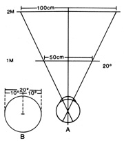
| Fig. 19. A. Diagram of tangent screens placed at 1- and 2-m viewing distances. B. A 20° diameter of central field is used as an example. Measured at the screens, the circle has a diameter of 50 cm at 1 m and a diameter of 100 cm when viewed from a distance of 2 m. The physiologic field of vision is actually a cone with the base outward. Visual field constrictions of functional origin show a tubular pattern with the patients failing to understand the effect of testing at variable distances, so that the field diameter is usually the same (or worse) at the more remote viewing distance. |