|
|
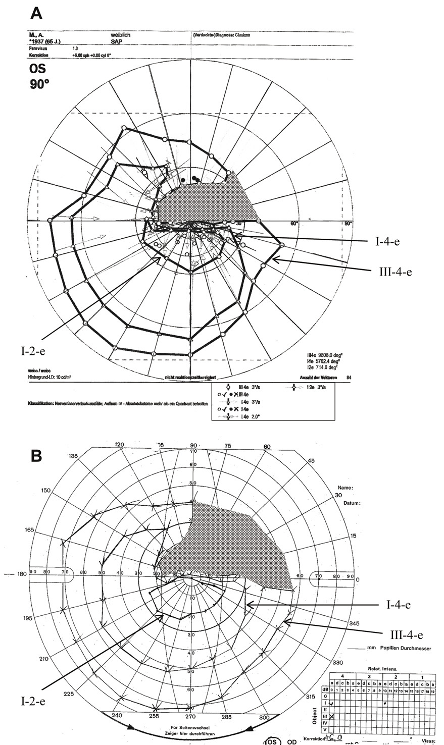
| Fig. 20. Visual field of the left eye of a 65-year-old patient with a superior arcuate scotoma from AION. Comparison of manual and computer-assisted kinetic perimetry. A. Visual field printout from the PKP (programmed kinetic perimetry) module on Octopus 101 instrument to three different stimuli (I-2, I-4, V-4). B. Manual kinetic perimetry on a Goldmann-type bowl perimeter performed on the same patient using the equivalent stimuli. In both A and B arrows labeling the isopters and shading highlighting the scotoma have been added to the original printouts. Note the somewhat smaller scotoma to the V-4 stimulus obtained with automated perimeter (see text). |