
|
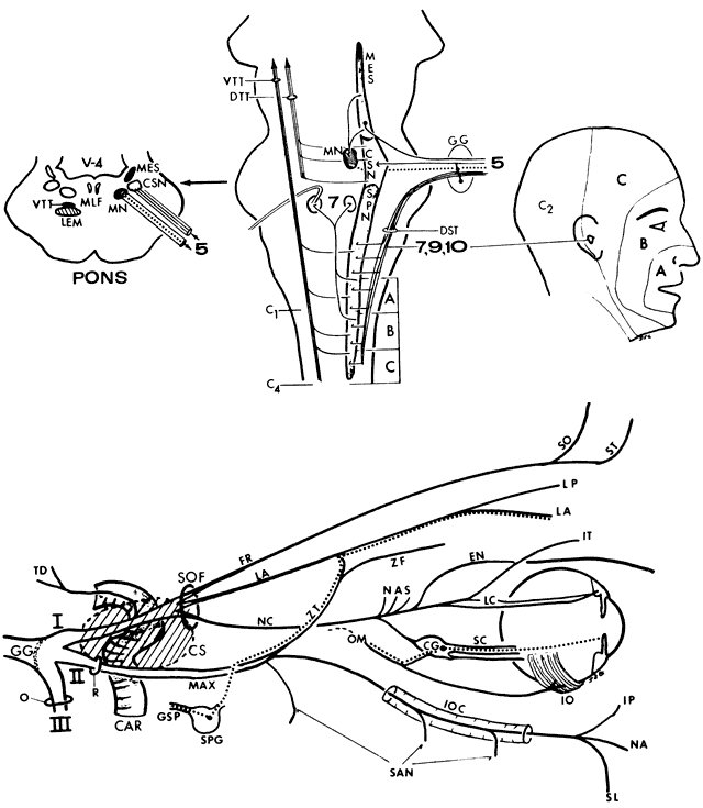
| Fig. 16. Top. Trigeminal-sensory complex. Ocular and facial sensation is mediated by the trigeminal nuclear complex, which extends from the midbrain downward into the upper cervical segments (C1 through C4). The rostral nucleus (MESencephalic) serves proprioception and deep sensation from the tendons and muscles of mastication. Axons of afferent cells also terminate in the motor nucleus (MN) of the trigeminal, which innervates the masticatory muscles (dotted line) via the mandibular division. The midportion of the trigeminal complex contains the chief sensory nucleus (CSN) located in the pons. This nucleus serves light touch from the skin and mucous membranes. Without a distinct transition, the nuclear complex continues caudally as the spinal nucleus (SPN), which receives pain and temperature afferents via the descending spinal tract (DST); cutaneous sensory components of 7, 9, and 10 also join the spinal tract, conducting sensation from the ear and external auditory meatus. Concentric areas of the face (A, B, and C) are projected on corresponding portions of the caudal aspect of the spinal nucleus. The corneal reflex is mediated via internuclear fibers to the facial nuclei. The fibers ascend to the thalamus via the ventral (VTT) and dorsal trigeminothalamic (DTT) tracts; an ipsilateral ascending DTT tract is not shown. The gasserian ganglion (GG) contains cell bodies of neurons mediating touch, pain, and temperature from three trigeminal divisions; cell bodies for proprioception and deep pain are contained in the MES. Cross-section of the pons at the level of the trigeminal roots includes the fourth ventricle (V-4), the median longitudinal fascicule (MLF), and the medial lemniscus (LEM). Bottom. Peripheral trigeminal distribution. Three major divisions arise from the gasserian ganglion (GG). The ophthalmic (I) enters the cavernous sinus (shaded area CS) lateral to the internal carotid artery (CAR), passes into the orbit via the superior orbital fissure (SOF), and divides into the frontal (FR), lacrimal (LA), and nasociliary (NC) branches. The supraorbital (SO) nerve supplies the medial upper lid and the conjunctiva, forehead, scalp, and frontal sinuses; the supratrochlear nerve (ST) supplies the conjunctiva, medial upper lid, forehead, and side of the nose. The lacrimal nerve (LA) serves the conjunctiva and skin in the area of the lacrimal gland (lateral palpebral branch, LP) and carries post-ganglionic parasympathetic fibers (dotted line) for reflex lacrimation. Pre-ganglionic fibers traverse the vidian canal with the greater superficial petrosal nerve (GSP) and enter the sphenopalatine ganglion (SPG), thence to the maxillary nerve, which transmits post-ganglionic fibers to the lacrimal nerve via an anastomosis, the zygomaticotemporal nerve (ZT). The nasociliary (NC) branch of the ophthalmic nerve supplies sensation to the globe. A series of nasal nerves (NAS) serves the mucosa of nasal septum, middle and inferior turbinates, and lateral nasal wall. An external nasal (EN) branch innervates skin of nose tip. The infratrochlear (IT) branch supplies the canaliculi, caruncle, lacrimal sac, conjunctiva, and skin of the medial canthus. Two long ciliary (LC) nerves carry sensory fibers from the ciliary body, iris, and cornea, and sympathetic innervation to the pupil dilator. Multiple short ciliary (SC) nerves transmit sensory fibers from the globe that pass through the ciliary ganglion (CG) and join the nasociliary nerve via its sensory root; short ciliaries also carry post-ganglionic parasympathetic fibers (dotted line) from the ciliary ganglion to the pupil constrictor and ciliary muscle. These fibers reach the ciliary ganglion via the inferior division of the oculomotor nerve (OM) destined to innervate the inferior oblique (IO) muscle. The maxillary division (MAX, II) does not usually traverse the cavernous sinus, but exits the skull through the foramen rotundum (R) into a variable “trigeminal” sinus, thence into the pterygopalatine fossa in relation to the sphenopalatine ganglion, thence into the inferior orbital fissure, continuing along the orbital floor into the infraorbital canal (IOC). The posterior, middle, and anterior superior alveolar nerves (SAN) supply the upper teeth, maxillary sinus, nasopharynx, tonsils, soft palate, and roof of the mouth. The infraorbital nerve exits onto face through infraorbital foramen, supplying the lower lid via the inferior palpebral (IP), side of the nose via the nasal (NA), and upper lid via the superior labial (SL) nerves. A zygomaticofacial (ZF) innervates side of cheek. The mandibular (III) is the only mixed motor-sensory division of the trigeminal. Both roots pass through the foramen ovale (O) into the infratemporal fossa; the motor branches supply the pterygoid, masseter, and temporalis muscles; the sensory branches supply the mucosa and skin of the mandible and lower lip, tongue sensation, external ear, and tympanum. A tentorial-dural branch (TD) arises from the intracavernous portion of the opthalmic, to supply sensation to the dura of the cavernous sinus, anterior fossa, sphenoid wing, petrous ridge, trigeminal cave (Meckel's), tentorium cerebelli, posterior aspect of falx cerebri, and dural venous sinuses. Sensation from cerebral veins and arteries, as well as nerves III, IV, and VI, may be mediated by these dural nerves. |