|
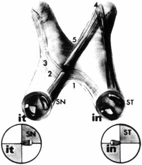
Fig. 14 Retinotopic
organization of visual fibers in the anterior visual pathways (after Hoyt).
Diagram of homonymous retinal quadrants and their fiber projections, anterior
aspect. it, inferior temporal; in, inferior nasal; SN,
superior nasal; ST, superior temporal. Note the following: the superior
fibers retain a superior course, and the inferior fibers retain an inferior
position; the anterior notch (1) is occupied by inferonasal (superior
temporal field) fibers; the inferonasal fibers bend slightly into the contralateral
nerve (2), Wilbrand's knee; inferior homonymous fibers converge in
the chiasm (3), but superior homonymous fibers converge beyond the
chiasm in the tract (4); the posterior notch (5) is occupied
by superior nasal (inferior temporal field) fibers, as well as macular fibers
(cf. Fig.
9). |