|
|
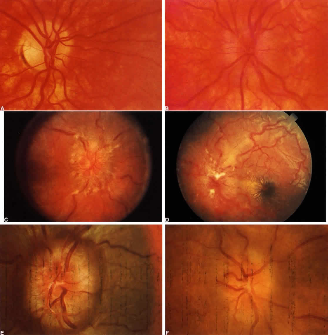
| Color Plate 5-4. A. Papilledema of raised intracranial pressure. In patient with frontal astrocytoma, right disk shows early edema of superior pole. B. Left disk of same patient shows more advanced edema, yet absence of hemorrahges, exudates, or engorgment. C. Fully developed papilledema in a case of pseudotumor cerebri. Multiple superficial infarcts of nerve fiber layer (“cotton-wool spots”). Veins are dilated and tortuous. The disk diameter appears enlarged by edema that spreads laterally into, and elevates, the retinal nerve fiber layer. Center of disk relatively spared. D. Severe papilledema associated with dural venous sinus thrombosis in young boy. Note exudative partial “star” figure at fovea. E. Chronic papilledema of many months duration. “Champagne cork” appearance after resolution of hemorrhages. F. Chronic papilledema after detumescence of edema, revealing pallor and formation of retinochoroidal venous shunts. |