|
|
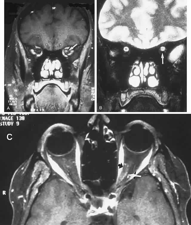
| Fig. 31. Optic neuritis. A 45-year-old woman with left eye acuity 20/100, eye movement pain, and normal optic disc: magnetic resonance imaging. A. Fat-suppression T1-weighted scan, with contrast, high-intensity signal of the left optic nerve (arrow). B. T2-weighted, FLAIR sequence shows a hyperintense left nerve (arrow); compare with the right nerve, with central dark nerve surrounded by hyperintense cerebrospinal fluid. C. Fat-saturated T1-weighted image with gadolinium shows a hyperintense signal of nerve (between arrows). |