|
|
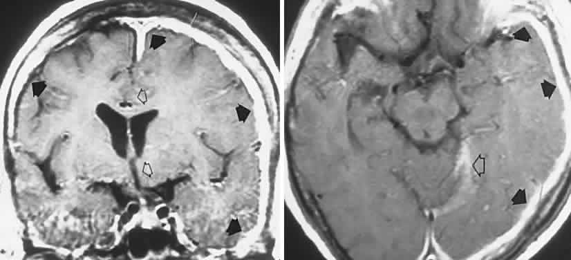
| Fig. 36. Chronic idiopathic pachymeningitis in a 64-year-old woman with headaches, bilateral visual loss, and left disc edema: magnetic resonance T1-weighted image with gadolinium. Left. Coronal section shows massive hypertrophic thickening of the meninges (black arrows), and rightward shift of the midline structures (open arrows) because of edema of the left hemisphere. Right. Axial section shows infiltration of the meninges and tentorium (open arrow); note also diffuse hemispheral edema. (From ref. 264) |