|
|
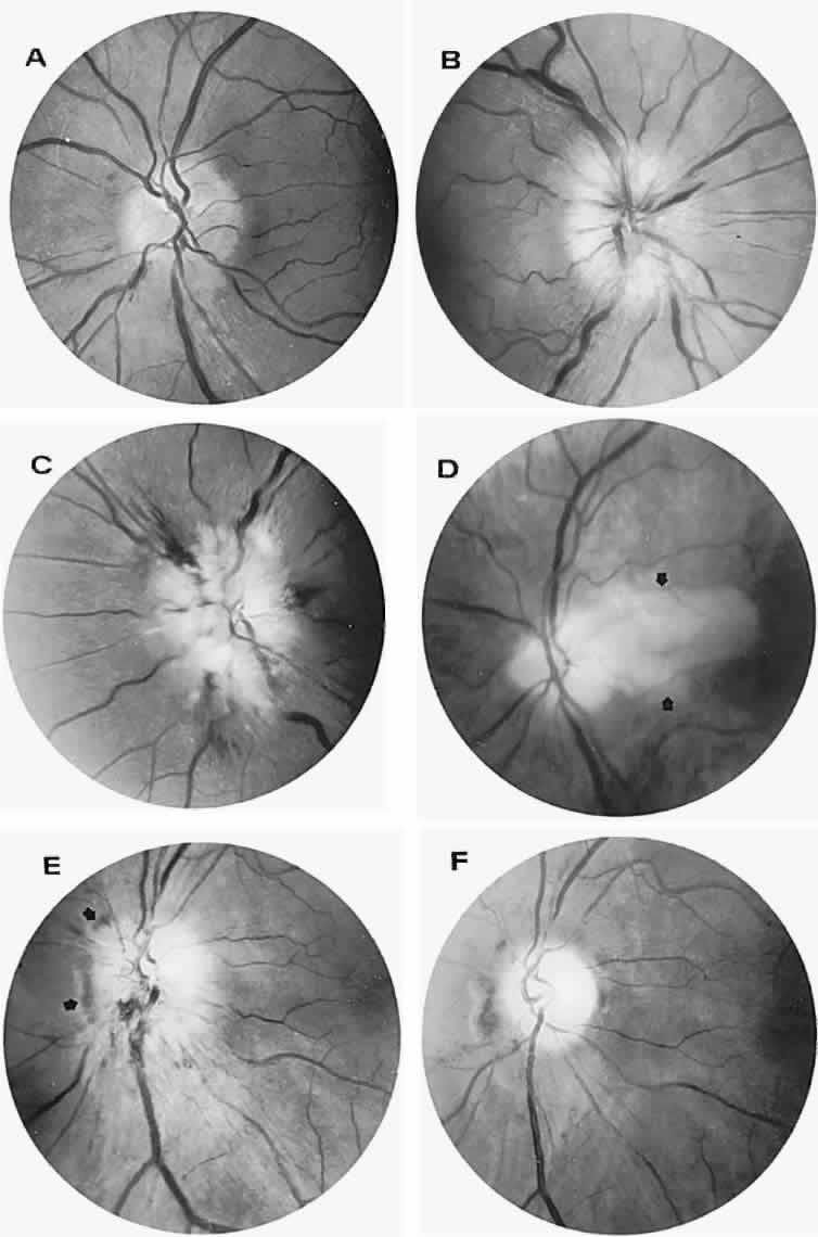
| Fig. 37. Ischemic optic neuropathy. A. Minimal swelling of inferior disc border with a few fine superficial linear hemorrhages. Arteries are narrowed. B. Diffuse disc swelling with pallid edema of the nerve fiber layer. Veins are slightly engorged. C. Massive disc swelling with hemorrhages and microinfarcts. D. Milky disc infarct without hemorrhages, extending into the retina in the distribution of the cilioretinal artery (arrows). The other eye was simultaneously involved, with similar fundus appearance. The patient had vision of hand movement in both eyes, erythrocyte sedimentation rate, 123 mm/hour, and a painful temporal artery. E. Acute infarction of the disc. Note subretinal hemorrhage at the nasal margin (arrows). F. Fundus in E 2 months later. The disc is atrophic, and the arteries are strikingly narrowed. The subretinal blood is incompletely resorbed. |