
|
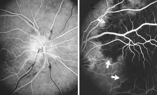
| Fig. 38. Fluorescein angiography in ischemic optic neuropathy (ION). Left. Common ION; at 16 seconds, arteriovenous phase shows telangiectasia of disc capillaries and normal choroidal background fluorescence. Right. Arteritis ION; even by 34 seconds, note dark, nonperfused choroid (arrows). |