
|
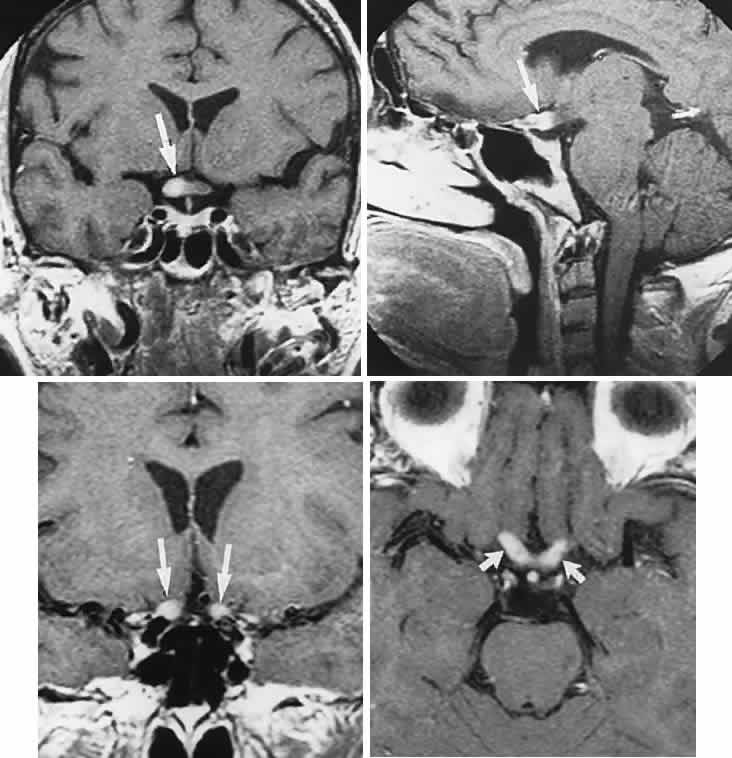
| Fig. 40. Vasculitis-induced optic neuropathies: magnetic resonance T1-weighted imaging with gadolinium enhancement. Top. A 54-year-old woman with rheumatoid arthritis and no light perception in the right eye. Left, coronal and right, sagittal sections show enlargement and contrast enhancement of the right optic nerve (arrow). Bottom. A 62-year-old woman with Sjögren's disease and visual loss. Left, coronal and right, axial sections, show contrast enhancement of both optic nerves. (Sklar EML, Schatz NJ, Glaser JS et al: MR of vasculitis-induced optic neuropathy. AJNR Am J Neuroradiol 17:121, 1996) |