
|
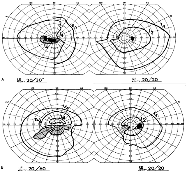
| Fig. 41. Central defects in glaucoma. A. A 62-year-old woman with slowly progressive visual loss in the left eye. Optic canal laminography were negative. Tonographic outflow values were 0.04 and 0.06. Note the centrocecal scotoma of the left eye and bilateral nasal defects. B. A 66-year-old man with long-standing reduced vision in left eye, previously diagnosed as amblyopia. Headache and left Gunn's pupil raised the question of a neurologic lesion. Findings were suggestive of glaucomatous discs. Fields show nasal step in the right eye, a central altitudinal nerve fiber bundle, and nasal step in the left eye. Co-efficients of outflow were 0.25 and 0.11. LE, left eye; RE, right eye. |