|
|
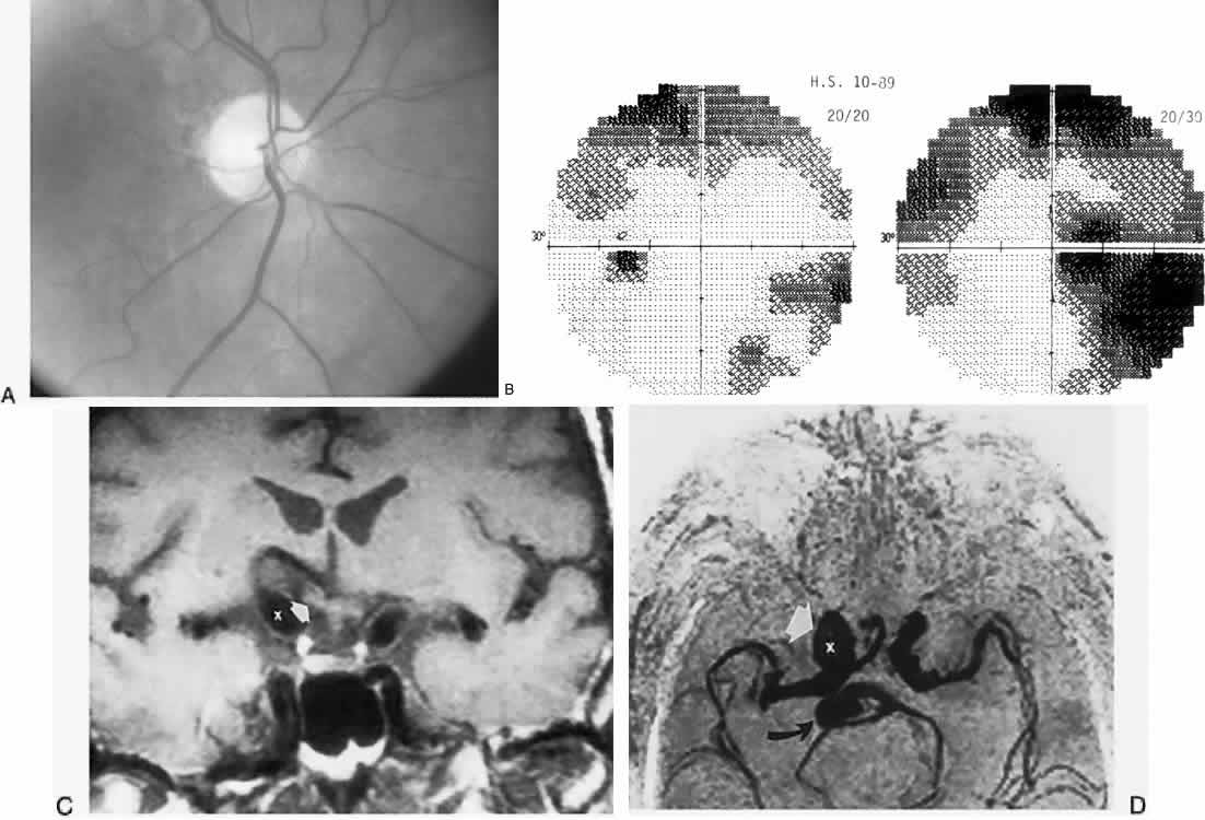
| Fig. 47. Optic neuropathy with carotid dolichoectasia in a 74-year-old man with progressive dimming of right eye vision: acuity of right, 20/30, and left, 20/20. A. Right optic disc shows a saucer-like excavation diagnosed as “normal tension glaucoma.” B. Visual fields. C. Magnetic resonance imaging shows elevation and distortion of the right side of the chiasm (arrows) by fusiform dilation of the internal carotid artery (x). D. Magnetic resonance angiogram demonstrates an ectatic right (x) carotid artery (arrow); the basilar artery (curved artery) is also ectatic. |