|
|
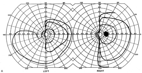
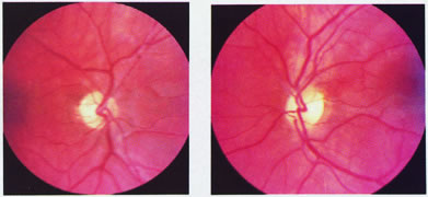
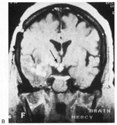
| Fig. 1. Optic tract lesion. A 53-year-old woman with central nervous system sarcoidosis diagnosed 7 years previously with focal motor seizures. She had no visual symptoms. Acuity was 20/40 in both eyes. A. There was RAPD in the left eye; visual fields showed incongruous left hemianopia. B. Fundoscopy showed temporal pallor of the right optic disc and “bow-tie” temporal and nasal pallor of the left optic disc. C. MRI showed enhancing lesions of right temporal lobe, infundibular region, and right optic tract (arrow). (From Rosen ES, Eustace P, Thompson HS, Cumming WJK [eds]: Neuro-Ophthalmology. London: Mosby, 1998.) |