
|
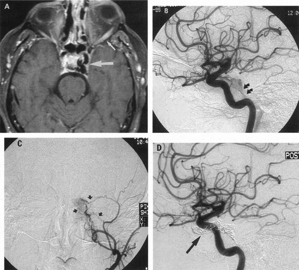
| Fig. 20. A 54-year-old man presented with worsening proptosis and chemosis of his left eye following endoscopic sinus surgery. A: Contrast-enhanced T1-weighted axial magnetic resonance imaging demonstrates an asymmetric dilation of veins (large arrow) in the left cavernous sinus; note enlarged lateral rectus muscle (small arrow). Lateral view of selective left internal carotid arteriogram (B) and frontal view of selective left external carotid arteriogram (C) demonstrate opacification of the cavernous sinus through numerous small vessel shunts; note opacification of the inferior petrosal sinus (small arrows in B), indicating a potential route for transvenous approach. The cavernous sinus was packed with fibered coils using transvenous access via the inferior petrosal sinus. D: Lateral view of post-embolization angiogram of the left internal carotid artery shows complete closure of the malformation. The mesh of packed coils is seen on the subtracted image (arrow). |