|
|
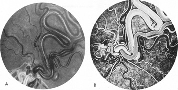
| Fig. 27. Racemose angioma of the retina. A: Knot of retinal tissues at the inferior aspect of the photograph represents the area of the optic disc. Visual acuity is finger-counting. B: Fluorescein angiogram demonstrates complex flow patterns, including rapid arteriovenous shunting. The patient had an arteriovenous malformation of the right maxilla. (Case reported as von Hippel's disease by LaDow CS, McFall TA. Central hemangioma of the maxilla, with von Hippel's disease: Report of case. J Oral Surg 22:252, 1964. Copyright by American Dental Association. Reprinted by permission. Photographs courtesy of Mr. John Justice) |