|
|
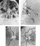
| Fig. 30. Orbital venous varix. A 27-year-old man was evaluated for two episodes of severe left orbital pain; the last episode was accompanied by spontaneous hemorrhage in the lower lid. Motility and vision were normal, but 3 mm of proptosis was present. Orbital venography was performed. The unsubtracted frontal projection (A) shows two lobulated venous varices (arrows), which are also demonstrated by the lateral subtracted view (B). C: Right fundus is normal. D: Left fundus shows anomalous dilated and tortuous veins. |