|
|
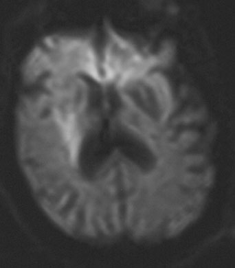
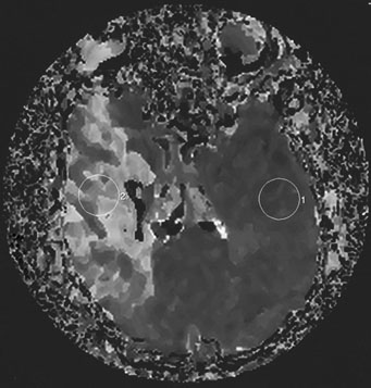
| Fig. 1. An 81-year-old man with atrial fibrillation developed acute onset of left-sided weakness and presented to hospital 3 hours and 40 minutes after onset of symptoms. A: The diffusion-sequence magnetic resonance imaging (MRI), with infracted tissue in the right basal ganglia in white. B: Decreased perfusion in the right middle cerebral artery distribution (white area). Given the relatively small diffusion abnormality and the large perfusion defect, it was decided to proceed with thrombolysis despite the time elapsed. |