|
|
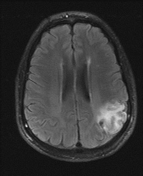
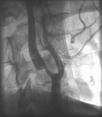
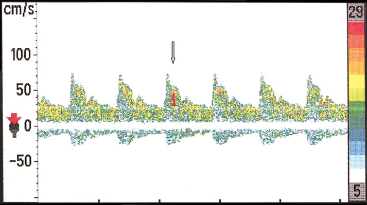
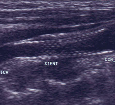
| Fig. 2. This 48-year-old diabetic man suddenly developed confusion and clumsiness of the right hand. On examination, he had inability to identify objects by touch and difficulty identifying the right from the left. A. Fluid attenuated inversion recovery (FLAIR) MRI shows a left parietal cortical infarct. Two potential embolic sources were noted: a left ventricular apical clot was seen on echocardiography from a presumed recent silent myocardial infarction, and ultrasound and subsequent angiography confirmed a significant left internal carotid stenosis. B: A 70% left internal carotid artery origin stenosis is shown on angiography. Monitoring both middle cerebral arteries with transcranial Doppler showed microembolic signals only over the left, suggesting that the carotid atherostenotic plaque was active or destabilized. C: An interruption (arrow) of the normal Doppler flow pattern of the middle cerebral artery is shown, representing a microembolic signal. The artery was stented rather than revascularized by endarterectomy because of the recent myocardial infarction. D: A patent revascularized carotid is revealed by ultrasound; note the struts of the stent. |