|
|
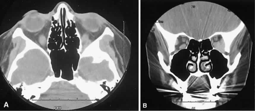
| Fig. 2. Orbital varix. A. Axial view shows a small soft tissue mass within the inferolateral left orbital apex. B. Coronal view with patient repositioned prone with neck extension. Increased venous pressure distends the varix, accounting for increased size. |