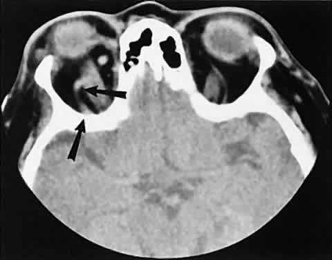
Fig. 3.
Axial view showing a dilated superior ophthalmic vein (SOV;
arrows
) secondary to dural-cavernous sinus fistula. Note the normal-sized SOV in the contralateral orbit.