|
|
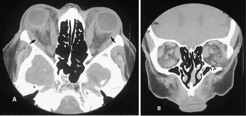
| Fig. 8. Low-grade lymphoma confined to orbit mistaken for Graves' ophthalmopathy in a 65-year-old man. A. On the axial view, orbital fat appears “dirty” with marked increase in soft tissue stranding. Muscles do not have a smooth appearance, lateral rectus muscles have lumpy appearance (arrow), and both lateral rectus muscles are disproportionately large for what typically is seen in Graves' orbitopathy. B. Coronal view also shows dirty orbital fat. Note left inferior rectus, which is small (arrowhead), and also is atypical in Graves' orbitopathy when there is enlargement of the other extraocular muscles. |