|
|
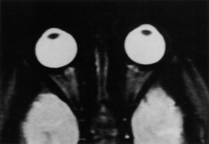
| Fig. 9. T2-weighted image of the orbits does not delineate normal anatomic details well. The lens can be visualized in the bright vitreous. (Dortzbach RK, Kronish JW, Gentry LR: Magnetic resonance imaging of the orbit. Part II. Clinical applications. Ophthal Plast Reconstr Surg 5:161, 1989) |