|
|
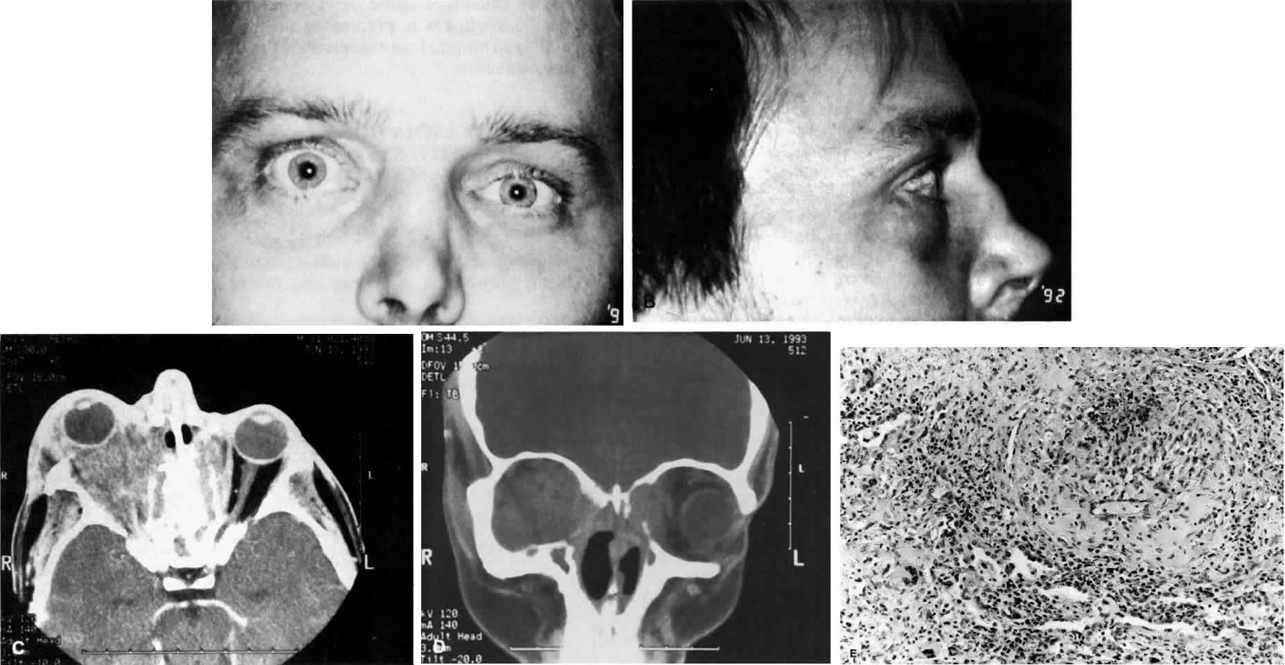
| Fig. 11. A. This 31-year-old man has had Wegener's granulomatosis for 2 years. His disease process is stable on chronic corticosteroid therapy. He has no evidence of systemic disease. Note right-sided proptosis and hyperglobus. B. Profile of same patient demonstrating collapse of nasal bridge from bony destruction secondary to Wegener's granulomatosis. Note presence of swelling in lower eyelid. C. Axial CT image from the same patient demonstrating significant bilateral disease and bony destruction. Despite the extent of the orbital process on the right, the patient does not have diplopia. D. Coronal CT image showing destruction of medial orbital walls, vomer, and orbital septum. E. Pulmonary biopsy specimen from patient with orbital signs contains an almost obliterated vessel to right of center and scattered giant cells on left (H&E, ×160). |