|
|
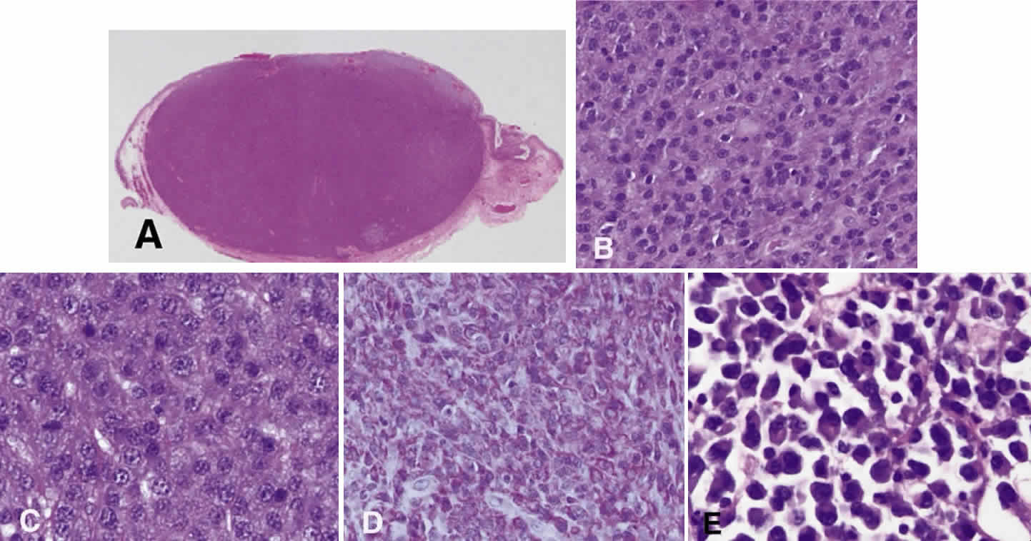
| Fig. 16. Plasmacytoma. A. A rounded conjunctival stromal nodule. The immediate subepithelial region contains a slightly more blue lymphoid infiltrate. The inferior portion of the lesion contains a recognizable lymphoid follicle within its substance (hematoxylin and eosin, × 1.25). B. A reasonably homogeneous population of large cells with pink cytoplasm and regular nuclei predominate in the nodule (hematoxylin and eosin, × 10). C. The typical “clock face” or “cartwheel” nuclei are visible on higher magnification as well as the abundance of pinkish blue cytoplasm (hematoxylin and eosin, × 40). D. A section stained with methyl green pyronin demonstrates the presence of red pyrinophilic cytoplasm typical of cells rich in ribosomes (methyl green pyronin, × 10). E. Plasmacytoid differentiation is apparent, with eccentrically placed nuclei and paranuclear cytoplasmic pallor (“hof”) (hematoxylin and eosin, × 40). (Courtesy of Dr. A. Mowat) |