|
|
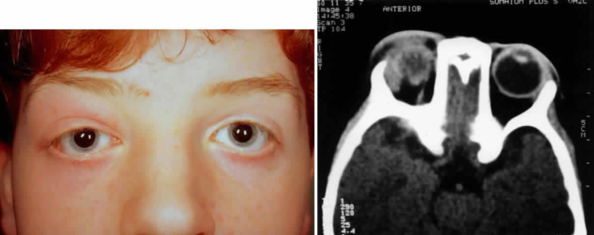
| Fig. 3. A 17-year-old male with a 1-week history of pain and swelling over the right lacrimal gland. A. Clinical photograph demonstrating inflammation of the right lacrimal gland with minimal erythema and edema of adjacent soft tissues. B. Axial computed tomograph demonstrating the poorly demarcated, oblong lesion associated with inflammation of the lacrimal gland. |