|
|
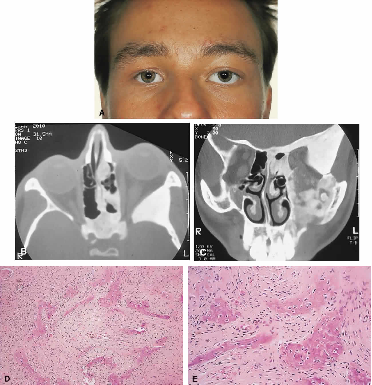
| Fig. 2. A. A 20-year-old man presented with a longstanding history of left proptosis and facial asymmetry. B and C. Bone window CT scan showed extensive fibrous dysplasia involving the greater wing of the sphenoid in a sclerotic fashion and a more pagetoid appearance in the maxillary and ethmoidal regions. D and E. Dominant histologic features consisted of irregular trabeculae of woven bone in a fibrous stroma with minimal osteoblastic activity (E) surrounding the osteoid (hematoxylin-eosin; D × 20, E × 50). |