|
|
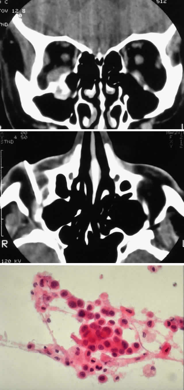
| Fig. 3. A 73-year-old man presented with a 2-year history of infraorbital numbness and burning sensation, which had progressed to include the supraorbital region, forehead, and lower face during the past 6 months. He had been treated with radiotherapy for prostate carcinoma 7 months before orbital presentation. Examination was significant for proptosis of 2 mm and dysesthesia, including corneal numbness, in all three divisions of cranial nerve V. CT scan revealed a soft tissue mass in the inferior orbit contiguous with the inferior rectus muscle (A). The lesion extended through the infraorbital canal to involve the pterygopalatine fossa and was associated with soft tissue hanging into the upper portion of the maxillary sinus. An axial CT scan-guided aspiration biopsy was performed (B) and revealed squamous cell carcinoma. Groups of cohesive malignant squamous epithelial cells (C) were noted to have pleomorphic nuclei and abundant eosinophilic to orange cytoplasm, with no features of mucinous differentiation (H & E, × 320). (C from White VA, Rootman J: Orbital pathology. In Albert DM, Jakobiec FA (eds): Principles and Practice of Ophthalmology, Vol 4, p 2342. Philadelphia, WB Saunders, 1994.) |