|
|
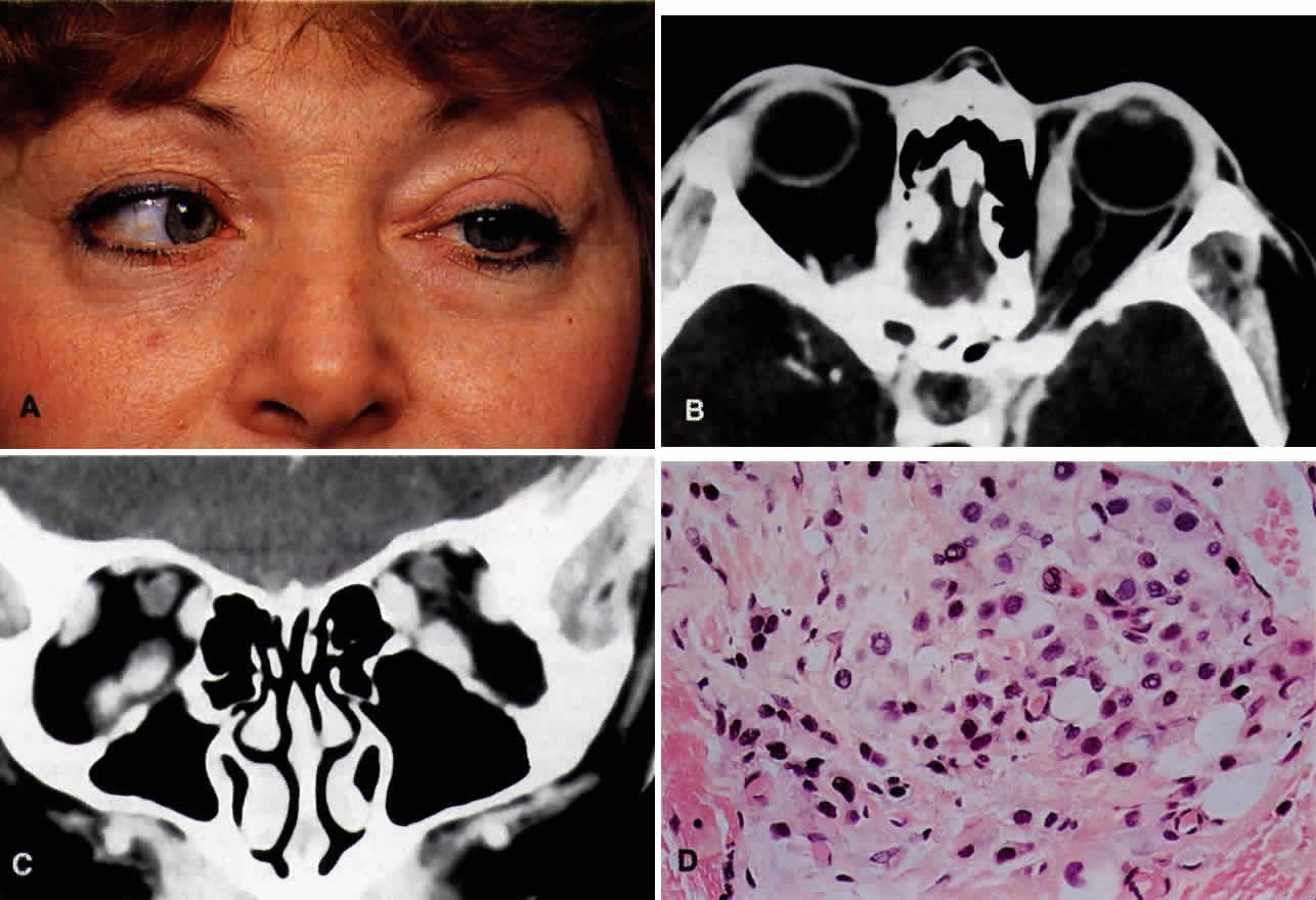
| Fig. 18. A 46-year-old white woman presented with a 3-month history of progressive horizontal diplopia on left gaze. She had a history of right breast carcinoma and had undergone mastectomy 5 years before the orbital presentation. She was orthophoric in primary gaze, with 1 mm of left enophthalmos; however, the left eye was limited to just 10° of abduction (A). Abduction on the right was also limited to 45°. CT scan revealed enlargement and enhancement of the left medial rectus greater than the right, and of both inferior recti (B and C). Orbital biopsy revealed metastatic adenocarcinoma, and on further examination she was found to have a left breast carcinoma with axillary lymphadenopathy. Routine histopathology (D) demonstrates a poorly differentiated adenocarcinoma with some “Indian file” configuration of tumor cells (H & E, × 320). |