Chapter 8
The Facial Nerve
STEVEN L. GALETTA and MARK MAY
Main Menu   Table Of Contents

Search

EMBRYOLOGY
ANATOMY
FACIAL NERVE FUNCTION AND ASSESSMENT
IDIOPATHIC (BELL'S) FACIAL PALSY
INFECTIOUS AND IMMUNE-MEDIATED NEUROPATHIES
TRAUMA
TUMORS
MISCELLANEOUS CONDITIONS
BILATERAL OR RECURRENT FACIAL NERVE PARESIS
FACIAL EMBRYOPATHIES AND CHILDHOOD PALSIES
HYPERKINETIC FACIAL DISORDERS
DISORDERS OF EYELID OPENING
MANAGEMENT OF FACIAL PALSY
REFERENCES

The facial nerve, by virtue of its unique neuroanatomy and physiology and its complex course across the skull base and face, is often involved in neuro-ophthalmologic problems. Abnormalities of lid position and defective tearing are regularly symptomatic, demanding ophthalmologic attention. This chapter provides both an anatomic and an etiologic approach to the spectrum of facial nerve disorders. The current management of facial nerve paresis and facial hyperkinetic syndromes is emphasized.
Back to Top
EMBRYOLOGY
Normal and abnormal functions of the facial nerve can be best understood through an awareness of its embryonic development. The complexity of the nerve's course, its branching patterns, and its anatomic relationships are established during the first 3 months of prenatal life.1 During this period, the muscles of facial expression also differentiate, become functional, and actively contract.2 Critical phases in facial nerve development occur throughout gestation, and the nerve is not fully developed until about 4 months after birth (Table 1).


TABLE 1. Time During Gestation When Anatomic Structures Appear


Week of GestationStructures Noted
3rdCollection of neural crest cells, anlage of VII cranial nerve nucleus (CNN)
5thChorda tympani, greater petrosal, VII motor nucleus
6thExternal genu, postauricular branch, branch to posterior belly of digastric
7thGeniculate ganglion, nervus intermedius
8thStapedius nerve, temporofacial and cervicofacial part of extracranial facial nerve
End of 8thTerminal branches of VII
7th to 8thMyoblasts that will form the facial muscles
12thAll facial muscles


INTRAMEDULLARY SEGMENT

The motor nuclei of the sixth and seventh cranial nerves are initially in close proximity in the pontine subdivision of the metencephalon. As the metencephalon elongates and expands, the facial nucleus migrates ventrolaterally in relation to the abducens nucleus, displacing facial motor axons that loop dorsally in the floor of the fourth ventricle to form the facial colliculus (Fig. 1). This intimate relationship between the abducens and facial motor nuclei is the anatomic substrate for the clinical findings in congenital Möbius' syndrome and in acquired inflammatory, vascular, and neoplastic lesions that involve the intramedullary segment of the facial nerve.

Fig. 1 Cross section of pons, with relationships of facial motor nucleus (VII), abducens nucleus (VI), corticospinal tract, trigeminal nucleus, and tract. Facial colliculus (arrow). (Carpenter M: Core Text of Neuro-Anatomy. 3rd ed. Baltimore: Williams & Wilkins, 1985:152)

EXTRAMEDULLARY SEGMENT

The facial nerve rootlets become distinguishable near the end of the seventh week of gestation, when the geniculate ganglion becomes well defined.1 The nervus intermedius, which contains afferent and general visceral efferent fibers, exits the brain stem between the facial motor root and the vestibular nerve. The afferent fibers arise from the geniculate ganglion, whereas the preganglionic parasympathetic fibers originate in the superior salivatory nucleus in the brain stem (Fig. 2). The fact that the geniculate ganglion and the parasympathetic fibers form independent of the motor pathways allows patients with congenital facial paralysis to enjoy intact tearing and taste, but with selective impairment of the motor neurons innervating the facial muscles.

Fig. 2 Brain stem components and peripheral branches of facial nerve. (Dejong RN: The Neurologic Exam. 4th ed. Hagerstown, MD: Harper & Row, 1979)

INTRATEMPORAL SEGMENT

Small but important ganglia are scattered along the course of the facial nerve through the temporal bone.3 These ganglia are likely composed of aberrant sensory neurons from the tympanic plexus that enter the facial nerve near the stapedial branch. Because most viruses have an affinity for afferent nerve fibers, such sensory fibers may play an important role in the development of viral-immune facial nerve palsies.

Of all the cranial nerves, the facial nerve has the largest number of communications with other nerves.4 This situation may account for the variety of neurologic signs sometimes seen with herpes zoster cephalicus. The cutaneous branches of the second and third cervical ganglia (part of the cervical plexus of spinal nerves) are the initial nerves that establish connections with the facial nerve at its emergence from the stylomastoid foramen. In the facial palsy of herpes zoster cephalicus, these communications predict the distribution of the cutaneous eruption that involves the angle of the jaw, the back of, the head, and the neck, thus following the pattern of the C2–C3 dermatome (see later).

ANOMALIES

The facial nerve develops within the second branchial arch during the same period that closely adjacent derivatives of the first arch, the first internal pouch and external groove, are forming the external and middle ear regions. Therefore, anomalies of the facial nerve within the temporal bone should be anticipated whenever there are associated malformations of the external or middle ear. In these cases, displacement of the facial nerve and complete absence of the bony facial canal are the most common anomalies observed. Typically, the facial nerve can be seen sagging inferiorly against the stapes bone.5

Back to Top
ANATOMY
Familiarity with the general anatomy of the neural pathways for facial function is essential for accurate diagnosis and appropriate management of the spectrum of facial neuropathies. Specific diagnostic possibilities can be implied by precise regional localization of typical lesions (Table 2).


TABLE 2. Topical Diagnoses of Lesions of the Facial Nerve at Various Levels


LevelSignsProbable Diagnosis
Supranuclear  
 Cortex and internal capsuleTone and upper face intact, loss of volitional movement with intact spontaneous expression, slurred speech (tongue weakness), hemiparesis on side of facial involvement. Paresis of upper extremity begins with involvement of thumb, finger, and hand movement.Lesion of motor cortex or internal capsule on opposite side of facial involvement
 ExtrapyramidalIncreased salivary flow, spontaneous facial movement impaired, volitional facial movement intact, masked face of parkinsonism or dystonia, progressive hemifacial spasmTumor or vascular lesion of basal ganglia: parkinsonism, Meige's syndrome (cervical facial dystonia)
 MidbrainOculomotor paresis on opposite side of lower facial paresis and hemiparesisSyndrome of Weber
 Pontine nucleusInvolvement of cranial nerves VII and VI on side of facial paresis; associated ataxia, cerebellovestibular signs, and contralateral hemiparesisInvolvement of pons at level of VII and VI nuclei by pontine glioma, multiple sclerosis, infection, infarction
 Cranial nerves VII and VI palsy noted from time of birth with or without other congenital anomalies. Facial motor involvement usually incomplete with sparing of corner of mouth or lower lip common. Another type of presentation is involvement of the lower lip with complete or partial sparing of upper face. Anomalies of the pinna, canal, or mandible associated with facial palsy indicate developmental defect of facial nerve.Developmental facial palsy (noted at birth), oculofacial syndrome, Möbius' syndrome, thalidomide toxicity. Nondevelopmental facial or abducens nerve anomalies are most often due to infranuclear lesions.
Infranuclear  
 Intracranial cerebellopontine angleImpairment of hearing, (especially discrimination out of proportion to pure tone scores), possible ataxia, abnormalities of tearing or taste, stapes reflex decay, decreased corneal sensation, facial motor deficit (late sign)Acoustic schwannoma
 Abnormalities in trigeminal, acoustic–vestibular, and facial nerve function, starting with facial pain or numbness. Lesion noted on CT (enhancement with contrast) may show evidence of calcification.Meningioma
 Abnormalities of facial and acoustic–vestibular nerve function, may start with facial twitching. Erosion or lytic area evident on CT of temporal bone.Cholesteatoma or facial schwannoma arising in temporal bone
 Abnormalities of cranial nerves VII, VIII, IX, X, XI, and XII; pulsatile tinnitus and purple–red pulsating mass bulging through the tympanic membraneGlomus jugulare tumor
 Abnormalities of abducens nerve in addition to aboveGlomus jugulare tumor extending to petrous apex to involve middle fossa
 Skull baseConductive or sensorineural hearing loss, acute or recurrent facial palsy, positive family history, abnormalities of bone density on CTOsteopetrosis
 Multiple cranial nerve involvement in rapid successionCarcinomatous meningitis, leukemia, Landry-Guillain-Barré, mononucleosis, diphtheria, tuberculosis, sarcoidosis, malignant external otitis
Transtemporal Bone  
 Internal auditory canal and labyrinthine segment of facial nerveEcchymosis around pinna and mastoid prominence (Battle's sign), hematotympanum with sensorineural hearing loss (tuning fork lateralizes to normal side), vertigo, nystagmus (fast component away from involved side), sudden complete facial paralysis after head trauma (usually associated with basilar skull fracture), loss of consciousness, and cerebrospinal fluid leak. Transection of facial nerve is more likely with this injury compared with longitudinal fracture.Temporal bone fracture (transverse, longitudinal, or combination)
 Geniculate ganglion Dry eye, decreased taste and salivation. Erosion of geniculate ganglion area or middle fossa is demonstrated by pluridirectional tomography and CT of temporal bone. Schwannoma, meningioma, cholesteatoma, hemangioma, arteriovenous malformation
  Ear pain, vesicles on pinna, dry eye, decreased taste and salivary flow, sensorineural hearing loss, nystagmus, vertigo, red chorda tympani nerve. Facial palsy may be complete or incomplete, or may progress to complete over 14 days. Herpes zoster cephalicus (Ramsey-Hunt syndrome)
  Same as above with vesicles; no other cause evident. Facial palsy may be complete or incomplete, or may progress to complete over 10 days. Idiopathic (Bell's) palsy (viral-inflammatory immune disorder)
  Same as above but no recovery in 6 months Tumor
  Ecchymosis around pinna and mastoid (Battle's sign), hematotympanum, conductive hearing loss (tuning fork lateralizes to involved ear), no vestibular involvement unless stapes subluxed into vestibule (causes fluctuating sensorineural hearing loss and vertigo with nystagmus) Longitudinal fracture of temporal bone; may be proximal or at geniculate ganglion (tearing symmetrical; tear test valid only in acute injury)
 Tympanomastoid Decreased taste and salivation, loss of stapes reflex and symmetrical tearing, sudden-onset facial palsy, which may be complete or incomplete or may progress to complete  
  Pain, vesicles, red chorda tympani Herpes zoster cephalicus
  Pain without vesicles, red chorda tympani Bell's palsy
  Red, bulging tympanic membrane, conductive hearing loss, usually history of upper respiratory infection. Lower face may be involved more than upper. Acute suppurative otitis media
  Pulsatile tinnitus, purple-red pulsatile mass noted through tympanic membrane Glomus tympanicum or jugulare
  Recurrent facial paralysis, positive family history, facial edema, fissured tongue; may present with simultaneous bilateral facial paralysis Melkersson-Rosenthal syndrome
Extracranial Incomplete facial nerve paresis; hearing balance, tearing, stapes reflex, taste, salivary flow spared Penetrating wound of face; sequelae of parotid surgery; malignancy of parotid, tonsil, or oronaso-pharynx, rarely with benign lesion of parotid gland compressing facial nerve
  Uveitis, salivary gland enlargement, fever Sarcoidosis (Heerfordt's syndrome), lymphoma
Sites Variable Bilateral facial paralysis from birth Möbius' syndrome
  Bilateral facial paralysis acquired Landry-Guillain-Barré syndrome, sarcoidosis, mononucleosis, leukemia, idiopathic (Bell's) palsy
  Facial paralysis, especially simultaneous bilateral facial paralysis with ascending areflexic quadriparesis; sensory changes usually mild, with spinal fluid typically showing albuminocytologic dissociation (elevated protein without cells) Landry-Guillain-Barré syndrome
  Deficits of cranial nerves VI and VII or VII, VI, and III, possibly in association with marked increase in jaw jerk or gag reflex Carcinoma of nasopharynx, metastatic carcinoma from breast, ovary, prostate; meningitis, leukemia, diabetes mellitus
Pseudobulbar Palsy Inappropriate or exaggerated laughing or crying; may be associated with marked increase in jaw jerk or gag reflex Demyelinating, degenerative, or vascular process involving bilateral corticobulbar pathways

CT, computed tomography


CORTEX AND SUPRANUCLEAR PATHWAY

The voluntary responses of facial muscles, such as smiling or grimacing on command, are dependent on discharges from the facial motor area situated in the precentral gyrus of the frontal cerebral cortex. The facial motor areas are represented with the forehead uppermost and the eyelids, midface, nose, and lips located sequentially below (Fig. 3). Supranuclear motor neurons from the cortical face area are carried as fascicles of the corticobulbar tract to the genu of the internal capsule, then via the cerebral peduncles to the lower pons or upper medulla. The portion of the facial nucleus that supplies the muscles of the upper face (frontalis, orbicularis oculi, and corrugator muscles) receives corticobulbar fibers from both right and left precentral motor cortices, but the supranuclear tracts innervating the lower face are crossed only. For this reason, the muscles that raise and wrinkle the forehead and close both eyes are bilaterally innervated. Thus, a unilateral lesion in the cortex or supranuclear pathways spares eyelid closure and forehead movement but results in paralysis of the contralateral lower face. This dissociation is characteristic of supranuclear lesions. Because the supranuclear pathways may descend to the ventromedial medulla before decussating to innervate the facial nucleus, low brainstem lesions may produce contralateral lower facial weakness.6 However, it is also possible to show upper facial sparing with lesions of the pontine facial nucleus, with selective defects within the temporal bone, or even with an injury to nerve rootlets within the parotid gland or facial musculature. This phenomenon is related to the general tendency for facial nerve function to be spatially dispersed, not only in its cortical distribution but also within the pontine nucleus and in the peripheral nerve.

Fig. 3 Organization of facial motor function. Note cortical distribution, pontine nuclear connections, and relationships to other cranial nerves on ventral view of brain stem.

Because preservation of forehead function is insufficient evidence of a cortical lesion, other neurologic signs should be sought. A cortical lesion that produces a contralateral lower facial palsy is usually associated with a motor deficit of the tongue and with weakness of the thumb, fingers, and hand on the same side as the hemifacial weakness, with lesser involvement of the leg. The cortical motor areas of the face, tongue, thumb, fingers, hand, and upper extremities lie near each other in the precentral territory nourished by the rolandic branch of the middle cerebral artery. Patients with such cortical lesions are unable to voluntarily smile, but facial expression is appropriate in response to an amusing story. Similar clinical findings may occur with lesions that involve the descending corticobulbar and corticospinal tracts.7

Although facial muscle tone is not significantly impaired with a supranuclear lesion, slight flattening of the nasolabial fold and drooping in the corner of the mouth may be detected contralateral to the cortical lesion.

EXTRAPYRAMIDAL SYSTEM

The extrapyramidal system (Fig. 4) consists of the basal ganglia and the descending motor projections other than the fibers of the corticospinal (pyramidal) tract. Anatomically, this system appears to be a diffuse, multisynaptic network that interconnects extensively with centrencephalic and brain stem structures. These pathways are not as well clarified as those of the primary (pyramidal) motor tracts. The extrapyramidal system is concerned with automatic and emotional facial language. The dull, expressionless face of parkinsonism is a well-known result of extrapyramidal pathway disease, whereas the spontaneous facial dystonia of Meige's syndrome is characterized by unilateral or, more often, bilateral blepharospasm associated with dystonic movements of the mouth and other lower facial muscles. Progression of this latter disorder may lead to chaotic contractions of the tongue and cervical muscles.

Fig. 4 Extrapyramidal connections for supranuclear facial pathways.

PONS

The facial motor nucleus contains about 7,000 motor nuclei 8 and is located in the ventrolateral angle of the lower pontine tegmentum (see Fig. 1). The facial nucleus can be divided into four separate cell groups that supply specific muscle groups: (1) dorsomedial (auricular and occipital muscles), (2) intermediate (frontalis, corrugator, and orbicularis oculi muscles), (3) ventromedial (platysma), and (4) lateral (buccinator and buccolabial).9 The motor axons exit the nucleus dorsally, loop around the abducens (VI) nucleus, and form the facial genu before emerging from the lateral aspect of the pons. The superior salivatory nucleus, which is located just rostral to the facial motor nucleus, is the origin of the parasympathetic fibers that supply the sublingual, submandibular, and lacrimal glands. These salivary and lacrimal fibers join the facial nerve as the nervus intermedius in the cerebellopontine angle.

Because the facial nucleus is located ventromedial to the cochlear nuclei and the spinal tract and nucleus of the trigeminal nerve, a lesion of the lateral pons may result in ipsilateral facial paresis, ipsilateral facial analgesia, ipsilateral Horner's syndrome, and ipsilateral deafness (Foville's syndrome). If the lesion extends further dorsally, an ipsilateral gaze paresis would result from involvement of the sixth nerve nucleus. The combination of a unilateral sixth nerve palsy, an ipsilateral seventh nerve palsy, and a contralateral hemiparesis is known as the Millard–Gubler syndrome. Intrinsic lesions of the brain stem are usually the result of infarction, hemorrhage, tumor, or demyelination.

CEREBELLOPONTINE ANGLE

The facial nerve, the nervus intermedius, and the eighth (vestibuloacoustic) cranial nerve exit together from the ventrolateral aspect of the pons, surrounded by a leptomeningeal covering. In the lateral pontine cistern, the anteroinferior cerebellar artery may loop between the seventh and eighth cranial nerves as the artery courses posteriorly to supply the dorsolateral pons and cerebellum.10 As the nervus intermedius approaches the internal auditory meatus, it joins the facial nerve. Because of the association of the facial nerve with the nervus intermedius and the vestibuloacoustic nerves at the level of the cerebellopontine angle and in the internal auditory canal, tearing, taste, submandibular saliva flow, hearing, and balance are disturbed with mass lesions at this level (Fig. 5).

Fig. 5 Anatomic relationships of facial nerve trunk and brain stem structure. Anterior view of ventral aspect of brain stem and clivus, especially cerebellopontine angle. The cerebral hemispheres were removed and the oculomotor and trigeminal nerves were divided to provide this view of the anterior surface of the brain stem. A. The superior cerebellar arteries (SCA) and posterior cerebral arteries (PCA) arise from the superior end of the basilar artery (BA). The oculomotor (III) and trochlear (IV) nerves are above and the trigeminal (V) nerves are below the superior cerebellar arteries. The right anterior inferior cerebellar artery (AICA) arises from the basilar artery, courses below the abducens nerve (VI), and passes between the nervus intermedius (VII NI) and the facial motor root (VII) anteriorly and the vestibulocochlear nerve (VIII) posteriorly. The left AICA is a duplicate artery. The rostral duplicate AICA (Dup. Ro. AICA) is not nerve related, but the caudal AICA (Dup. Ca. AICA) is nerve related and passes between the facial and vestibulocochlear nerves. The left posteroinferior cerebellar artery (PICA) arises from the left vertebral artery (VA). The hypoglossal (XII), glossopharyngeal (IX), vagus (X), and spinal accessory (XI) nerves are lateral to the vertebral arteries. The carotid arteries (CA) and the pituitary stalk (Stalk) have been divided. The premeatal segments (Pre. Mea. Seg.) approach the nerves, the meatal segments (Mea. Seg.) pass between the nerves, and the postmeatal segments (Post. Mea. Seg.) pass above the flocculus. A recurrent perforating artery (RPA) and an internal auditory artery (IAA) arise from the right AICA. B. Enlarged anterosuperior view of the right cerebellopontine angle. The premeatal segment passes below the abducens nerve, the meatal segment passes between the eighth cranial nerve and the nervus intermedius, and the postmeatal segment passes above the flocculus. A recurrent perforating artery and an internal auditory artery arise from the meatal segment. Pontine arteries arise from the right side of the basilar artery. (Martin RG, Grant JL, Peace D: Microsurgical relationship of the anterior inferior cerebellar artery and the facial-vestibulocochlear nerve complex. Neurosurgery 6:483, 1980)

From the brain stem to the internal auditory canal, the facial nerve is covered only by a thin layer of glia, which makes it quite vulnerable to any type of surgical manipulation but quite resistant to a slow process of stretching or compression, as might occur with an acoustic schwannoma. Large tumors that fill the cerebellopontine angle compress neighboring cranial nerves and cause defects of the 5th and, later, the 9th, 10th, and 11th cranial nerves. Lesions that occur in this area include temporal bone fractures, acoustic neuromas (schwannomas), meningiomas, and primary cholesteatomas. Hyperkinetic disorders are attributed to vascular compression of the root of the facial nerve.

TEMPORAL BONE

The motor portion of the facial nerve and the nervus intermedius are loosely joined together as they enter the internal auditory meatus with the acoustic nerve. In this region, the facial nerve and nervus intermedius course superiorly to the vestibuloacoustic nerve. As the facial nerve emerges from the internal auditory meatus, it departs from the vestibuloacoustic nerve to enter the fallopian (facial) canal.

The course of the facial nerve through the fallopian (facial) canal is unique; no other nerve traverses so long a distance through a canal (28 to 30 mm). The nerve follows a remarkable Z-shaped course in its intratemporal portion (Fig. 6).11 Furthermore, it incorporates a sensory ganglion, the geniculate. In the fallopian canal, the nerve trunk can be divided into labyrinthine, tympanic, and mastoid segments. The labyrinthine segment includes the geniculate ganglion, and it is at this level that the first branch of the facial nerve arises, the greater superficial petrosal nerve. This nerve traverses the dura of the floor of the middle cranial fossa, synapsing in the sphenopalatine ganglion; postganglionic secretory nerve fibers travel with the zygomaticotemporal nerve of the fifth nerve (V-2) and eventually join the lacrimal nerve of V-1 to innervate the lacrimal gland. Involvement of the greater superficial nerve in the middle fossa (e.g., from neoplastic invasion, inflammatory processes, and trauma) impairs reflex tear secretion. When defective tearing accompanies abducens or trigeminal nerve palsy, a lesion in the middle cranial fossa is indicated. Although lacrimal fibers are classically carried by the nervus intermedius, parasympathetic neurons variably reach their destination by way of branches of the fifth or ninth cranial nerves, because there are ample opportunities for intermingling among these nerves (Fig. 7).

Fig. 6 Z-shaped course of right facial nerve, anterolateral view. CPA, cerebellopontine angle segment; IAC, internal auditory canal segment; Tymp., tympanic segment in middle ear; Mast., mastoid segment; SMF, stylomastoid foramen. Segment lengths as indicated. (Adapted from Guerrier Y: Surgical anatomy, particularly vascular supply of the facial nerve. In Fisch U (ed): Facial Nerve Surgery. Birmingham, AL: Aesculapius, 1977:12–23)

Fig. 7 Peripheral communications of the facial nerve. Note interconnections with cranial nerves V, IX, and X and with the cervical plexus. (Modified from Gray's Anatomy, 36th British Edition. Philadelphia: WB Saunders, 1980:1068)

At the geniculate ganglion, the facial nerve makes a sharply angled turn posteriorly, forming a knee (genu) to enter the tympanic, or horizontal, portion of the fallopian canal. The distal tympanic segment emerges from the middle ear between the posterior wall of the auditory canal and the horizontal semicircular canal, just beneath the short process of the incus. At this point, the fallopian aqueduct makes another turn downward, forming the second genu. This marks the beginning of the mastoid segment. The nerve continues vertically downward on the anterior wall of the mastoid process to the stylomastoid foramen. The chorda tympani is the terminal branch of the nervus intermedius and usually arises from the distal third of the mastoid segment of the facial nerve. The chorda tympani nerve contains secretory motor fibers to the submaxillary and sublingual salivary glands. It also carries special sensory afferents from the anterior two-thirds of the tongue (taste) and somatic sensory fibers from the posterior wall of the external auditory meatus (pain and temperature). The afferent fibers for taste synapse in the rostral nucleus solitarius of the medulla, whereas those somatic sensory fibers from the periauricular region terminate in the nucleus of the spinal tract of the fifth nerve.

EXTRACRANIAL SEGMENT

As the nerve exits the stylomastoid foramen behind the mandibular angle, and before it bifurcates, motor branches are given off to the posterior belly of the digastric, stylohyoid, and posterior auricular muscles. The main trunk of the facial nerve enters the substance of the parotid gland and then bifurcates into an upper and lower division (Fig. 8). These divisions can be further subdivided into the temporal, zygomatic, buccal, mandibular, and cervical branches. After emerging from the parotid gland, the facial nerve passes over the fascia of the masseter muscle. Although the course in this region is variable, there are some relationships that are relatively constant. There are communications between the upper and lower divisions that form a variety of patterns.12 This rich plexus of nerve filaments in the peripheral zone, just before entering the undersurface of the facial muscles, permits extensive intermingling between peripheral branches of the upper and lower divisions. These anastomoses provide the substrate for misdirected peripheral regeneration that may follow a facial nerve palsy. Such inappropriate axonal sprouting accounts for spontaneous lower facial movement upon blinking or an eye closure provoked by smiling (facial synkinesis). Considering the number of possible routes available to each interrupted neuron, it is truly remarkable that any patients are able to voluntarily control appropriate individual muscle movement. Similarly, “crocodile tears” (see later discussion) are the result of faulty regeneration of parasympathetic fibers that mistakenly innervate the lacrimal gland instead of the salivary glands. Thus, increased ipsilateral lacrimation associated with eating may occur after a denervating lesion of the facial nerve appears at or above the site of the geniculate ganglion or along the course of the greater superficial petrosal nerve.

Fig. 8 Patterns of peripheral distribution of facial nerve. (Modified from Davis RA, Anson BJ, Puddinger JM, Kurth RE: Surgical anatomy of the facial nerve and parotid gland based upon a study of 350 cervical facial halves. Surg Gynecol Obstet 102:385, 1956)

BLOOD SUPPLY

The cortical motor area of the face is nourished by the rolandic branch of the middle cerebral artery. Within the pons, the facial nucleus and its motor axons receive their blood supply primarily from a combination of the anteroinferior cerebellar artery and the short and long circumferential arteries.

The extramedullary blood supply to the facial nerve as described by Nager and Nager13 is derived from three sources: the anteroinferior cerebellar artery, which enters the internal auditory meatus in close association with the seventh and eighth cranial nerves; the petrosal branch of the middle meningeal artery, which accompanies the greater petrosal nerve; and the stylomastoid branch of the posterior auricular artery, which enters the facial canal at the stylomastoid foramen. The territories supplied by the three arteries tend to overlap at any given level. Despite the richness of the blood supply to most segments of the facial nerve, vascular compromise is likely a factor in the pathogenesis of facial palsies. The area proximal to the geniculate ganglion is especially vulnerable to ischemic compression, not only because this is the narrowest part of the fallopian canal but also because there are no anastomoses between the arterial systems immediately proximal to the geniculate ganglion.14,15

Back to Top
FACIAL NERVE FUNCTION AND ASSESSMENT

MOTOR EVALUATION

Assessment of facial nerve motor function begins by observing the patient at rest and noting any asymmetries of the face or of blink pattern. Most supranuclear and infranuclear facial nerve palsies are associated with a flattened nasolabial fold and a slightly widened palpebral fissure on the paretic side. Facial movement in response to emotional stimuli and voluntary command should also be assessed. A dissociation in response of spontaneous and voluntary movements is suggestive of supranuclear defects. Disease of the corticobulbar tracts tends to spare emotional facial responses, whereas disease of the basal ganglia preserves voluntary movements. Preservation of forehead wrinkling, seen best in attempted upward gaze, is also characteristic of supranuclear lesions (Fig. 9). Forced eyelid closure should be performed, and asymmetries in eyelash burying or lagophthalmos (i.e., partial or total inability to close the lids) should be noted.

Fig. 9 Right peripheral facial palsy. A. Note paralysis of right face, including lowered eyebrow, ectropion of right lower lid with widened palpebral fissure, and flattening of nasolabial fold. B. During upward gaze, there is failure of the frontalis muscle to elevate eyebrow. C. Cotton swab is applied to anterior portion of tongue for taste testing with saline (or sugar) solution. D. Schirmer's test paper strips are applied to test reflex tearing (no topical anesthesia) after conjunctival tear lakes are dried.

TASTE

Taste receptors are distributed over the tongue and pharynx. Axons with cell bodies in the geniculate ganglion receive stimuli from receptors and project postganglionic afferents back to the nucleus tractus solitarius in the medulla. The seventh nerve carries taste fibers from the anterior two-thirds of the tongue; taste from the posterior third of the tongue is supplied by the ninth nerve. Taste is best tested with a cotton swab dipped in a sour, sweet, or bitter solution (see Fig. 9). Unilateral ageusia (loss of taste) may be useful in identifying a facial lesion as peripheral, but it should be noted that bedside tests of taste are crude and often unreliable.

TEAR FUNCTION

Evaluation of tear function by observation alone, without actual testing, may lead to erroneous impressions (e.g., that a patient with a facial palsy is tearing excessively).

Testing for tearing is of limited diagnostic and prognostic value, unless tear production is drastically reduced or absent on the involved side. In addition, the results of actual tear testing may be similarly misleading. Increased tear flow (epiphora) noted by history can be due to exposure irritation, paralytic ectropion, or failure of the lacrimal pump apparatus of the lower lid; it is not likely to be related to an irritative lesion of the greater superficial petrosal nerve. Similarly, decreased tearing, rather than suggesting a destructive process involving, for example, the greater superficial petrosal nerve, may be due to corneal hypesthesia.

Tear testing usually involves Schirmer paper strips placed in the inferior conjunctival cul-de-sacs. To avoid erroneous results from pooled tears, the conjunctival tear lake is dried before insertion of the paper strips (see Fig. 9). A Schirmer strip is inserted into the conjunctival sac of the uninvolved eye, and moments later a strip is placed in the involved eye. By following this sequence, the reflex blepharospasm and tearing provoked by stimulating the normal eye do not influence results in the contralateral eye.

About 10% of patients with Bell's palsy have decreased or absent corneal sensation, usually from exposure hypesthesia or as a form of adaptation.16 In such cases, a topical anesthetic is instilled into both eyes before the Schirmer strips are placed, to eliminate the problem of selectively stimulating the eye with normal corneal sensation. The length of moistened paper is compared on the two sides after a period of 5 minutes. In the event that one of the filter strips becomes completely moistened before 5 minutes, both strips are removed and compared for results. Less than 5 mm of wetting is highly suggestive of a tear deficiency, but this finding should be viewed in the context of the patient's clinical history and the results of the slit lamp examination.

BLINK REFLEX

Stimulation of either cornea with a cotton wisp or tissue corner will cause a bilateral blink. The ophthalmic division of the fifth nerve (V-1) is the afferent limb of the blink reflex, with first-order neurons synapsing primarily in the chief sensory nucleus within the pontine tegmentum. Second-order neurons project from the chief sensory nucleus to both facial nerve nuclei. Thus, if the left ophthalmic division is defective, neither eye will blink to left corneal stimulation. If the right cornea is stimulated in this setting, both eyes will blink. This scenario must be contrasted with the case of a left facial nerve palsy in which only the right eye will blink fully, regardless of which cornea is stimulated.

The neural control of eyelid function, and especially of the blink reflex, is the subject of an extensive monograph by Schmidtke and Buttner-Ennever,17 and electromyographic studies of eyelid movements are reviewed by Evinger and colleagues.18

PHYSIOLOGIC FACIAL SYNKINESIS

Classically, Bell's phenomenon results in the upward and outward deviation of each eye during lid closure against resistance. This palpebral–oculogyric reflex is particularly obvious in patients with lower motor neuron facial paresis and lagophthalmos (i.e., incomplete eye closure). Although the precise neural pathway is unknown, connections between the seventh- and third-nerve nuclei are implicated by this phenomenon. Francis and Loughhead19 have found a wide variability in the character of Bell's phenomenon in normal subjects. In their series, many patients showed responses that did not conform to the typical “up and out” eye movement pattern. In addition, on repeated testing, subjects showed variable responses. Clinically, Bell's phenomenon is most useful in distinguishing infranuclear and supranuclear ocular palsies. Typically, upward deviation of the eyes with forced eyelid closure is preserved in supranuclear lesions.

In some normal persons, the external ear retracts and flattens against the mastoid with conjugate lateral gaze. This is known as the oculogyric auricular reflex and is usually greater in the ear opposite the direction of lateral gaze. The presumed neural mechanism involves proprioceptive input from the extraocular muscles to the facial nuclear complex.

In the nasolacrimal reflex, the secretion of tears may be induced by chemically stimulating the nasal mucosa by sniffing dilute solutions of ammonia or formaldehyde. The neural pathway for this reflex results from connections of the trigeminal nerve (V1) to the greater superficial petrosal nerve. There are numerous other facial reflexes, including blinking during the sudden introduction of a bright light or loud noise, but a full description of these phenomena is beyond the scope of this text.

Back to Top
IDIOPATHIC (BELL'S) FACIAL PALSY
Peripheral facial paralysis is a diagnostic challenge. Every effort must be made to uncover the cause, because often a treatable lesion can be found. The causes of facial paralysis diagnosed and managed over a 24-year period are listed in Table 3. Of 2,406 patients seen during this time, no specific cause for the paralysis could be found in 53%. Although it is tempting to label all acute facial palsies as “idiopathic” (i.e., Bell's palsy), 10% of the patients referred with a diagnosis of Bell's palsy were found to have a treatable, progressive, or life-threatening lesion. It must be emphasized that Bell's palsy is a diagnosis of exclusion, reserved for cases in which all other causes of acute acquired, isolated peripheral facial paralysis have been considered and investigated if necessary.


TABLE 3. Causes of Facial Nerve Disorders in 2406 Patients Seen Over 24 Years by One Clinician


 Patients
CauseNo.%
Bell's palsy127253
Trauma50221
Herpes zoster cephalicus1738
Tumor1557
Infection884
Birth (cogenital and acquired)783
Hemifacial spasm542
Central nervous system (axial) disease271
Other572
 Total2406100


Although Bell's palsy is a term reserved to designate an acute peripheral facial palsy of unknown cause, accumulating evidence supports a viral inflammatory–immune mechanism. In about 60% of cases, Bell's palsy is associated with a viral prodrome. The disorder is self-limiting, is nonprogressive, is not life threatening, and spontaneously recovers; at this time it can be neither prevented nor cured. Incidence varies between 15% and 40% per 100,000 population annually.20–23

Subjective complaints include pain around the ear (50%), facial numbness (40%), changes in taste (50%), and numbness of the tongue (20%).16 A family history of facial palsies is noted in 14% of patients, and the syndrome is recurrent in 12%. Of those with a history of recurrence, the same side is involved in 36%. Disturbances of the stapes reflex (dysacusis; failure to dampen the vibrating ear ossicles, as determined by middle ear function studies), loss of taste of the anterior two-thirds of the tongue, and decreased sublingual and submandibular salivary secretion are most suggestive of a lesion in the tympanomastoid portion of the facial nerve.16

The onset of facial palsy is not in itself diagnostic. Tumors, like Bell's palsy, may present with incomplete, complete, sudden, slowly progressive, or recurrent ipsilateral peripheral facial palsy. However, when a facial nerve palsy progresses for more than 3 weeks, a tumor must be excluded. In some cases of otherwise uncomplicated Bell's palsy, examination of the spinal fluid reveals a pleocytosis and an increase in protein, without a microorganism being disclosed. Nonetheless, the presence of a CSF pleocytosis should prompt consideration of HIV infection, Lyme disease, sarcoidosis, and herpes infection24,25

In a case-control study,26 24.8% of patients with Bell's palsy had diabetes, compared with an age-matched control group who had a 13.1% incidence of diabetes. This difference is highly significant and implies a direct relationship between diabetes and Bell's palsy. Preservation of taste was significantly more common in patients with diabetes than in nondiabetics with Bell's palsy. This finding in diabetic patients is in accordance with previously reported studies27 and suggests a lesion distal to the chorda tympani branch of the facial nerve.

Bell's palsy appears to have a higher incidence during pregnancy. In one study,28 the calculated frequency in pregnant women was 45.1/100,000 births, compared with 17.4/100,000 per year in nonpregnant women of the same age group. Over 75% of the palsies occurred in the third trimester of pregnancy, and there was no apparent relationship between toxemia, primiparity, and hypertension.

Finally, there appears to be a genetic predisposition to Bell's palsy. The incidence of a positive family history for Bell's palsy in our patients was 14%. The reported frequency of a positive family history for idiopathic palsy has ranged from 2.4% to 28.6%.29,30

When one considers the degree of palsy and uses electromyographic data, the prognosis for recovery of facial function can be predicted with a high degree of accuracy. Ninety percent of patients will have a satisfactory recovery if the palsy is incomplete and the response to evoked electromyography (performed with supramaximal stimulation of the facial nerve at the stylomastoid foramen) remains greater than 10% of normal beyond the first 14 days after onset. Patients who do not fulfill these criteria nonetheless have at least a 50% chance of satisfactory recovery31 (i.e., complete or near complete return of facial function) (Table 4).32


TABLE 4. Classification System for Reporting Results of Recovery*


Degree of InjuryGradeDefinition
Normal (1°)INormal symmetric function in all areas
Mild dysfunction (barely noticeable) (1° or 2°)IISlight weakness noticeable only on close inspection; complete eye closure with minimal effort; slight asymmetry of smile with maximal effort; synkinesis barely noticeable, contracture, or spasm absent
Moderate dysfunction (obvious difference) (2° or 3°)IIIObvious weakness, but not disfiguring; may not be able to lift eye-brow; complete eye closure and strong but asymmetric mouth movement with maximal effort; obvious, but not disfiguring, synkinesis, mass movement, or spasm
Moderately severe dysfunction (3°)IVObvious disfiguring weakness; inability to lift brow; incomplete eye closure and asymmetry of mouth with maximal effort; severe synkinesis, mass movement, spasm
Severe dysfunction (3° to 4°)VMotion barely perceptible; incomplete eye closure, slight movement of corner of mouth; synkinesis, contracture, and spasm usually absent
Total paralysisVINo movement, loss of tone, no synkinesis, contracture, or spasm

(House JW: Facial nerve grading system. Laryngoscope 93:1056, 1983)
*Recovery results noted 1 year or longer after onset.


Peitersen22 studied the natural history of over 1,000 patients with Bell's palsy, seen over a 15-year period, and found that in 84% recovery was satisfactory; 71% recovered without sequelae, and 13% had defects that were barely noticeable. In the remaining 16% of patients with unsatisfactory recovery of facial function, the sequelae were “crippling” in only 4% (House grade IV or worse; see Table 4). There was not a single patient without some recovery, and 85% began to recover facial function within 3 weeks of onset of the palsy. Peitersen concluded that the sooner recovery is noted, the better the prognosis for satisfactory function.

A variety of viral agents have been associated with idiopathic facial palsy, but the herpes group of viruses has been the one most often implicated.33 On this basis, some authors have recommended the routine use of acyclovir in Bell's palsy patients.34 A review of the literature on this issue found seroconversion rates of just 9.3% for varicella zoster virus and 3.7% for herpes simplex virus in patients with Bell's palsy.33 A Swedish study of 147 patients with acute facial palsy found elevated viral titers in 9% of patients and elevated titers to Borrelia burgdorferi in another 11%. Despite extensive serologic testing, 67% of isolated facial palsy cases remained unexplained.35 However, a recent study of Bell's palsy and herpes simplex virus supports a stronger viral relationship.36 By utilizing polymerase chain reaction techniques on endoneurial fluid and posterior auricular muscle tissue, Murakami and colleagues found herpes simplex type genomic material in 79% of patients tested.

The diagnostic evaluation of patients with acute facial palsy requires consideration of entities such as Lyme disease, HIV infection, sarcoid, herpes infection, syphilis, and a variety of meningeal processes. Magnetic resonance imaging (MRI) is not routinely performed in the evaluation of patients with Bell's palsy. However, nonspecific gadolinium enhancement of the facial nerve is often observed. The severity of the facial palsy has no relationship to the findings on MRI, and the unaffected facial nerve may also show pathologic enhancement.37 Imaging should be performed in patients with associated vestibular symptoms, hearing loss, or if the palsy fails to improve after 6 months of observation.38

Treatment for Bell's palsy is supportive, involving heat, massage, and facial exercises. Decompressive surgery has not been shown to alter the natural history of Bell's palsy, and the use of steroids is controversial. For instance, there is no large, well-controlled study that unequivocally establishes the efficacy of steroids.39–41 At present, the decision regarding the use of steroids should be individualized. Considerations should include the patient's age, the patient's general medical condition, the duration and the completeness of the palsy, and the presence of pain. We do not use corticosteroids if it is possible that the facial palsy is caused by Lyme disease, because their administration may render this condition refractory to future antibiotic treatment. Although data is limited, the routine use of acyclovir in the treatment of Bell's palsy is becoming more widely accepted. A recent small double-blind study of Bell's palsy supports the combination of acyclovir and prednisone over prednisone alone.42 At this time, many experts favor the use of corticosteroids and acyclovir for the treatment of Bell's palsy, however, further study is necessary to determine the optimal treatment.

Back to Top
INFECTIOUS AND IMMUNE-MEDIATED NEUROPATHIES

HERPES ZOSTER CEPHALICUS (RAMSAY HUNT SYNDROME)

Hunt first described the syndrome of herpes zoster cephalicus, which is characterized by a viral prodrome followed by severe pain in and around the ear, with vesicles involving the external canal and pinna.43,44 Vesiculation may involve the ear, face, neck, tongue, larynx, or buccal mucosa. The distribution of the vesicles depends on which sensory fibers are infected. Any of the nerve branches that communicate with the facial nerve may be involved, including cranial nerves V, VIII, IX, and X, and cervical nerves II through IV (Fig. 10). In the mildest form, neurologic signs are absent, whereas in severe cases there may be accompanying sensorineural hearing loss, disturbed vestibular function, and even viral encephalitis. Herpes zoster cephalicus is characterized by vesicles, a high incidence of eighth cranial nerve involvement, postherpetic pain, and a poorer prognosis for recovery of the facial palsy. The presence of hearing loss in a patient with suspected idiopathic facial palsy should strongly suggest varicella zoster virus infection.

Fig. 10 Herpes zoster cephalicus, with Ramsay Hunt syndrome and cutaneous eruption in distribution of second, third, and fourth cervical nerves.

The natural history of herpes zoster differs from that of Bell's palsy in several ways, perhaps reflecting the difference in behavior between herpes simplex type I and the varicella-zoster viruses. Bell's palsy recurs in some 12% of cases, but herpes zoster cephalicus rarely recurs. In addition, the acute phase of the infection, as measured by electrical response and progression of facial weakness, peaks at 5 to 10 days with Bell's palsy, but at 10 to 14 days with herpes zoster cephalicus. Lastly, 84% of persons suffering from Bell's palsy have a satisfactory recovery of facial function, in contrast to 60% of those with herpes zoster cephalicus.

Treatment of herpes zoster is similar to that of Bell's palsy, but with the addition of therapeutics to control pain and vesicular eruption. Often, narcotics are required. Several studies have supported the efficacy of acyclovir in the treatment of herpes zoster cephalicus.45–47 One study also suggested that the combination of acyclovir and corticosteroids was superior to a regimen of corticosteroids alone.48

POLYRADICULOPATHY (GUILLAIN-BARRÉ SYNDROME)

Guillain-Barré syndrome (GBS) is an acute inflammatory polyradiculopathy evolving as a paralytic disease. It is of unknown cause but is distinctly immune mediated.49 Approximately one-third of patients may have evidence of Campylobacter infection. The characteristic pathologic features of GBS are lymphocytic cellular infiltration of peripheral nerves and destruction of myelin. Therefore, the major complaint is weakness, the severity of which covers a wide spectrum ranging from mild ataxia to total paralysis of any or all motor and cranial nerves. In most instances, symptoms occur first in the legs, but they can begin in the arms, and tendon reflexes are abolished in the affected areas. Facial nerve paralysis occurs in about half the cases and is usually bilateral (Fig. 11).50 Weakness can evolve to total motor paralysis, and when the diaphragm and chest muscles become involved, respiratory embarrassment may lead to death. Abnormal cerebrospinal fluid (CSF) findings are characteristic of this disorder, although in the first few days, the results may be normal. After several days, the protein value begins to rise and may become very high, peaking at about 4 to 6 weeks after the onset of clinical symptoms. Cells in the CSF are typically absent, but in a small percentage of patients a mild pleocytosis may exist.50 In the latter cases, it is important to exclude HIV infection, lymphoma, and vasculitis as possible etiologies. In the variant known as the Miller Fisher syndrome, facial weakness may be present in association with ophthalmoparesis, ataxia, and areflexia. Many of these patients will have GQ1b autoantibodies in their serum. There is evidence that GQ1b-like lipopolysaccharides on Campylobacter jejuni may be the immunogens for the synthesis of these antibodies.51

Fig. 11 Bulbar polyneuropathy (Guillain-Barré syndrome) with facial diplegia. A. At rest. B. Attempted smile shows some retained function on right. C. Attempted eye closure is incomplete (lagophthalmos).

It has been suggested that Bell's palsy and Guillain-Barré syndrome represent a continuum of a clinical entity ranging from idiopathic unilateral facial paralysis to severe generalized polyneuropathy.52 Evidence for this theory comes from the work of Abramsky and associates.49 In patients with Bell's palsy and Guillain-Barré syndrome, a strong similarity in their lymphocyte response to the peripheral nerve basic protein P1L was observed. Neither disorder showed a lymphocyte response to the other neural antigens (P2, BE, AChR), which are commonly used to study experimental autoimmune neurologic diseases. In addition, a control group of 26 patients with a wide spectrum of neurologic disorders showed no response to the neural antigens. In our experience, the prognosis for spontaneous facial nerve recovery in Guillain-Barré syndrome is the same as for idiopathic palsy.

INFECTIOUS MONONUCLEOSIS

Infectious mononucleosis (IM) is characterized by fluctuating fever, sore throat, and lymphadenopathy. Uncommonly, unilateral, recurrent, and simultaneous bilateral facial paralysis has been caused by this disorder. The syndrome of infectious mononucleosis caused by Epstein-Barr virus has a classical presentation and can often be diagnosed on clinical grounds. The prodrome lasts from 3 to 5 days and consists of headache, malaise, myalgia, and fatigue. Sore throat occurs in the first week and is the most common feature of IM. A grayish white exudative tonsillitis is practically pathognomonic, persists for 7 to 10 days, and is present in about 50% of cases. Palatine petechiae located near the border of the hard and soft palates are observed in about one-third of patients toward the end of the first week of illness. Lymph node enlargement is a hallmark of IM. The onset is gradual, and anterior and posterior cervical lymph node chains are the most commonly involved. IM resembles a number of febrile disorders characterized by fever, sore throat, adenopathy, and lymphocytosis. It may be difficult to distinguish from the early stages of other forms of febrile exudative pharyngotonsillitis, such as streptococcal infections and exudative tonsillitis of viral etiology. The differentiation depends on the results of throat cultures as well as on hematologic and serologic features characteristic of IM. An absolute increase in lymphocytes and monocytes exceeding 50% or more and 10% atypical lymphocytes in the peripheral blood suggest IM. Positive results of a monospot serologic test, a rising titer for heterophil antibodies, and the development of persistent antibody against Epstein-Barr virus confirm the diagnosis.

LYME DISEASE

Lyme disease may cause unilateral or bilateral facial paralysis (Fig. 12).53 This disease is characterized by erythema chronicum migrans, tick-borne meningopolyneuritis, myocardial conduction abnormalities, and Lyme arthritis. The disorder was first recognized in 1975 by close geographic clustering of children with arthritis in the small community of Lyme, Connecticut. The spirochete B. burgdorferi is transmitted by an arthropod vector (the deer tick, Ixodes dammini). The skin lesion begins as a red macule or papule and expands to form a large red ring with partial central clearing. The lesion typically lasts about 3 weeks or longer (Fig. 13). Associated symptoms include malaise, fatigue, chills, fever, headache, myalgias, nausea, vomiting, and sore throat. Some patients develop a spectrum of neurologic symptoms and a clinical picture suggesting collagenosis, syphilis, or multiple sclerosis.

Fig. 12 Axial T1-weighted, gadolinium-enhanced MRI showing bilateral proximal facial nerve enhancement (arrows) in patient with Lyme disease and facial diplegia.

Fig. 13 Cutaneous lesion, erythema chronicum migrans, of tick-borne Lyme disease. (MacDonald AB: Lyme disease: A neuro-ophthalmologic view. J Clin Neuro-Ophthalmol 7:187, 1987)

Cranial neuropathies occur frequently in Lyme disease; facial nerve palsy is the most common of these. Unilateral or bilateral facial nerve palsies occurred in 11% of patients with Lyme disease in one series.53 In the United States, facial palsy is observed in half the patients with Lyme meningitis. The prognosis for facial nerve recovery with or without therapy is excellent, with the majority of patients achieving satisfactory facial function. Despite the frequent spontaneous resolution of the facial paresis, therapy should be administered to prevent the late neurologic and arthritic complications that can occur. Interestingly, facial nerve paralysis occurs with or without CSF pleocytosis. Distinguishing Lyme-associated facial palsy from idiopathic (Bell's) palsy is essential. Lyme disease is suggested when there is coexisting multiorgan involvement such as cardiac disease or arthritis. Fever, enlarged lymph nodes, pharyngalgia, and headache are more common with Lyme disease than with Bell's palsy.54 Nontender swelling and erythema of the face before the onset of the facial palsy may also be a distinguishing feature of Lyme disease.55

Serologic titers using the enzyme-linked immunosorbent assay (ELISA) and Western blot techniques may help confirm the diagnosis of Lyme disease. However, both false-positive and false-negative Lyme titers are often observed.56 Because the ELISA and Western blot techniques are not standardized, the reliability of these tests varies widely. In one study, 9 of 40 patients with Lyme disease showed reactivity to the fluorescent treponemal antibody absorption (FTA-ABS) test for syphilis at a 1:5 dilution,57 but in these patients the Venereal Disease Research Laboratories (VDRL), rapid plasma reagin (RPR), and microhemagglutination assay–Treponema pallidum (MHATP) tests were negative. Successful treatment of early Lyme disease has been achieved with either doxycycline or penicillin. When the facial palsy is associated with meningeal inflammation, a 2- to 4-week course of intravenous ceftriaxone is recommended.

EAR INFECTIONS

Despite of the frequency of acute otitis media, particularly in children, associated facial paralysis is quite uncommon. In these cases, the facial nerve is most vulnerable because it traverses the tympanic portion of the fallopian canal. The infectious process may track along the chorda tympani nerve, the stapedius nerve, or the posterior tympanic artery to reach the facial nerve within the fallopian canal. The presence of a congenitally narrow fallopian canal is an important risk factor for the development of a facial palsy in acute otitis media.58 Delayed facial palsy occurring several weeks after a bout of acute otitis media suggests a secondary mastoiditis. A spontaneous and satisfactory recovery is the usual course after treatment with appropriate antibiotics and myringotomy. Surgical therapy is indicated if the infection does not respond to these measures.

Chronic suppurative infection of the middle ear has a different natural history and does call for immediate surgical intervention when associated with a peripheral facial paralysis. Often the pathologic process involves compression of an exposed nerve by cholesteatoma or chronically infected granulation tissue.59 Abscess and osteitis are not unusual findings at the time of surgery.

In cases of complicated otitis media, localized inflammation of the petrous apex may occur, resulting in the so-called Gradenigo's syndrome.60 This entity is characterized by facial pain associated with trigeminal, abducens, and facial nerve palsies. Neuroradiologic imaging should be performed to exclude the presence of an extradural abscess or mass lesions that might mimic Gradenigo's syndrome, such as invasive nasopharyngeal carcinoma. Treatment consists of appropriate antibiotic coverage and possible surgical debridement of the petrous bone.

A closely related disorder is the malignant external otitis syndrome. This infectious disorder is usually seen in elderly patients with diabetes and begins in the external auditory canal. The offending organism is Pseudomonas aeruginosa. The facial nerve may be involved at the level of the stylomastoid foramen by an associated necrotizing osteomyelitis, which may spread to involve the occipital bone, clivus, and contralateral petrous pyramid. Other complications include venous dural thromboses, meningitis, and brain abscess. Treatment should consist of broad-spectrum antibiotics effective against all strains of P. aeruginosa. Facial nerve paralysis is reversible until the nerve itself becomes necrotic.61,62

OTHER INFECTIONS AND POSTIMMUNIZATION

Facial nerve paresis has been observed in a variety of other infectious processes, including chickenpox,63 mumps,63 influenza,64 brain stem encephalitis,64 polio,65 enterovirus,66 leprosy,67 tuberculosis,68 mucormycosis,69,70 syphilis,71 tetanus,72 diphtheria,73 Bartonella henselae,74 and human monocytic ehrlichiosis75 Facial paresis has also occurred after vaccination76–78 and after the administration of tetanus antiserum.79 Unilateral or bilateral facial palsy has been observed in HIV infection. The facial palsy associated with early HIV may spontaneously resolve and has been documented to occur at the time of HIV seroconversion.80 In contrast, the appearance of a facial palsy in the advanced stages of HIV infection should prompt a search for other etiologies, such as meningeal lymphoma, herpes zoster, and cryptococcal meningitis.

Back to Top
TRAUMA
Facial nerve trauma may be accidental or iatrogenic, such as facial paralysis occurring after ophthalmologic surgical procedures in which local injections are used to block the upper division of the facial nerve to achieve akinesia of the orbicularis oculi muscles.81 The mechanism may be either direct infiltration of the nerve with the anesthetic or precipitation of an inflammatory immune disorder similar to Bell's palsy. The onset after manipulation is obvious; an unresolved facial paralysis after injection most likely is related to mechanical trauma, whereas a palsy that develops days later is likely to be of the inflammatory–immune type. Treatment is the same as for Bell's palsy (see earlier). Prognosis depends on the completeness of the palsy, electrostimulation results, and the onset of recovery.

Facial palsies often are produced by closed-head trauma, especially when lateral skull compression has occurred. The resulting temporal bone fractures may be longitudinal or transverse to the axis of the petrous bone (Fig. 14). Longitudinal fractures are more common and usually spare the facial nerve, but if the facial nerve is impaired, the longitudinal fracture usually involves the segment just distal to the geniculate ganglion.82 Paralysis typically results from compression and ischemia rather than direct injury.83 Transverse fractures are associated with facial palsy in about 50% of cases; the facial nerve is usually impaired in the labyrinthine segment proximal to the geniculate ganglion. In transverse fractures, the paralysis is typically more severe and immediate.83

Fig. 14 Skull base showing transverse and longitudinal axes (arrows) of petrous fractures. (Gray H: Anatomy of the Human Body. 26th ed. Philadelphia, Lea & Febiger, 1955)

In a series of 90 cases of temporal bone fractures reported by Cannon and Jahrsdoerfer,83 ecchymosis over the mastoid (Battle's sign) was present in 8 patients. This sign is usually seen when the skull base is fractured and results from blood extravasated along the course of the posterior auricular artery. In longitudinal fractures, rupture of the tympanic membrane is common with associated CSF otorrhea and hemotympanum. Transverse fractures can also cause hemotympanum but are rarely associated with rupture of the tympanic membrane and bleeding from the external canal. The facial nerve is also subject to trauma as it exits the stylomastoid foramen, where it may be impaired by blunt force, knife wounds, or local infiltration of an anesthetic.

Surgical exploration and possible repair in acute traumatic paralysis are indicated in selected cases when the nerve has been crushed, stretched, or transected. Such an injury is likely in cases of temporal bone fracture that have a sudden and complete onset of paralysis, that have displacement of the temporal bone fragments noted by computed tomography (CT) scan, and that have lost the electrical response to stimulation by the fifth day. Surgical exploration in such cases should be undertaken as the patient's condition permits. If an injury occurs accidentally during surgery, repair should be performed at that time.

Back to Top
TUMORS
Tumors of the head and neck may envelop or invade the facial nerve in its course from the pons through the temporal bone, middle ear, and parotid gland. Intra-axial lesions involving facial function include pontine gliomas and metastatic lesions. The nerve traverses the subarachnoid space, where it may be invaded by tumor or by lymphoma that has infiltrated the meninges. In the cerebellopontine angle, the facial nerve is most commonly compromised by acoustic neuromas, facial neuromas, metastatic lesions, and meningiomas.84 Other lesions that may affect the nerve in the temporal bone are glomus tumors85 and epidermoids.86 In its extracranial course through the parotid gland, the facial nerve may be involved by malignant tumors of the parotid gland; the two most common tumors are mucoepidermoid and adenoid cystic carcinoma. A facial nerve palsy is rarely associated with a benign parotid tumor.87

ACOUSTIC NEUROMA

Most acoustic neuromas arise from the vestibular division of the eighth nerve within the internal auditory canal. When the tumor is confined to this canal, hearing loss is the main sign; few patients have obvious involvement of the facial nerve. As the tumor expands and extends to the cerebellopontine angle, facial weakness may occur from stretching of the facial nerve. Other findings include nystagmus, decreased corneal reflex, and facial hypesthesia. In a series of 53 acoustic neuromas, only five patients had facial weakness at presentation.88 When a patient presents with bilateral acoustic neuromas, the diagnosis of neurofibromatosis should be considered.

On CT scan, acoustic neuromas may be seen as enhancing masses in the internal auditory canal or cerebellopontine angle. For smaller tumors, gadolinium-enhanced MRI may be very helpful. On T1-weighted gadolinium images, neuromas may appear as uniformly enhancing masses, effacing the brain stem and cerebellum with extension into the internal auditory meatus or canal. On T2-weighted images these lesions appear hyperintense (Fig. 15).89 Treatment usually consists of microsurgical excision with the use of techniques that may provide complete removal of tumor and preservation of the facial nerve. Intraoperative facial nerve monitoring during acoustic neuroma surgery may improve the preservation of facial nerve function.90,91 Spontaneous recovery of facial function usually begins by 3 to 4 months after surgery and is typically finished by 1 year. Those palsies persisting beyond one year are likely to be permanent.92

Fig. 15 Magnetic resonance imaging of acoustic neuroma. A. Coronal section through pons showing bilateral acoustic neuromas in a patient with neurofibromatosis. B. Normal study of same area.

FACIAL NEUROMA

The facial paralysis associated with facial nerve neuromas usually has a gradual onset, but it may be more rapid, simulating idiopathic facial paralysis. Indeed, the pareses may fluctuate or may be associated with hemifacial spasm. In some cases, hearing loss precedes the onset of facial weakness, thereby simulating an acoustic neuroma.93 CT scan typically shows a uniformly enhancing mass in the fallopian canal. T2-weighted MRI images may show a hyperintense mass in the facial canal89 that enhances with gadolinium on T1-weighted images (Fig. 16). The ultimate diagnosis of facial neuroma requires surgical exploration and biopsy. Biopsy usually results in facial paralysis. This possibility should be discussed with the patient before surgery. Facial function recovery after resection of tumor and grafting, although never normal, may include restored tone, symmetry, and weak voluntary movement. The more facial function present before surgery, the better the results with grafting. This observation must be shared with the patient, because he or she might elect to wait until facial function is lost before consenting to surgical removal. When the tumor is located eccentrically, removal is possible with preservation of facial function.94

Fig. 16 Patient with facial neuroma. A. Coronal CT scan demonstrating enlarged stylomastoid foramen and facial nerve mass (arrow). B. Note size of canal in Figure A (black arrow) compared with normal sized canal (open arrow). C. Axial MRI scan showing an enhancing mass of tympanic segment (arrow). D. Coronal gadolinium-enhanced MRI section demonstrating same mass in mastoid segment (arrow).

METASTATIC LESIONS

A history of cancer (particularly involving the breast, lung, thyroid, kidney, ovary, or prostate) associated with a rapidly progressive facial paralysis strongly suggests a metastatic lesion. The facial nerve may be involved by a bony metastasis or by meningeal infiltration. Neuroimaging studies are indicated to search for the primary site and to localize the site of facial nerve involvement (Fig. 17). If these are unrevealing, serial lumbar punctures may be necessary to exclude meningeal carcinomatosis. In some cases, surgical exploration of the temporal bone or of the extracranial course of the facial nerve is recommended to locate the lesion. In one study of meningeal carcinomatosis, the seventh nerve was affected in 15 of 90 patients.95 Facial nerve involvement is often unilateral, but it occurs bilaterally in about 10% of such patients.96

Fig. 17 Axial MRI scan discloses idolinium enhancement and enlargement of right facial nerve (arrow) in patient with meningeal carcinomatosis.

Back to Top
MISCELLANEOUS CONDITIONS

SARCOIDOSIS

Infiltrations of the parotid glands may affect branches of the seventh nerve in their intraglandular course. In other patients, the facial nerve is involved within the subarachnoid space.97 Facial palsy is the most common neurologic manifestation of sarcoidosis.98 In a patient presenting with facial paralysis and uveitis, sarcoidosis should be strongly suspected.99 Sarcoidosis is a granulomatous disease of undetermined origin that involves multiple organ systems. Although there is no single laboratory test that is diagnostic, sarcoidosis is characterized by an elevation in serum and urinary calcium levels, an increase in serum globulin, and an elevated serum angiotensin-converting enzyme level. Chest films commonly reveal hilar adenopathy or diffuse pulmonary infiltrates, and examination of the eyes may indicate chronic anterior or posterior uveitis. Body positron emission tomography (PET) and gallium scanning may also show evidence of systemic inflammation. The diagnosis is made on the basis of clinical findings together with biopsy of involved tissue, which typically shows noncaseating granuloma with giant cells.

ACUTE PORPHYRIAS

Bilateral facial paralysis is rarely caused by the acute porphyrias.100 These are disorders characterized by various abnormalities in the synthesis of heme that result in an accumulation of heme precursors. Clinical manifestations usually include abdominal pain as the initial and most prominent symptom. In addition, photosensitivity and often an acute neurologic crisis may ensue, characterized by seizures, mental disturbances, cranial nerve palsies, autonomic dysfunction, and peripheral neuropathy.101 These crises may be precipitated by medications, including sulfonamides and barbiturates. In one series of acute intermittent porphyria,102 the seventh nerve was affected in about 50% of patients with neuropathic crises. The diagnosis is confirmed by noting elevated urinary and stool porphyrins and a markedly elevated urinary porphobilinogen level.

MYASTHENIA GRAVIS

Myasthenia gravis is an autoimmune disorder with antibodies directed against the postsynaptic membrane of the neuromuscular junction. It is characterized clinically by fluctuating weakness of the skeletal muscles, but particularly affected are the ocular, facial, and bulbar muscles. Facial weakness may be unilateral, but it is often bilateral, manifested by paresis of both orbicularis oculi muscles and a weak, “transverse” smile in which the corners of the mouth are turned downward.

Osher and Griggs103 have described a “peek” sign as a manifestation of orbicularis oculi weakness: with attempts at sustained gentle eyelid closure, the palpebral fissure opens slightly as the orbicularis muscle fatigues and exposes sclera, appearing as if the patient is “peeking.” The diagnosis of myasthenia can be confirmed by an edrophonium (Tensilon) test. The noninvasive ice pack and sleep tests are other bedside tools that may be used to establish the diagnosis of myasthenia gravis. In the ice pack test, a bag or glove of ice is placed over the ptotic lid for one minute, and then the response is assessed. In the sleep test, the patient is allowed to sleep for 30 minutes to see if rest improves the neurologic function. Other useful ancillary tests include electromyography, which characteristically shows a decremental response to both low and high rates of stimulation. Single-fiber electromyography of the orbicularis oculi is now the most sensitive test to confirm the facial weakness associated with myasthenia gravis.104 Serum titers for antiacetylcholine receptor antibodies can also be obtained. When facial weakness is coupled with ptosis and external ophthalmoparesis, the diagnosis of myasthenia gravis should be strongly considered.

BOTULISM

Bilateral facial paralysis may be caused by botulism, but this is uncommon.105 This disease can be recognized clinically by a red parched tongue, oropharynx, hypopharynx, and larynx, associated with bilateral cranial nerve deficits. The disorder results from a neuromuscular transmission blockade by toxin that interferes with acetylcholine release at nerve terminals. Early diagnosis is critical because respiratory collapse may be imminent. The diagnosis is confirmed by isolating botulinum toxin from contaminated food or from a stool specimen from the patient. Toxin can also be demonstrated in the patient's serum by the mouse neutralization test and immunofluorescent techniques. The absence of sensory findings and the presence of normal CSF are characteristic. Electromyography typically shows an increase of the motor unit action potential amplitude to high rates of repetitive stimulation. Treatment is supportive and involves administration of the botulism equine trivalent antitoxin, and gastric lavage in early cases. Recovery is usually complete, but some patients may have residual weakness secondary to denervation atrophy.73

MYOTONIC DYSTROPHY

Myotonic dystrophy is a progressive distal myopathy associated with weakness of the muscles of the face, jaw, neck, and levators of the eyelids. It is caused by an unstable cytosine-thymine-guanine trinucleotide (CTG) repeat on chromosome 19. Children with congenital myotonic dystrophy usually present at birth with facial diplegia, although without abducens paralysis; only later is the progressive nature of the myopathy evident.106 Myotonia is conspicuously absent in the neonatal period but becomes manifest later in life. Unlike Möbius' syndrome, there is progressive muscle wasting, particularly of the sternocleidomastoid, temporal, and facial muscles. This creates an expressionless face, the so-called myopathic facies (Fig. 18). Other nonmuscular dystrophic anomalies, such as cataract, premature frontal baldness, and testicular atrophy, are also present. The neck is usually described as swanlike; this is due to wasting of the masticatory and sternocleidomastoid muscles.

Fig. 18 Myotonia dystrophica. The expressionless face and swanlike neck are due to atrophy of masticatory, facial, and sternocleidomastoid muscles.

PROGRESSIVE HEMIFACIAL ATROPHY (PARRY-ROMBERG SYNDROME)

Hemifacial atrophy is an unusual condition of unknown etiology. It is characterized by spontaneous and slowly progressive atrophy of the skin and subcutaneous tissue on one side of the face and scalp (Fig. 19). There is a slight female predominance, and the disorder typically begins within the first two decades of life. Involvement of the soft tissues surrounding the orbit and in the orbit is common, such that progressive unilateral enophthalmos occurs107 Other findings include focal alopecia, loss of lashes and eyebrow hairs, linear scarring of the scalp (en coupe de sabre), exophthalmos, poliosis, ptosis, miosis, mydriasis, iris heterochromia, uveitis, motility disturbances, corneal opacities, refractive error, and optic nerve atrophy. The presence of subtle fundus abnormalities, including choroidal atrophy, retinal telangiectasis, and retinal pigment epithelium changes, has been documented.108 Besides migraine headache, the most common neurologic complication is seizures, which may be generalized or focal in nature.109 Other reported neurologic associations include hemiparesis, hemianesthesia, hemianopia, and aphasia. Chung and colleagues110 reported a case of epilepsy with frontal lobe leptomeningeal sclerosis subjacent to forehead scleroderma. There is no treatment for this disorder, and reconstructive surgery is best delayed until progression ceases.

Fig. 19 Hemifacial atrophy (Parry-Romberg syndrome). A. Arrows outline depression of forehead due to loss of subcutaneous tissue; atrophy also extends to left cheek. Note loss of eyebrow hair and lashes from left upper lid. B. Spontaneous scalp creasing extending to forehead and upper brow (scleroderma en coupe de sabre). Lateral eyebrow is absent and lid lashes are false. Note slight left enophthalmos.

Back to Top
BILATERAL OR RECURRENT FACIAL NERVE PARESIS
Bilateral simultaneous facial nerve pareses present a special diagnostic and therapeutic challenge and may be a medical emergency. In our series, the onset of facial diplegia was acute in 12 cases, and the most common cause was the Guillain-Barré syndrome. Other causes of acute or chronic bilateral facial palsy include leukemia, meningeal carcinomatosis, idiopathic (Bell's) palsy, sarcoidosis, skull fracture, myasthenia gravis, polio, porphyria, Lyme disease, Möbius' syndrome, and myotonic dystrophy.

Recurrent facial paralysis has been noted to occur in an idiopathic form, the Melkersson-Rosenthal syndrome, and even with tumors.111 The recurrence rate in our experience with idiopathic palsy was 12%. In contrast to recurrent facial paralysis on the same side, contralateral recurrence is almost always due to idiopathic palsy; alternating facial paralysis has been noted only rarely with other disorders.

Melkersson-Rosenthal syndrome (Fig. 20) is the most common example of a rare disorder that features recurrent alternating facial palsy; recurrent edema of the lips, face, and eyelids; cheilitis; and fissured tongue. Most authors agree that the presence of any two of these four manifestations permits the diagnosis.112 It has been suggested that facial swelling is the most consistent finding in Melkersson-Rosenthal syndrome.113 Perhaps the diagnosis of this syndrome should be reserved for only those patients with the finding of facial edema, because there is recurrence in 12% of Bell's palsy patients, and a fissured tongue can be seen in otherwise normal persons. The etiology of Melkersson-Rosenthal syndrome is unknown, although biopsy findings of buccal mucosa have shown noncaseating granulomas. Associated ophthalmic findings have included retrobulbar neuritis, lagophthalmos, corneal opacities, and keratoconjunctivitis sicca. Treatment has largely been unsuccessful and has included trials of antibiotics, antihistamines, irradiation, steroids,113 and more recently, thalidomide.

Fig. 20 Melkersson-Rosenthal syndrome. Patient with recurrent, alternating facial palsy. A. Facial swelling B. Fissured tongue.

Back to Top
FACIAL EMBRYOPATHIES AND CHILDHOOD PALSIES
Facial disorders in childhood constitute a special diagnostic category and should not be attributed so readily to the idiopathic category. The information presented here is based principally on the diagnosis and management of facial paralysis in 332 patients, newborn to 18 years. Although the causes of facial palsy in these children were generally similar to those in adults, the exceptions were neonatal paralysis and cases due to acute otitis media (Table 5). The principles of management of facial paralysis in children are similar to those in adults, with few exceptions.


TABLE 5. Causes of Facial Palsy in 332 Patients Newborn to 18 Years Old


CausesNo. of PatientsPercent
Bell's palsy13039
Herpes zoster cephalicus134
Birth6118
 Developmental (45)  
 Traumatic (16)  
Trauma5617
 Accidental (31)  
 Iatrogenic (22)  
 Surgical (3)  
Infection3811
 Acute otitis media (27)  
 Chronic otitis media (4)  
 Cholesteatoma (1)  
 Chickenpox (3)  
 Mononucleosis (1)  
 Mumps (1)  
 Diphtheria (1)  
Tumor155
Tumor suspect31
Other165
 Melkersson-Rosenthal syndrome (4)  
 Poliomyelitis (5)  
 Guillain-Barré syndrome (3)  
 Hypothyroidism (1)  
 Sickle cell crisis (1)  
 Myotonic dystrophy (1)  
 Sarcoidosis (1)  
Total332 


The differential diagnosis and treatment of facial paralysis in the newborn have been reviewed by May and associates114 and Harris and co-workers.115 The two main differential diagnostic possibilities are developmental and traumatic (Table 6). Traumatic neonatal facial palsies are usually unilateral and may result from pressure on the infant's mastoid area from the maternal sacral prominence.116 Although Hepner117 found the incidence of facial paralysis to be the same for natural and forceps deliveries, more recent studies implicate obstetric forceps as a risk factor for neonatal facial palsy. Among 44,292 babies delivered over a 5-year period, 92 were found to have facial palsy.118 Eighty-one of these palsies were believed to be related to birth trauma, most notably forceps use. The remaining 11 instances were developmental. Nearly 90% of the facial palsies related to birth trauma had complete spontaneous recovery by 1 year.118


TABLE 6. Facial Palsy at Birth: Differential Diagnosis


 DevelopmentalTraumatic
HistoryNo recovery after birth. Family history of facial and other anomalies.Total paralysis at birth with some recovery noted subsequently
PhysicalOther anomalies, bilateral palsy, lower lip or upper face palsy. Other cranial nerve deficits.Hemotympanum, ecchymosis, synkinesis
Radiograph of temporal boneAnomalous external, middle, or inner ear, or mandible; vertical segment of facial nerveFracture
Maximal stimulation/evoked electromyographyResponse decreased or absent, without change on repeat testingNormal at birth, then decreasing to possible loss of response
ElectromyographyReduced or absent response, no evidence of degenerationNormal at birth, then loss of spontaneous motor units; 10 to 21 days later, appearance of fibrillation and giant motor unit potentials
Auditory brain stem responseAbnormality in cranial nerves III to VNormal, provided hearing is normal


The most common finding associated with developmental facial paralysis is the presence of one or more other congenital anomalies. Weakness of the lower lip has particular significance in that it may be associated with multiple congenital anomalies. In one study of asymmetric facies, noted especially during crying, associated congenital anomalies were found in the skeletal, genitourinary, respiratory, and cardiovascular systems119 Developmental bilateral facial palsy is often incomplete, with the lower portion of the face usually less affected than the upper part. This distinguishes it from facial palsy due to trauma, which is rarely bilateral and shows equal involvement of the lower and upper face. Bilateral immobility of the face may not be apparent at birth and may be manifested by incomplete eyelid closure when asleep, by a gaping mouth, and by an inability to suck.

Möbius' syndrome is a rare congenital disorder that usually includes bilateral hypoplasia of facial muscles, unilateral or bilateral horizontal gaze palsy, anomalies of the extremities, absence of chest muscles, and involvement of other cranial nerves, especially the hypoglossal. Neuroimaging may demonstrate brainstem hypoplasia and calcification. Many pathologic findings have been described in Möbius' syndrome, including nuclear aplasia and neuronal degeneration (Fig. 21) (see elsewhere in these volumes).120

Fig. 21 Möbius' syndrome. A. Child born with bilateral facial weakness. B. Asymmetry of orbicularis function and relative sparing of lower face. This patient also showed horizontal gaze paresis. C. Moderate atrophy of the left side of the tongue, suggesting XII nerve involvement. D. Congenital finger anomalies, including webbed bases.

There is no effective way to restore facial function in the newborn or young child with a facial paralysis due to a congenital anomaly. Management should be directed toward preventing complications and performing reanimation procedures as the patient approaches the teens. Children with facial paralysis from birth usu-ally do not have problems with keratitis and corneal scarring unless there is a poor Bell's phenomenon, decreased tearing, or entropion with irritation of the globe from eyelashes rubbing against the cornea. The child should undergo slit lamp evaluation periodically, and if there is any evidence of frank keratitis, medical and perhaps surgical measures should be considered to correct the deformities.

Back to Top
HYPERKINETIC FACIAL DISORDERS
A wide variety of spontaneous, anomalous facial movements occur with surprising frequency. These hyperkinesia syndromes include primary and secondary blepharospasm, hemifacial spasm, facial synkinesis following recovery of facial palsies, tonic contracture, and myokymias.

BLEPHAROSPASM

This condition is characterized by involuntary, bilateral spasmodic eye closure. Forceful contraction of the orbicularis oculi muscle is present, accompanied by tonic depression of the eyebrows (Fig. 22). Unlike hemifacial spasm, which is unilateral, blepharospasm tends not to involve the lower face or platysma. Secondary blepharospasm is the result of ocular irritation and may be seen in cases of keratitis, scleritis, or uveitis or after the application of topical ocular medications. Once ocular irritation has been excluded, primary blepharospasm should be considered in a variety of extrapyramidal disorders, including Parkinson's disease, postencephalitic parkinsonism, Huntington's chorea, bilateral basal ganglia infarction,121 and Meige's syndrome (blepharospasm and oral facial dystonia). In some patients, blepharospasm may have a multifactorial etiology including a combination of altered brain inhibition and local ocular disease.122 Functional MRI studies suggest that benign essential blepharospasm is produced by an inappropriate activation of the striatum, particularly a subregion of the putamen.123 Putamen dysfunction could lead to an exaggerated compensatory response to dry eyes or excessive blinking even in the absence of an ocular disease.124 Blepharospasm should also be considered as a side effect of dopamine agonists and antagonists.125 For the most part, medical therapy of blepharospasm is disappointing. Pharmacologic therapy with dopamine-depleting agents, neuroleptics, sedatives, centrally acting cholinergic drugs, and gamma-aminobutyric acid agonists has had variable success. Psychotherapy and biofeedback also have been helpful in a limited number of cases.125 Surgical therapy is a last resort and has included procedures directed at weakening the facial nerve126 or stripping the orbicularis oculi muscle.127 These procedures are often limited by complications such as facial weakness, ectropion, and recurrent blepharospasm.

Fig. 22 Essential blepharospasm. A. Forceful spontaneous contraction of orbicularis oculi muscle. B. Meige's syndrome with blepharospasm and orofacial dystonia. (May M: The facial nerve. New York: Thieme Stratton, 1986)

Many studies strongly support the use of botulinum, a presynaptic blocking agent injected subcutaneously into the orbicularis oculi muscles.128–130 Most patients enjoy a significant reduction in the facial spasm within a few days of the injection, and these effects last several months. Complications are usually mild and transient, including ptosis, exposure keratopathy, lower facial weakness, and ecchymosis around the injection sites. Recurrence of facial spasm occurs in almost all patients within several months, but repeated injections are well tolerated.

HEMIFACIAL SPASM

Hemifacial spasm is the result of unilateral, hyperactive facial nerve dysfunction and is characterized by the spontaneous onset of unilateral intermittent spasms of the orbicularis oculi muscle. These spasms gradually increase in severity and frequency and spread downward to involve the muscles of facial expression, including the platysma (Fig. 23). Hemifacial spasm should be distinguished from facial tics that begin in childhood and can be suppressed voluntarily for a period of time. Focal epilepsy involving the face can also be distinguished from hemifacial spasm by the presence of an abnormal electroencephalogram or the appearance of a postictal facial paralysis (Todd's paralysis). The spasms are usually brief, lasting only seconds, and may persist during sleep. Rarely is a specific cause of hemifacial spasm uncovered, but vascular compression of the facial nerve at its exit from the brain stem has been frequently implicated and may be demonstrated by high-resolution magnetic resonance tomographic angiography (MRTA).131

Fig. 23 Hemifacial spasm. A. At rest. B. During involuntary facial contraction.(May M: The facial nerve. New York: Thieme Stratton, 1986)

Although the underlying pathophysiology of this disorder is unknown, the concept of ephaptic transmission remains a dominant theory.132–134 Ephaptic transmission refers to a lateral spreading of neural impulses by damaged axons that excite adjacent nerve fibers. This leads to the spontaneous and simultaneous contraction of adjacent facial muscles. Anomalous facial motor nucleus firing is an alternative theory. Ferguson135 has suggested that damage to the facial nerve near its root entry zone, or in the brain stem, leads to deafferentation of the facial motor nucleus. This would result in augmented and automatic firing of the facial motor nucleus, presumably by disinhibition and reorganization of the central nuclear pool. Other cases are recorded after idiopathic palsy and with extramedullary compression by tumor.136 Neuroradiologic imaging, especially MRI of the posterior fossa, should be performed to exclude compression of the facial nerve by tumor (Fig. 24). magnetic resonance tomographic angiography is currently the most sensitive test to detect vascular compression of the seventh nerve at its exit zone.131,137

Fig. 24 Patient with left hemifacial spasm. A. CT with contrast shows dolichoectatic vertebrobasilar artery (arrow) abutting left facial nerve. B. Similar finding confirmed by axial T2-weighted MRI scan.

Neurosurgical treatment is directed toward decompression of the facial trunk at its exit from the brain stem in proximity to a tortuous or dolichoectatic anteroinferior cerebellar, posteroinferior cerebellar, vertebral, or basilar artery (see Fig. 24).10 The surgical approach is by way of a suboccipital craniotomy with placement of a sponge prosthesis between the facial nerve and the offending artery. In a series of 54 patients, Auger and colleagues138 reported complete relief in 70% of patients who underwent microvascular decompression for hemifacial spasm; 11% had initial improvement followed by a recurrence of facial spasm within 2 years, whereas another 9% had improvement without total resolution. Only 9% of patients showed no benefit. The mean follow-up period was 3.9 years. Complications included unilateral hearing loss, transient and permanent facial weakness, facial numbness, and unsteady gait. In a series of 310 patients treated by microvascular decompression of the facial nerve, over 90% had complete relief of spasm with a late recurrence of only 1%.139 Other surgical approaches include unilateral myectomy 140 and facial neurotomy.124

Medical therapy with carbamazepine141 and baclofen142 has also been successful in relieving hemifacial spasm in some patients. Injection of botulinum A toxin into the orbicularis oculi muscle has become standard treatment of hemifacial spasm.128

FACIAL SYNKINESIS

When a patient suffers a seventh nerve paresis, he or she may subsequently demonstrate ipsilateral involuntary narrowing of the palpebral fissure upon volitional contraction of the orbicularis oris (Fig. 25) and other facial muscles (i.e., during pursing the lips, forceful opening of the mouth, smiling, or chewing with the mouth closed). In addition, lower facial muscles may contract during volitional eye closure. This phenomenon is due to aberrant regeneration of the seventh nerve with sprouting of axons to supply more than one muscle group. Frueh143 used electromyography to show that the narrowing of the palpebral fissure was secondary to contraction of the ipsilateral orbicularis oculi muscles and not due to inhibition of the levator superioris. At present, there is no effective way to prevent intrafacial synkinetic phenomena. Injections of botulinum toxin into the orbicularis oculi muscle may provide temporary relief.128

Fig. 25 Patient with facial synkinesis following Bell's palsy. Note contraction of left eyelid when she smiles.

Facial synkinesis should be distinguished from the rare congenital inverse jaw-winking phenomenon, in which contraction of the trigeminal innervated pterygoid muscle induces narrowing of the ipsilateral palpebral fissure. In this condition, there is a mild ptosis at rest, which increases upon opening the mouth or with lateral deviation of the jaw. Electromyography has shown inhibition of the levator muscle without any change in the firing of the orbicularis oculi muscle.144 This synkinesis has been attributed to abnormal central connections between the trigeminal and oculomotor nuclei, resulting in ipsilateral levator superioris inhibition.

GUSTATORY TEARING (CROCODILE TEARS)

Persons who have undergone a peripheral facial palsy may experience uncontrollable ipsilateral tearing while eating or even in anticipation of a meal. This psychic or gustatory lacrimation phenomenon is the result of an aberrant resprouting of salivary fibers that gain access to the ipsilateral lacrimal glands. In such cases, the facial nerve is denervated at or above the level of the geniculate ganglion or along the course of the greater superficial petrosal nerve. This anomaly is known as “crocodile tears” and is reminiscent of the myth that crocodiles are said to tear while eating their prey.145,146 Injection of botulinum toxin into the affected lacrimal gland may be an effective treatment for this disorder.

GUSTATORY SWEATING

Unilateral profuse sweating in the preauricular area of the face while eating is known as gustatory sweating or Frey's syndrome.147 This disorder usually occurs several months to years after injury to the facial nerve in the parotid gland.148 Presumably, misdirected salivary fibers supply the sweat glands. This is a rare condition that often remits spontaneously. Subcutaneous botulinum injections into the affected area is a useful remedy for this disorder.149

FACIAL AND EYELID MYOKYMIA

Facial myokymia consists of fine fibrillary or undulating movement of the facial muscles. These contractions are unilateral, are intermittent, and may ripple across the face. This condition has been associated with several etiologies, including multiple sclerosis,150 intrinsic brain stem tumors,151 extramedullary compression,152 brain stem infarction,153 Guillain-Barré syndrome,154 toxins,155 anoxia,156 and obstructive hydrocephalus.142 This disorder may represent enhanced irritability of the facial motor unit, starting with the supranuclear pathways and ending at the neuromuscular junction of the facial nerve.154 When facial myokymia is associated with ipsilateral tonic facial contracture and paresis (spastic-paretic facial contracture) (Fig. 26), the lesion is most likely to be within the pons.157

Fig. 26 Spastic-paretic facial contraction due to pontine glioma. Note constant spasm of left facial musculature. (May M: The facial nerve. New York: Thieme Stratton, 1986)

In a study of facial myokymia and multiple sclerosis, MRI demonstrated a high signal abnormality in the pontine tegmentum in the region of the postgenu segment of the facial nerve in 11 of 12 (90%) patients.158 Facial myokymia should be distinguished from benign eyelid myokymia, a disorder that is transient and of no pathologic significance. Benign eyelid myokymia is seen in well persons and consists of fine twitching of the upper and lower eyelids on one side. Episodes last from several hours to several days or even weeks and are commonly associated with fatigue, anxiety, nicotine, or excessive caffeine. Most patients with benign eyelid myokymia will respond to simple reassurance that the myokymia will spontaneously resolve. A small number of persistent cases will require treatment with botulinum injections.

Back to Top
DISORDERS OF EYELID OPENING
The hyperkinetic facial disorders that narrow or close the lid aperture are to be distinguished from mechanical or neural causes of ptosis (e.g., senile or traumatic levator dehiscence, myasthenia, oculomotor palsy) and also from rather rare supranuclear defects of eyelid opening.

Apraxia of eyelid opening is a supranuclear disorder characterized by an inability to voluntarily open closed lids. This can be distinguished from blepharospasm by the absence of visible orbicularis oculi and upper facial contractions, and it can be distinguished from bilateral cerebral ptosis by its intermittent nature. Some patients may suffer from a combination blepharospasm and apraxia of eyelid opening.159 Electromyography in apraxia of eyelid opening may demonstrate subclinical activity in the septal and pretarsal portions of the orbicularis oculi. Lid opening can be triggered by backward head thrusts or extensive frontalis and brow elevation.160 This defect of eyelid opening can be seen in patients with extrapyramidal dysfunction, including progressive supranuclear palsy, Parkinson's disease, the Shy-Drager syndrome, and Wilson's disease.161 Lepore and Duvoisin162 object to the term apraxia because of the associated extrapyramidal dysfunction seen in these patients. They prefer to regard the disorder as involuntary levator inhibition of supranuclear origin. It is now believed that apraxia of eyelid opening may result from a variable combination of involuntary levator inhibition and contraction of the pretarsal orbicularis oculi.163 Although technically difficult, electromyography may help distinguish the contribution of these two factors. Rarely, apraxia of eyelid opening is seen in patients with cortical lesions, particularly those involving the right hemisphere.164 Two patients with isolated apraxia of eyelid opening had complete resolution of their eyelid dysfunction with the administration of levodopa.165 Treatment of the condition is vexing, but botulinum injections can also be tried.

There is also a supranuclear disorder characterized by an inability to voluntarily close the lids. Such patients have intact reflex eye closure and sleep with the eyes closed. This condition should be distinguished from motor impersistence, in which the lids can be closed to command but eye closure is not maintained. Failure to maintain or initiate voluntary lid closure is most often seen with bilateral frontal lobe disease. It has also been documented in several cases of Jakob-Creutzfeldt disease.166 Lessell167 believes that this disorder represents actual motor dysfunction rather than “apraxia” because there is no evidence for disconnection between the language and motor areas.

A study has been reported of bilateral cerebral ptosis occurring after acute right hemisphere damage.168 All 13 patients showed a conjugate deviation of the eyes to the right and ptosis of at least 4 mm. Some cases had nearly complete ptosis. Asymmetric upper lid drooping was seen in 10 patients and may have resulted from associated left frontalis weakness. However, in 5 patients the ptosis was greater on the right side. This finding might imply the existence of unequal supranuclear innervation to the levator nuclear complex. Contralateral lower facial weakness was seen in 5 patients, and both upper and lower facial weakness was seen in the remaining 8 patients. One patient showed both bilateral cerebral ptosis and apraxia of eyelid opening. The predominance of bilateral cerebral ptosis with right hemisphere lesions implies functional or anatomic asymmetry in hemispheric control of eyelid opening. Also, bilateral central ptosis is reported in AIDS encephalopathy involving the periaqueductal midbrain.169

Recent experimental and clinical evidence supports a premotor eyelid control center that is dorsal to the oculomotor nucleus and is known as the supraoculomotor area or supra III. This area may be under the ultimate control of the nucleus of the posterior commissure. Lesions in this region have been implicated in cases of supranuclear ptosis and eyelid retraction and lag.17,170

Back to Top
MANAGEMENT OF FACIAL PALSY
Efforts should be directed toward keeping the eye moist to prevent exposure keratitis and corneal epithelial breakdown. The patient can manually close the eyelids on the involved side whenever the eye feels irritated or burns. In addition, artificial tears and a moisture chamber can be used during the day, and Lacri-Lube ointment and patching are used at night. If there is significant incomplete eyelid closure, the lower lid may need to be elevated to protect the cornea. Transpore tape works quite well for this purpose. The end of the tape is applied to the center of the lower lid with the upper edge about ¼ inch below the lashes. The tape is used like a pull-tab, with the tension directed up and laterally; it is secured laterally to the orbital rim (Fig. 27). In this fashion, the irritation to the palpebral conjunctiva, caused by ectropion, will be reduced. The palpebral aperture can be further narrowed by limiting the opening of the upper lid. A crescentic piece of tape resembling a half moon can be placed overriding the tarsal fold in the upper lid (Fig. 28).

Fig. 27 Patient with Bell's palsy and BAD syndrome (inadequate Bell's phenomenon, Anesthesia of cornea, Dry eye). A. Corneal stain with fluorescein. B. Application of tincture of benzoin. C. Lower lid splinted with Transpore tape. D. Enhanced eye closure.

Fig. 28 Technique for improving upper lid position with tape. A. Note widened right palpebral fissure due to orbicularis weakness. B. Incomplete eyelid closure. C. Crescent of tape is cut. D. Tape is placed to override lid fold to assist lid closure. E. Appearance with eyes opened.

When the eyelids are difficult to tape shut, temporary tarsorrhaphy using lid sutures or botulinum A toxin-induced ptosis may be preferable.171 The latter technique involves injection of botulinum toxin into the levator palpebral muscle on the side of the facial paresis. This induced ptosis protects the cornea from further exposure and promotes healing of damaged epithelium for several weeks. These two techniques are reserved for those patients who are not responding to the methods already mentioned; they are the last resorts before considering more complicated surgical techniques. Surgery to reanimate the paralyzed eyelids should be considered if medical treatment is ineffective, especially for patients who lack Bell's phenomenon, have corneal anesthesia, and have dry eye—the BAD syndrome. A number of surgical procedures are available to achieve eyelid closure, including the use of a palpebral spring172–174 or a gold weight175,176 to correct upper lid lagophthalmos. The lower lid can also be elevated surgically to prevent corneal exposure. These techniques are preferable to permanent tarsorrhaphy, which may lead to a deformed lid configuration and a constricted visual field from a narrowed palpebral aperture. Finally, hypoglossal-facial anastomosis177 and temporalis muscle transposition are useful procedures when lower facial movement is desired and direct anastomosis of the facial nerve cannot be performed within 30 days.

Back to Top
REFERENCES

1. Gasser RF: The development of the facial nerve in man. Ann Otol Rhinol Laryngol 76:37, 1967

2. Gasser RF: The development of the facial muscles in man. Am J Anat 120:357, 1967

3. Goycoolea MV, Paparell MM, Carpenter AM: Ganglia and ganglion cells in the middle ear. Arch Otolaryngol 108:276, 1982

4. Bischoff EPE: Microscopic analysis of anastomoses between the cranial nerves. In Sachs E, Valtin EW (eds): The facial nerve. Hanover, NH: University Press of New England, 1977: 43–76

5. Jahrsdoerfer RA: The facial nerve in congenital middle ear malformations. Laryngoscope 91:1217, 1981

6. Terao S, Miura N, Takeda A, et al: Course and distribution of facial corticobulbar tract fibers in the lower brainstem. J Neurol Neurosurg Psych 69:262, 2000

7. Crosby EC, DeJonge BR: Experimental and clinical studies of the central connections and the central relations of the facial nerve. Ann Otol 72:735, 1963

8. Van Buskirk C: The seventh nerve complex. J Comp Neurol 82:303, 1945

9. Carpenter M: Core Text of Neuro-Anatomy, Baltimore: Williams & Wilkins, 1985: 151

10. Martin RG, Grant JL, Peace D: Microsurgical relationship of the anterior inferior cerebellar artery and the facial vestibulocochlear nerve complex. Neurosurgery 6:483, 1980.

11. Guerrier Y: Surgical anatomy, particularly vascular supply of the facial nerve. In Fisch U (ed): Facial Nerve Surgery. Birmingham: Aesculapius Publishing Co., 1977: 12–23

12. Davis RA, Anson BJ, Puddinger JM, Kurth RE: Surgical anatomy of the facial nerve and parotid gland based upon a study of 350 cervical facial halves. Surg Gynecol Obstet 102:385, 1956

13. Nager GT, Nager N: The arteries of the human middle ear, with particular regard to the blood supply of the auditory ossicle. Ann Otol Rhinol Laryngol 62:923, 1953

14. Blunt MJ: The possible role of vascular changes in the etiology of Bell's palsy. J Laryngol 70:701, 1956

15. Donath T, Lengyel I: The vascular structure of the intrapetrosal section of the facial nerve, with special reference to peripheral facial palsy. Acta Med Acad Sci Hung 10:249, 1957

16. May M, Hardin WB: Facial palsy: interpretation of neurologic findings. Trans Am Acad Ophthalmol Otolaryngol 84:710, 1977

17. Schmidtke K, Buttner-Ennever JA: Nervous control of eyelid function. A review of clinical, experimental and pathologic data. Brain 115:227, 1992

18. Evinger C, Manning KA, Sibony PA: Eyelid movements. Mechanisms and normal data. Invest Ophthalmol Vis Sci 32:387, 1991

19. Francis IL, Loughhead JA: Bell's phenomenon. A study of 508 patients. Aust J Ophthalmol 12:15, 1984

20. Hauser WA, Karnes WE, Annis J: Incidence and prognosis of Bell's palsy in the population of Rochester, MN: Mayo Clin Proc 46:258, 1971

21. Adour KK, Byl FM, Hilsinger RL: The true nature of Bell's palsy: Analysis of 1000 consecutive patients. Laryngoscope 88:787, 1978

22. Peitersen E: The natural history of Bell's palsy. Am J Otol 4:107, 1982

23. Katusic SK, Beard CM, Wiederholt WC: Incidence, clinical features and prognosis in Bell's palsy. Ann Neurol 20:622, 1986

24. Kohler A, Chofflon M, Sztajzel R, et al: Cerebrospinal fluid in acute peripheral facial palsy. J Neurol 246:165, 1999

25. Park HW, Watkins AL: Facial paralysis: Analysis of 500 cases. Arch Phys Med 30:749, 1949

26. Paolino E, Granieri E, Tola MR: Predisposing factor in Bell's palsy: A case control study. J Neurol 232:363, 1985

27. Pechet P, Schattner A: Concurrent Bell's palsy and diabetes mellitus: A diabetic mononeuropathy? J Neurol 232:363, 1985

28. Hilsinger RL, Aduor KK, Doty HE: Idiopathic facial paralysis, pregnancy and the menstrual cycle. Ann Otol Rhinol Laryngol 84:433, 1975

29. Alter M: Familial aggregation of Bell's palsy. Arch Neurol 8:557, 1963

30. Takahash A, Fujiwara R: Familial Bell's palsy. Report of seven families. Clin Neurol (Tokyo) 11:454, 1971

31. May M: Muscle transposition for facial reanimation. Indications and results. Arch Otolaryngol 110:184, 1984

32. Dumitru D, Walsh NE, Porter LD: Electrophysiologic evaluation of the facial nerve in Bell's palsy: A review. Am J Phys Med Rehabil 67:137, 1988

33. Morgan M, Nathwani D: Facial palsy and infection: The unfolding story. Clin Infect Dis 14:263, 1992

34. Adour KK: Medical management of idiopathic Bell's palsy. Otolaryngol Clin North Am 24:666, 1991

35. Hyden D, Roberg M, Forsberg P: Acute idiopathic peripheral facial palsy: Clinical, serological, and cerebrospinal fluid findings and effects of corticosteroids. Am J Otolaryngol 14:179, 1993

36. Murakami S, Mizobuchi M, Nakashiro Y, et al: Bell palsy and herpes simplex virus: Identification of viral DNA in endoneural fluid and muscle. Ann Intern Med 124:27, 1996

37. Kohsyu H, Aoyagi M, Tojima H: Facial nerve enhancement in Gd-MRI in patients with Bell's palsy. Acta Otolaryngol 511(suppl):165, 1994

38. Knox G: Treatment controversies in Bell's palsy. Arch Otolaryngol Head Neck Surg 124:821, 1998

39. May M, Klein SR, Taylor FH: Idiopathic (Bell's) facial palsy: Natural history defies steroid or surgical treatment. Laryngoscope 95:406, 1985

40. Stankiewicz JA: A review of the published data on steroids and idiopathic facial paralysis. Otolaryngol Head Neck Surg 97:481, 1987

41. Grogan PM, Gronseth GS: Practice parameter: Steroids, acyclovir, and surgery for Bell's palsy (an evidence-based review). Report of the Quality Standards Subcommittee of the American Academy of Neurology. Neurology 56:830, 2001

42. Adour KK, Ruboyianes JM, Von Doersten PG, et al: Bell's treatment with acyclovir and prednisone compared with prednisone alone: A double-blind randomized controlled trial. Ann Otol Rhinol Laryngol 105:37, 1996

43. Hunt JR: On herpetic inflammations of the geniculate ganglion. A new syndrome and its implications. J Nerve Ment Dis 34:73, 1907

44. Hunt JR: A further contribution to herpetic inflammations of the geniculate ganglion. Am J Med Sci 136:226, 1916

45. Portenoy RK, Duma C, Foley KM: Acute herpetic and postherpetic neuralgia. Clinical review and current management. Ann Neurol 20:651, 1986

46. Stafford FW, Welch AR: The use of acyclovir in Ramsay Hunt syndrome. J Laryngol Otol 100:337, 1986

47. Inamura H, Aoyagi M, Tojima H, Koike Y: Effects of acyclovir in Ramsay Hunt syndrome. Acta Otolaryngol 446(suppl):111, 1988

48. Kinishi M, Amatsu M, Mohri M, et al: Acyclovir improves recovery rate of facial nerve palsy in Ramsay Hunt syndrome. Ausis, Nasus, Larynx 28(3):223, 2001

49. Abramsky O, Webb C, Tietelbaum D: Cellular immune response to peripheral nerve basic protein in idiopathic facial paralysis (Bell's palsy). J Neurol Sci 26:13, 1975

50. Asbury AK: Diagnostic considerations in Guillain-Barré syndrome. Ann Neurol 9(suppl):1, 1981

51. Ohtsuka K, Nakamura Y, Hashimoto M: et al.: Fisher syndrome associated with IgG antiGQ1b antibody following infection by a specific serotype of Campylobacter jejuni. Ophthalmology 105: 1281, 1998.

52. Charous DI, Saxe BI: Landry-Guillain-Barré syndrome: Report of an unusual case with a comment on Bell's palsy. N Engl J Med 267:1334, 1962

53. Clark JR, Carlson RD, Sasak CT: Facial paralysis in Lyme disease. Laryngoscope 95:1341, 1985

54. Peltomaa M, Pyykko I, Seppala I, et al: Lyme borreliosis and facial paralysis-a prospective analysis of risk factors and outcome. Am J Otolaryngol 23(3), 125, 2002.

55. Markby DP: Lyme disease facial palsy: Differentiation from Bell's palsy. Br Med J 299:605, 1989

56. Rawlings JA, Fournier PU, Teltow GJ: Isolation of Borrelia spirochetes from patients in Texas. J Clin Microbiol 25:1148. 1987

57. Hunter EF, Russel H, Farstly CE: Evaluation of sera from patients with Lyme disease in the fluorescent treponemal antibody-absorption test for syphilis. Sex Transm Dis 13:232, 1986

58. Kamitsuka M, Feldman R, Richardson M: Facial paralysis associated with otitis media. Pediatr Infect Dis 6:682, 1985

59. Yetiser S, Tosun F, Kazkayasi M, et al: Facial nerve paralysis due to chronic otitis media. Otology and Neurotology 23:580, 2002

60. Gradenigo G: A special syndrome of endocranial otitic complications. Ann Otol Rhinol Laryngol 13:637, 1904

61. Kohut RF, Lindsay JR: Necrotizing (malignant) external otitis histopathologic processes. Ann Otol 88:714, 1979

62. Naldol JB: Histopathology of Pseudomonas osteomyelitis of the temporal bone starting as malignant external otitis. Am J Otolaryngol 115:359, 1980

63. McGove FH: Bilateral Bell's palsy. Laryngoscope 75:1070, 1965

64. Dreifus FE, Martin JD, Green RC: Brainstem encephalitis. Va Med Month 91:15, 1964

65. Yasui I, Miyasaki T: Case of poliomyelitis due to virus type I manifested only by right facial paralysis. J Jpn Assn Infect Dis 36:427, 1962

66. Sklar VEF, Patriarca PA, Onorato IM: Clinical findings and results of treatment in an outbreak of acute hemorrhagic conjunctivitis is southern Florida. Ain J Ophthalmol 95:45, 1983

67. Bosher SK: Leprosy presenting as facial palsy. J Laryngol 76:827, 1962

68. Lucente FE, Tobias GW, Parisier SC: Tuberculous otitis media. Laryngoscope 88:1107, 1978

69. Bergstrom L, Hemenway WG, Barnhardt RA: Rhinocerebral and otologic mucormycosis. Ann Otol 79:70, 1970

70. Gussen R, Canalis RF: Mucormycosis of the temporal bone. Ann Otol 91:27, 1982

71. Verduijn PG, Bleeker JD: Secondary syphilis of the facial nerve. Arch Otolaryngol 48:675, 1939

72. Dastur FD, Shahani MT, Dastoor DH, et al: Cephalic tetanus: demonstration of a dual lesion. J Neurol Neurosurg Psychiatry 40:782, 1977

73. Harrison TR. Principles of internal medicine. 12th ed. New York: McGraw-Hill, 1987

74. Thompson PK, Vaphiades MS, Saccente M, et al: Cat scratch disease presenting as neuroretinitis and peripheral facial palsy. J Neuro-Ophthalmology 19:240, 1999

75. Grant AC, Hunter S, Partin WC: A case of acute monocytic ehrlichiosis with prominent neurologic signs. Neurology 48:1619, 1997

76. Sahludovich S: Accidents due to antirabies vaccine: pseudoperitoneal syndrome followed by bilateral paralysis: Case. Ed Dia Medico (Buenos Aires) 18:1454, 1946

77. Jappich G: Effects and side effects of oral poliomyelitis vaccinations. Monatsschr Kinderheilkd 112:112, 1964

78. Danforth HB: Familial Bell's palsy. Ann Otol 73:179, 1964

79. Lerond J: Ascending paralysis after tetanus antiserum's rapid regression in member lingering facial paralysis. Bull Mem Soc Med Hop Paris 50:1695, 1926

80. Wechsler AF, Ho DD: Bilateral Bell's palsy at the time of HIV seroconversion. Neurology 39:747, 1989

81. Wright RE: Blocking the main trunk of the facial nerve in cataract operations. Arch Ophthalmol 55:555, 1926

82. Lambert PR, Brackman DE: Facial paralysis in longitudinal temporal bone fractures: A review of 26 cases. Laryngoscope 94:1022, 1984

83. Cannon CR, Jahrsdoerfer RA: Temporal bone fractures: Review of 90 cases. Arch Otolaryngol 109:285, 1983

84. Hitselberger WE, House WF: Tumors of the cerebellopontine angle. Arch Otolaryngol 80:720, 1964

85. Kettel K: Peripheral facial palsies due to tumors: Pathology and clinical picture: Review of the literature and a report of three cases of intratemporal tumors of the facial nerve. Arch Otolaryngol 69:276, 1959

86. Cawthorne T, Griffith A: Primary cholesteatoma of the temporal bone. Arch Otolaryngol 73:252, 1961

87. Koide C, Imai A, Nagaba A: Pathological findings of the facial nerve in a case of facial palsy associated with benign parotid tumor. Arch Otolaryngol Head Neck Surg 120:410, 1994

88. Pulec JL, House WF: Facial nerve involvement and testing in acoustic neuromas. Arch Otolaryngol 80:685, 1964

89. Lee SH, Rao K. Cranial computed tomography and MRI. 2nd ed. New York: McGraw-Hill, 1987

90. Harner SG, Daube JR, Beatty CW, Ebersold MJ: Intraoperative monitoring of the facial nerve. Laryngoscope 98:209, 1988

91. Uziel A, Benezech J, Frerebeau P: Intraoperative facial nerve monitoring in posterior fossa acoustic neuroma surgery. Otolaryngol Head Neck Surg 108:126, 1993

92. Kunihiro T, Kanzaki J, Shiobara R, et al: Long-term prognosis of profound facial nerve facial nerve paralysis secondary to acoustic neuroma resection. J Oto-Rhinolaryngol and Related Specialties 61:98, 1999

93. Pillsbury HE, Price HC, Gardner LH: Primary tumors of the facial nerve. Laryngoscope 93:1045, 1983

94. Sherman JD, Dagnew E, Pensak ML, et al: Facial nerve neuromas: Report of 10 cases and review of the literature. Neurosurgery 50:450, 2002

95. Wasserstorm WR, Glass PT, Posner JB: Diagnosis and treatment of leptomeningeal metastasis from solid tumors. Cancer 49:759, 1982

96. Olson ME, Chernik NC, Posner JB: Infiltration of the leptomeninges by systemic cancer. Arch Neurol 30:122, 1974

97. Cohen JP, Lachman LJ, Hammerschlag PE: Reversible facial paralysis in sarcoidosis. Confirmation by serum angiotensin-converting enzyme assay. Arch Otolaryngol 109:832, 1983

98. Stern BT, Krumholz A, Johns C: Sarcoidosis and its neurological manifestations. Arch Neurol 42:909, 1985

99. Facial palsy in Heerfordt's syndrome: Electrophysiological localization of the lesion. Muscle and Nerve 22(9): 1279, 1999

100. Lewis M, Kallenbach J, Hockman M: Otolaryngologic complications of acute porphyria. Laryngoscope 93:483, 1983

101. Suarez JI, Cohen ML, Larkin J, et al: Acute intermittent porphyria: Clinicopathologic correlation: report of a case and review of the literature. Neurology 48:1678, 1997

102. Ridley A: The neuropathy of acute intermittent porphyria. Q J Med 151:301, 1969

103. Osher RH, Griggs RC: Orbicularis fatigue: The “peek” sign of myasthenia gravis. Arch Ophthalmol 97:677, 1979

104. Milone M, Monaco ML, Evoli A: Ocular myasthenia: Diagnostic value of single fiber EMG in the orbicularis oculi muscle. J Neurol Neurosurg Psychiatry 56:720, 1993

105. Shugar MA, Granich MS, Reardon EJ: The otolaryngologic presentation of botulism. Laryngoscope 91:121, 1981

106. Hanson PA, Rowland LP: Möbius' syndrome and facioscapulohumeral muscular dystrophy. Arch Neurol 24:31, 1971

107. Bandello F, Rosa N, Ghisolfi F: New Findings in the Parry Romberg syndrome: A case report. Eur J Ophthalmol 12: 556, 2002

108. Miller MT, Sloane H, Goldberg MF: Progressive hemifacial atrophy (Parry-Romberg disease). J Pediatr Ophthalmol Strabismus 24:27, 1987

109. Stone J: Parry-Romberg syndrome: A global survey of 205 patients using the Internet. Neurology 61:674, 2003

110. Chung MH, Sum J, Morrell MJ, Horoupian DS: Intracerebral involvement in scleroderma en coup de sabre: Report of a case with neuropathologic findings. Ann Neurol 37:679, 1995

111. Saberman MN, Tenta LT: The Melkersson-Rosenthal syndrome. Arch Otolaryngol 84:292, 1966

112. Chen C, Huigol SC, James C, et al: Melkersson-Rosenthal syndrome presenting with upper lid edema and facial palsy. Can J Ophthalmol 37(6):361, 2002

113. Minor MW, Fox RW, Bukantz SL: Melkersson-Rosenthal syndrome. J Allergy Clin Immunol 80:64, 1987

114. May M, Fria TJ, Blumenthal F: Facial paralysis in children: Differential diagnosis. Otolaryngol Head Neck Surg 89:841, 1981

115. Harris JP, Davidson TM, May M: Evaluation and treatment of congenital facial paralysis. Arch Otolaryngol 109:145, 1983

116. McHugh HE: Facial paralysis in birth injury and skull fractures. Arch Otol 78:443, 1963.

117. Hepner WR: Some observations on facial paresis in the newborn infant: Etiology and incidence. Pediatrics 8:494, 1951

118. Falco NA, Eriksson E: Facial nerve palsy in the newborn: Incidence and outcome. Plast Reconstr Surg 85:1, 1990

119. Pape KE, Pickering B: Asymmetric crying facies: An index of other congenital anomalies. J Pediatr 81:21, 1972

120. Towfighi T, Marks K, Palmer E: Möbius' syndrome: neuropath observations. Acta Neuropathol 48:11, 1979

121. Keane JR, Young JA: Blepharospasm with bilateral basal ganglia infarction. Arch Neurol 42:1206, 1985

122. Hallet M: Blepharospasm: recent advances. Neurology 59:1306, 2002

123. Schmidt KE, Linden DEJ, Goebel R, et al: Striatal activation during blepharospasm revealed by fMRI. Neurology 60:1738, 2003

124. Evinger C, Perlmutter JS: Blind men and blinking elephants. Neurology 60:1732, 2003

125. Jankovic J, Havins WE, Wilkins RB: Blinking and blepharospasm: Mechanism, diagnosis and management. JAMA 284:3160, 1982

126. Reynolds DH, Smith JL, Walsh TH: Differential sections of the facial nerve for blepharospasm. Trans Am Acad Ophthalmol Otolaryngol 71:656, 1967

127. Fox SA: Essential blepharospasm. Arch Ophthalmol 76:318, 1966

128. Biglan A, May M: Treatment of facial spasm with Oculinum. J Pediatr Ophthalmol Strabismus 23:216, 1986

129. Engstrom P, Arnoult J, Malow M: Effectiveness of botulinum toxin therapy for essential blepharospasm. Ophthalmology 94:971, 1987

130. Jordan DR, Patrinely JR, Anderson RL: Essential blepharospasm and related dystonias. Surv Ophthalmol 34:123, 1989

131. Fukuda H, Ishikawa M, Okumura R: Demonstration of neurovascular compression in trigeminal neuralgia and hemifacial spasm with magnetic resonance imaging: Comparison with surgical findings in 60 consecutive cases. Surg Neurol 59(2):93, 2003

132. Jannetta PT, Abbasy M, Marion JC: Etiology and definitive microsurgical treatment of hemifacial spasm: Operative techniques and results in 47 patients. J Neurosurg 47:321, 1977

133. Gardner WJ, Sava GA: Hemifacial spasm: A reversible pathophysiologic state. J Neurosurg 19:240, 1962

134. Nielsen VK: Pathophysiology of hemifacial spasm: A reversible pathophysiologic state. Neurology 34:418, 1984

135. Ferguson JH: Hemifacial spasm and the facial nucleus. Ann Neurol 4:97, 1978

136. Davis WE, Luterman BF, Pulliam MW: Hemifacial spasm caused by cholesteatoma: Am J Otol 2:272, 1981

137. Adler CH, Zimmerman RA, Savino PJ: Hemifacial spasm: Evaluation by magnetic resonance imaging and magnetic resonance tomographic angiography. Ann Neurol 32:502, 1992

138. Auger R, Piepras D, Laws E: Hemifacial spasm: Results in microvascular decompressions of the facial nerve in 54 patients. Mayo Clin Proc 61:650, 1986

139. Huang CI, Chen IH, Lee LS: Microvascular decompression of hemifacial spasm: Analysis of operative findings and results in 310 patients. Neurosurgery 30:53, 1992

140. Garland PE, Patrinely JR, Andreson RI: Hemifacial spasm: Results of unilateral myectomy. Ophthalmology 94:288, 1987

141. Alexander GE, Moses H: Carbamazepine for hemifacial spasm. Neurology 32:286, 1982

142. Sandyk R: Facial myokymia. J Neurosurg 59:1108, 1983

143. Frueh BR: Associated facial contracting after seventh nerve palsy mimicking jaw-winking. Ophthalmology 90:1105, 1983

144. Lubkin V: The inverse Marcus Gunn phenomenon. Arch Neurol 35:249, 1978

145. Chorobski J: Syndrome of crocodile tears. Arch Neurol Psychiatry 65:299, 1951

146. Axelsson A, Laage-Hellman JE: The gustolachrymal reflex: The syndrome of crocodile tears. Acta Otolaryngol 54:239, 1962

147. Frey L: Syndrome of auriculotemporal nerve. Rev Neurol 2:97, 1923

148. Laage-Hellman JE: Gustatory sweating and flushing. Aetiological implications of latent period and mode of development after parotidectomy. Acta Otolaryngol 49:366, 1958

149. Guntinas-Luchius O: Increased botulinum toxin type A dosage is more effective in patients with Frey's syndrome. Laryngoscope 112:746, 2002

150. Andermann F, Cosgrove JBR, Lloyd-Smith DL: Facial myokymia in multiple sclerosis. Brain 84:31, 1961

151. Tenser RB, Corbett JJ: Myokymia and facial contraction in brainstem glioma: An electrographic study. Arch Neurol 30:425, 1974

152. Espinosa RE, Lambert EH, Klass DW: Facial myokymia affecting the electroencephalogram. Mayo Clin Proc 42:258, 1967

153. Radu EW, Skorpil V, Raeser HE: Facial myokymia. Eur Neurol 13:499, 1975

154. Wasserstrom WR, Starr A: Facial myokymia in the Guillain-Barré syndrome. Arch Neurol 35:576, 1977

155. Feldman RG, Lessell S, Travers PM: Neuro-ophthalmologic and neuropsychological effect of trichloroethylene intoxication: 18 year follow-up. Neurology 34:242, 1984

156. Morris HH, Esters MC: Bilateral facial myokymia following cardiopulmonary arrest. Arch Neurol 38:393, 1981

157. Waybright EA, Gutman L, Chou SM: Facial myokymia. Pathological features. Arch Neurol 36:244, 1979

158. Jacobs L, Kaba S, Pullicino P: The lesion causing continuous facial myokymia in multiple sclerosis. Arch Neurol 51:1115, 1994

159. Tozolovanu V, Forget R, Iancu A, et al: Prolonged orbicularis oculi activity: A major factor in apraxia of lid opening. Neurology 57(6):1013, 2001

160. Goldstein JE, Cogan DG: Apraxia of lid opening. Arch Ophthalmol 73:155, 1965

161. Keane JR: Lid opening apraxia in Wilson's disease. J Clin Neuro-Ophthalmol 8:31, 1988

162. Lepore F, Duvoisin R: “Apraxia” of eyelid opening: An involuntary levator inhibition. Neurology 35:423, 1985

163. Boghen D: Apraxia of lid opening: a review. Neurology 48:1491, 1997

164. Johnston J, Rosenbaum DM, Picone CM: Apraxia of eyelid opening secondary to right hemisphere infarction. Ann Neurol 25:622, 1989

165. Dewey RB, Maragonore DM: Isolated eyelid opening apraxia: Report of a new levodopa-responsive 
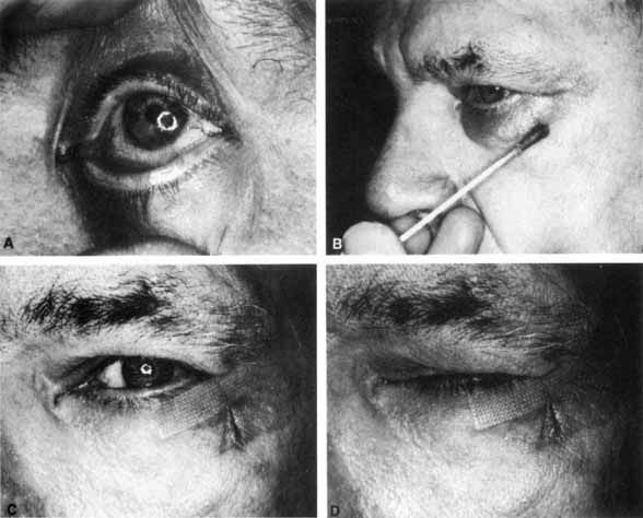
syndrome. Neurology 44:752, 1994

166. Ross Russell RW: Supranuclear palsy of eyelid closure. Brain 103:71, 1980

167. Lessell S: Supranuclear paralysis of voluntary lid closure. Arch Ophthalmol 88:241, 1972

168. Lepore F: Bilateral cerebral ptosis. Neurology 37:1043, 1987

169. Barton JS: Bilateral central ptosis in acquired immunodeficiency syndrome. Can J Neurol Sci 22:52, 1995

170. Galetta SL, Gray LG, Raps EC, Schatz NJ: Pretectal eyelid retraction and lag. Ann Neurol 33:554, 1993

171. Adams GG, Kirkness JP: Botulinum toxin A induced protective ptosis. Eye 1:603, 1987

172. Morel-Fatio D, Lalardrie JP: Palliative surgical treatment of facial paralysis. The palpebral spring. Plast Reconstr Surg 33:446, 1964

173. Morel-Fatio D, Lalardrie JP: Le ressort palpebral: contribution a l'etude de la chirurgie plastique de la paralysie faciale. Neurochirurgia 11:303, 1965

174. Levine RE, House WF, Hitselberger WE: Ocular complications of seventh nerve paralysis and management with the 
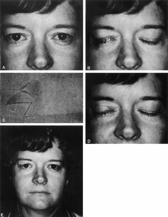
palpebral spring. Am J Ophthalmol 73:219, 1972

175. Jobe RP: A technique for lid-loading in the management of lagophthalmos in facial palsy. Plast Reconstr Surg 53:29, 1974

176. Sansone V, Boyton J, Palenski C: Use of gold weights to correct lagophthalmos in neuromuscular disease. Neurology 48:1500, 1997

177. Tankere F, Bernat I, Vitte E, et al: Hypoglossal-facial nerve anastomosis: Dynamic insight into the cross-innervation phenomenon. Neurology 61:693, 2003

Back to Top