Chapter 14 Orbital Diseases and Neuro-ophthalmology Table Of Contents |
Part I: An Overview |
History-Taking Orbital Examination Diagnostic Considerations Graves' Disease Inflammations Vascular Lesions Neoplasms Trauma Diagnostic Procedures Surgery of the Orbit
That diseases of the orbit may be conjoined with neuro-ophthalmology should come as no surprise. These two domains of ophthalmologic interest are integrated by common symptoms, signs, and anatomic structures. The orbit shares bony partitions with the sinuses medially and inferiorly, with the anterior cranial fossa above, with the superior orbital fissure and cavernous sinus at the orbital apex, and with the sphenoid complex and middle cranial fossa posteriorly (Figs. 1 and 2). Moreover, there are congenital, inflammatory, neoplastic, vascular, and traumatic processes that bridge the orbito-cranial junction. Clinically, orbital lesions that cause double vision are regularly mistaken for intracranial problems. In evidence, one need look no further than the single most common cause of spontaneous diplopia in the adult, that is, the restrictive myopathy of Graves' disease, which is so frequently misdiagnosed as cranial nerve palsies or myasthenia. And while proptosis is so compelling an indicator of orbital masses, passive orbital congestion with exophthalmos is also a sign of arteriovenous fistula, of intracranial obstruction of venous flow, or of tumor growth in the middle cranial fossa or paranasal sinuses. This chapter elucidates the communal turf shared by diseases of the orbit and neuro-ophthalmologic disorders, with emphasis on the clinical means by which strictly neurologic dilemmas may be distinguished from pathologic processes predominantly of the orbit. The material here is intended to provide a pragmatic approach to diagnosis of orbital disease, with special accent on the physical examination; orbital lesions causing diplopia are discussed at length in Chapter 12. Neither the specifics of orbito-cranial anatomy (Figs. 1 and 2) nor an exhaustive commentary on all orbital diseases is included, but common surgical procedures are discussed in Chapter 14, Part II. The reader is referred to the several excellent anatomic atlases available, and especially to Rootman's1 Diseases of the Orbit. |
HISTORY-TAKING |
As with other fields of clinical medicine, accuracy in diagnosis and appropriateness
of management depend on an orderly protocol that begins
with historical documentation (Table 1). Review of the personal medical history is an essential part of the assessment
of orbital and neuro-ophthalmologic problems. For example, failure
to uncover a past history of dysthyroidism is to ignore a substantial
clue to the single most common cause of uni- or bilateral proptosis. Likewise, a
rapidly progressive orbital congestive syndrome with
ophthalmoplegia, occurring in a patient with diabetes, should suggest
the possibility of an opportunistic fungus such as mucormycosis. Clearly, any
previous tumor surgery is suspect, although metastases constitute
only 3% to 7% of biopsied orbital masses2; nasopharyngeal and sinus neoplasms accounted for 23% of secondary orbital
masses in the British Columbia series.3 A history of cranial or facial trauma likewise must be assessed, but is
usually not obscure or forgotten.
TABLE 1. History of Illness: Orbit Medical history Dysthyroidism Family history Dysthyroidism Onset Insidious Symptoms Proptosis Progression Rapid
In a similar vein, family medical history is important and occasionally of diagnostic value. It is recognized, for instance, that optic glioma is a relatively frequent manifestation of neurofibromatosis (see Volume 2, Chapter 5). Therefore, in a child with unexplained chronic proptosis, the occurrence of skin lesions (birthmarks, skin lumps, or tumors), seizures, or central nervous system (CNS) masses in blood relatives is critical information. Ideally, such family members should be examined or further details obtained. Dysthyroidism also has a distinct familial predilection. The sudden onset of rapidly progressive proptosis in childhood (Fig. 3) should be considered an orbital emergency, the clinician's responsibility being to rule out a life-threatening tumor such as rhabdomyosarcoma or metastatic neuroblastoma. A similar picture may evolve with acute orbital congestion associated especially with ethmoidal or maxillary sinusitis, in children with or without fever.4,5 In the adult, with the exception of metastatic tumors, most orbital neoplasms produce insidiously progressive exophthalmos. Inflammatory orbital “pseudotumor”6 is the only common cause of relatively abrupt, usually painful, proptosis with diplopia in the otherwise well adult, and myositis occurs in children or adults.7 In contrast, intermittent painful proptosis, at times accompanied by spontaneous subconjunctival hemorrhage or lid ecchymoses, is practically pathognomonic of venous varices or lymphangiomas.8 An acute phase of Graves' disease may mimic other orbital congestive syndromes (see below), but usually this most common orbitopathy is chronic and not characterized by significant pain. Over a wide age spectrum, with severe coughing or prolonged retching, or during protracted obstetrical labor, pressure in intraorbital veins may be momentarily raised to the point of rupture with formation of a usually painful retrobulbar hematoma.9,10 These Valsalva orbital hemorrhages usually resolve spontaneously. In situations where the duration of proptosis is not clear, review of antecedent photographs (driver's license, family snapshots, etc.) may be extremely helpful. The clinician should be aware that proptosis may be suddenly discovered rather than actually occurring rapidly, so that the evidence provided by review of previous photographs is helpful in dating true onset. Such photographs may be scrutinized with the illumination and magnification provided by the slit lamp. As a rule, insidiously progressive orbital masses that produce axial (straightforward) displacement of the globe tend not to produce diplopia until proptosis is relatively marked. Inflammatory pseudotumor is an exception, because of the propensity for infiltration of extraocular muscles (myositis),7,11 and the restrictive myopathy of congestive Graves' disease regularly results in concomitant diplopia. Tumors located superiorly in the orbit commonly produce deficits in upward gaze, nasal masses produce adduction deficits, and temporal masses produce abduction deficits. Under ideal conditions these physical observations should be confirmed by enhanced computed tomography (CT) or magnetic resonance imaging (MRI); standardized ultrasonography is an ideal complementary procedure. Defects in visual function are due to compression of the optic nerve by intra- or extra-conal masses but, as with neural structures elsewhere, if tumor growth is insidious, remarkably large masses are compatible with normal nerve function. Retrobulbar masses that indent the posterior pole of the globe may induce relative hyperopia, requiring additional plus lenses to improve acuity. Transient blurring of vision in extremes of gaze, especially abduction or noted during reading,12 can be produced by any orbital mass, including dysthyroidism. This phenomenon may be related to compression of the optic nerve as it is dynamically stretched over a mass, compressed by contracting muscles, as the posterior wall of the globe becomes deformed, or as vascular flow is compromised (or combinations of these mechanical effects). Proptosis with true orbital pain (as opposed to ocular irritation or foreign body sensation) is relatively rare, with the following exceptions: acute orbital inflammation, including myositis; metastases; elevated venous pressure due to arteriovenous fistulas or malformations; acute thrombosis of venous varices; and acute thrombosis of enlarged orbital veins associated with arteriovenous fistulas or vascular malformations. Of course, orbital and eye pain may be a symptom associated with the ophthalmoplegia of orbital apex and cavernous sinus syndromes (see Chapter 12), or referred from dural structures. Atta et al13 have elaborated a syndrome of venous stasis orbitopathy, composed of proptosis, ophthalmoplegia, and injected conjunctival vessels, of vascular (arteriovenous fistulas) and nonvascular etiologies. These authors note the usefulness of standardized echography, especially with measurement of extraocular muscle diameters, to distinguish fistulas from mass lesions. |
ORBITAL EXAMINATION | ||||
The ophthalmologist is secure in his or her ability to directly visualize
the anterior segment of the globe, and the fundus in most instances
may be similarly brilliantly illuminated and observed. But the orbit
is a bête noire (or better yet, a “black box”) that
does not allow the direct inspection that the transparent ocular media
so easily permit, and if the ophthalmologist is uncomfortable in orbital
diagnosis, how much more so the neurologist? Nonetheless, office evaluation
of the orbit is not as limited as one might think. In addition
to standard assessment of visual function and ocular motility, the
physical examination of the orbit may be conducted as outlined in Table 2.
TABLE 2. Orbit: Physical Examination Lids Position: ptosis, retraction Conjunctiva Vessels Globe Position: proptosis, displacement Palpation of anterior orbital tissues Auscultation Valsalva maneuver Forced (passive) ductions Intraocular tension Fundus
Since Graves' disease represents the most common orbital ailment, the lids must be carefully evaluated for spontaneous retraction (Fig. 4; see also Volume 2, Chapter 3, Fig. 11) or for induced lag on slow ocular pursuit movements from upward to downward. Preliminary observations of the lids should be made during history-taking, before the patient's attention is called to the actual examination; some patients will stare during intense concentration or conscious effort to cooperate. True stare should be spontaneous, and lag reproducible. Lid position and movement defects must be assessed before sympathomimetic agents are instilled for pupil dilation; for example, phenylephrine (Neosynephrine) will induce moderate to marked lid retraction that masks pathologic lid position.
Anomalies of lid position and movement need not be bilateral. Ptosis is quite rare in Graves' disease, but is fairly common with inflammatory pseudotumor. Ptosis, in the absence of pain or other congestive orbital signs, should always bring to mind the possibility of myasthenia. Unilateral ptosis may account for contralateral lid retraction; thus, a patient with right partial ptosis may show relative retraction of the left upper lid, due to increased effort in an attempt to overcome the ptosis (Hering's law of equivalent innervation is applicable to the two levator palpebrae).14 If the eye with ptosis is occluded, or the lid mechanically raised, the opposite retracted lid will assume a normal position. The causes of lid retraction are the following:
Lid swelling regularly results from the congestive edema of Graves' disease, but also typically occurs in preseptal or orbital cellulitis, inflammatory pseudotumor, arteriovenous fistulas, and with acute viral conjunctivitis. For obscure reasons, chronic orbital neoplasms may cause intermittent lid edema. Most lid edema is accentuated by sleep, during which time the head is maintained in a relatively dependent position. Palpation of a discrete mass in the lids is exceedingly helpful, since potential biopsy is facilitated. Veins may be visible within the thin lid tissues, either passively dilated by diffusely increased orbital pressure, or engorged by arterialization of orbital and adnexal vessels fed by arteriovenous communications. In the latter case, audible bruits and palpable thrills are evidence of turbulent, increased blood flow. Abnormalities of the conjunctiva may serve as clues to orbital diagnosis. In general, edema (chemosis) is too nonspecific a finding to be very helpful, being common in Graves' disease, inflammatory pseudotumor, and arteriovenous fistulas. Any retrobulbar mass may produce chronic or intermittent chemosis, presumably by interfering with venous drainage in the orbit. In Graves' ophthalmopathy, the vessels overlying the insertions of especially the medial and lateral recti muscles are commonly enlarged, and indeed the muscle insertions themselves may be visibly hypertrophied (Fig. 5). These two conjunctival signs are exceedingly useful in identifying Graves' orbitopathy and should be sought in cases of unexplained proptosis and/or diplopia. Also, engorgement of orbital vessels may be reflected as hypervascularity of conjunctival and scleral vessels, which take on a more-or-less specific pattern in the presence of arteriovenous fistula (see Volume 2, Chapter 17, Fig. 14). Diffuse or focal hyperemia of scleral and episcleral vessels (especially in the superior lateral quadrant of the globe) is seen in anterior scleritis (episcleritis), or as an anterior component of posterior scleritis that accompanies the painful ophthalmoplegia syndrome of idiopathic orbital inflammation (Fig. 6). The position of the globes relative to the orbital rims is subject to considerable individual and racial variations. Not the least problem is accuracy and reproducibility of measurements as obtained by a number of exophthalmometric techniques, of which the Hertel exophthalmometer is the most common, and probably the most accurate (Fig. 7). In a study of 681 normal adults ranging in age from 18 to 91 years, mean normal protrusion values were 15.4 mm in white women, 16.5 mm in white men, 17.8 mm in black women, and 18.5 mm in black men; upper limits of normal were 20.1, 21.7, 23.0, and 24.7 mm, respectively.15 No normal individual showed more than 2 mm of asymmetry. To reiterate, there is no clinically precise technique for quantitating proptosis, as minor deviations in positioning of exophthalmometers at the lateral orbital rim result in gross variations in readings. Depending on technique and experience, substantial interobserver variation is demonstrable.16 Subtle degrees of proptosis are difficult to detect and more difficult still to measure. The following techniques are useful in determining the presence of relative unilateral proptosis: viewing the position of the globes and lids from above the brows (Fig. 8, top); insertion of fingertips between inferior orbital rim and globe; and simultaneous palpation of corneal apices (Fig. 8, bottom). The clinician should be aware of several situations in which proptosis is more apparent than real, that is, a condition of pseudo-exophthalmos: unilateral lid retraction, wherein the homolateral eye appears larger; unilateral mild ptosis, wherein the contralateral eye appears larger; asymmetry of facial bones including orbital rims; and unilateral enophthalmos,17 wherein the normal contralateral eye appears prominent (Fig. 9). The differential diagnosis of enophthalmos is included in Table 3.
TABLE 3. Causes of Enophthalmos Senile orbital fat atrophy *Data from Miller MT, Spencer MA: Progressive, hemifacial atrophy: A natural history study. Trans Am Ophthalmol Soc 93:203, 1995 †Data from Soparkar CNS, Patrinely JR, Cuaycong MJ et al: The silent sinus syndrome: A cause of spontaneous enophthalmos. Ophthalmology 101:772, 1994
In a small number of patients, differences in the axial lengths of the globes may account for relative unilateral proptosis. This situation is resolved by finding differences in the amount of myopia (i.e., greater myopia in the apparently proptosed globe). Precise determination of axial globe lengths is afforded by ultrasonography. There is little evidence to support the claim that a complete third nerve palsy results in sufficient relaxation of the muscle cone to produce detectable proptosis. As a rule, any significant degree of proptosis should not be attributed to extraocular muscle weakness. This dictum may be confirmed by performing exophthalmometry on patients with vascular-diabetic oculomotor palsies; one finds no meaningful difference between the two eyes at the time of most profound ophthalmoplegia, and no changes in exophthalmometry readings following complete recovery. Moreover, irreducible proptosis (increased resistance to retropulsion of the globe through closed lids) in the presence of motor deficits signifies Graves' disease, or a mass at the orbital apex or superior orbital fissure. If oculomotor nerve palsy could result in proptosis, there would be no resistance to globe retropulsion, there being no mass or congestion present. Pulsation of the globe is encountered most commonly with acquired carotid-cavernous fistulas, and rarely in other conditions.20 Causes of pulsation are listed in Table 4.
TABLE 4. Causes of Globe Pulsation Congenital sphenoidal dysplasia with partial orbitocranial encephalocele (in
neurofibromatosis) *Data from Allen SJ, Naylor D: Pulsation of the eyeballs in tricuspid regurgitation. Can Med Assoc J 133:119, 1983
Biomicroscopical examination usually reveals even minimal pulsation, best seen during measurement of intraocular tension by Goldmann applanation tonometry. Cotton-swab sticks may be placed tangentially across the corneal apices, such that transmitted pulsations are amplified by the length of the swab (Fig. 10).
Exploration of the orbital rim by fingertip palpation (Fig. 11) may reveal masses lying in the tissues of the lids and also in the anterior portions of the orbit. Discovery of such masses is extremely helpful since biopsy via simple transseptal anterior orbitotomy is the most rapid route to tissue identification.
Auscultation of the globe and face is a useful maneuver to confirm the presence of vascular bruits, as encountered in acquired arteriovenous fistulas or congenital vascular malformations. Bruits may be more intense over zygoma or mastoid bones, where the diaphragm of the stethoscope is more effective than the bell, with which the globe itself is better auscultated (Fig. 12). Symmetrical cranial or ocular bruits are commonly present in normal children and therefore must be evaluated with caution. Forced expiration against resistance (Valsalva maneuver) raises venous pressure in the neck, face, and head, such that orbital masses with significant draining veins will increase in volume, evidenced by transient increase in proptosis (Fig. 13). This phenomenon is typically demonstrable in the presence of congenital venous varices or arteriovenous malformations, but may also be seen with acquired carotid-cavernous fistula or with primary or secondary bony defects that permit transmission of intracranial pressure to orbital contents. Crying infants with such lesions may show this sign spontaneously or with head-hanging. Where accompanying physical signs are absent or minimal, the single most useful technique in distinguishing neural ophthalmoplegia (cranial nerve palsies, myasthenia, chronic progressive external ophthalmoplegia) from local orbital disease (e.g., Graves' ophthalmopathy) is the forced (passive) duction test. In 1967, Stephens and Reinecke22 reported a method for quantitation of the forced duction test, but no standardized accessible and practical instrument is currently available. There are various techniques to test for mechanical resistance to rotation of the globe; the test described here is that preferred by this author (see Volume 2, Chapter 3, Fig. 6). Following topical conjunctival anesthesia with, for example, proparacaine (Ophthetic) or tetracaine (Pontocaine), cotton-tipped swabs are saturated with 10% aqueous cocaine and applied for about 1 minute to the area of the insertions of the four recti muscles of each eye. It is important to compare corresponding muscles in both eyes. For example, if the right eye lags in upward gaze, the insertion of the inferior rectus is grasped and, while the patient looks upward, an attempt is made to gently rotate the right globe upward. For comparison, the insertion of the left inferior rectus is then grasped, and the left globe rotated upward. With care, there is only slight discomfort, and the conjunctiva is minimally traumatized; minor subconjunctival hemorrhage is of no great consequence. Alternatively, the conjunctiva at the limbus may be grasped, or a cotton swab tip may be braced against the corneoscleral junction and used to “push the eye.” The forced duction test will be positive, that is, there will be resistance to mechanical rotation of the globe, in the following situations: Graves' restrictive myopathy, inflammatory pseudotumor (myositis), infiltrating carcinoma, incarceration of extraocular muscles and their surrounding soft tissue attachments that herniate into orbital floor, and medial wall fractures. Because of secondary fibrotic contractures of extraocular muscles, on rare occasions the forced duction test will be positive in the chronic fixed form of ocular myasthenia, in advanced chronic external ophthalmoplegia, or with extremely long-standing sixth or third nerve palsies. In situations where the globe is mechanically restricted (again, Graves' disease is typical) intraocular tension may be spuriously elevated, or pressure may inordinately rise on attempted upward gaze.23 There seems to be a correlation with higher ocular tensions and degree of proptosis; tension decreases in patients undergoing orbital decompression.24 However, it must be recognized that intraocular pressure increases linearly with vertical excursions of the globe, changes of as much as 7 mmHg being recorded in normal subjects.25 Indeed, this phenomenon in normal subjects brings into question the validity of this procedure as an adjunct in the diagnosis of Graves' orbitopathy.26 Carotid cavernous or dural arterial fistulas also typically elevate intraocular tension by raising episcleral venous drainage pressure. The ocular fundus may be altered by any retrobulbar mass in the following ways: indentations of the posterior wall of the globe produces chorioretinal striae (Fig. 14), while compression at the equator of the globe and beyond results in a more diffuse flattening, best appreciated by indirect ophthalmoscopy, and accentuated by rotation of the eye toward the quadrant(s) of the orbit occupied by the tumor; dilation and tortuosity of retinal veins (venous hemorrhages or occlusions suggest relatively high pressures, as encountered with arteriovenous fistulas); retinal arterial occlusions, especially in orbital phycomycoses such as mucormycosis; edema or frank elevation of the optic nerve head; optic atrophy in chronic compression; optociliary shunt vessels of the disk, especially with perioptic meningioma; retinal detachment or choroidal suffusion with inflammatory lesions or scleritis. De La Paz and Boniuk27 have extensively reviewed the fundus manifestations of orbital disease.
Optic disc swelling (Table 5) does not necessarily suggest actual infiltration of the nerve or its meninges, this fundus finding being rather nonspecific and observed potentially with any increase in retrobulbar mass. Indeed, in orbital context, disc edema is seen most commonly with Graves' orbitopathy. On rare occasions optic gliomas produce a picture of disc swelling with or without venous occlusion, and perioptic meningiomas may be characterized by a clinical triad of slowly progressive visual loss, pallor admixed with disk swelling, and papillary retinociliary venous shunts (see Chapter 5, Part II).
TABLE 5. Optic Disc Swelling with Orbital Lesions Graves' orbitopathy *Proptosis may be minimal or absent.
|
DIAGNOSTIC CONSIDERATIONS | ||||||||||||||||||||||||||||||||||||||||||||||||||||||||||||||||||||||||||||||
After history-taking and thorough physical examination as outlined in the
preceding sections, the clinician should be able to make at least a
tentative but rational diagnosis, even before special diagnostic studies
are undertaken. Excluding congenital dysostoses, malformations and
cysts, and traumatic fractures and hematomas, which rarely cause diagnostic
dilemmas, all orbital disease may in essence be classified into
only four common types: (1) Graves' ophthalmopathy, (2) idiopathic inflammations, (3) vascular
malformations and fistulas, and (4) true neoplasms. Large
series of patients with orbital disorders show variable
specific incidence rates, depending on referral patterns and age groups. Graves' orbitopathy
may be underestimated since many clinicians do
not regularly refer uncomplicated cases; the same is true for trauma and
congenital anomalies (Table 6).
TABLE 6. Causes of Orbital Disease*
*Based on approximately 1400 cases. Adapted from Rootman J: Diseases of the Orbit. Philadelphia, JB Lippincott, 1988
GRAVES' DISEASE Graves' congestive orbitopathy is the single most common orbital disorder, with annual incidence rates ranging from 12 to 20 per 100,000 population, and with higher prevalence rates (42%) among Caucasian (European) groups than in Asians (8%). The age-specific incidence rates are greatest among middle-aged patients, and are approximately four times higher in women than in men. In patients with hyperthyroidism, ophthalmopathy is evident in 25% to 50%, but severe complications evolve in just 3% to 5%. Several studies have linked tobacco smoking to increased severity of Graves' orbitopathy,28 possibly related to alterations in immunoregulatory cell function induced by smoking, and increased synthesis of glycosaminoglycans by orbital fibroblasts. Thyroid eye disease is usually associated with hyperthyroidism, and less frequently with Hashimoto's thyroiditis, euthyroid states, thyroid carcinoma, or primary hyperthyroidism. About 80% of patients will develop signs of ophthalmopathy either during the year before or the year after a diagnosis of thyroid malfunction.29 Tallstedt et al30 demonstrated a two- to threefold increase in risk of developing orbitopathy when the thyroid disease was managed with radioactive iodine treatment as opposed to surgical or medical treatment; this effect may be related to the release of thyroid antigens and to subsequent enhancement of autoimmune response directed toward antigens shared by the thyroid and the orbit. Evolution of ophthalmopathy after radioiodine therapy is often transient and may be ameliorated by the administration of prednisone.28,31 The principal signs are lid retraction and edema, proptosis, and diplopia. If congestive signs are slight, the ocular motor defects are regularly misdiagnosed, and unsuitable studies for intracranial disease follow. Eye movement defects with Graves' disease are discussed in detail in Volume 2, Chapter 12. Infrequently Graves' disease presents in an acute inflammatory form that mimics orbital cellulitis or idiopathic pseudotumor, including pain, lid swelling with ptosis, diplopia and infrequently visual loss.32 Proptosis and restrictive myopathy also may occur in children with hyperthyroidism.33 Obscured by the more obvious external congestive signs and by symptoms of diplopia, the complication of compressive optic neuropathy may be overlooked. Although its incidence is said to be less than 5% among patients with typical thyroid disease, Graves' optic neuropathy is a treatable cause of potentially disabling visual loss (see also Volume 2, Chapter 5, Part II). Congestive symptoms always precede visual loss, which is usually gradual in onset and bilateral in most patients, but may occasionally be acute and asymmetrical. Presenting acuities are poorer than 20/60 in 50% of cases; central scotomas, at times combined with inferior field depression, are the predominant field defects. Congestive signs are usually of moderate intensity without severe proptosis or exposure keratopathy. Bilateral and symmetrical ductional restriction is the most commonly associated motility disturbance. Oral corticosteroids are often effective in restoring visual function, but steroid-unresponsive neuropathy may be improved promptly by supervoltage orbital irradiation or surgical decompression.34,35 The medical management of Graves' disease is complex and demands a comprehensive approach, often requiring a variety of surgical (see below) and radiotherapeutic interventions.36,37 It is worth noting that Graves' ophthalmopathy frequently presents or worsens weeks to months after radioactive iodine ablative therapy, and that concomitant corticosteroid therapy may ameliorate this effect.28,31,38 Char39 has reviewed the immune mechanisms by which intrathyroidal clonally restricted B-cells secrete autoantibodies, with extraocular muscle and orbital tissues, including fibroblasts, acting as antigenic targets; fibroblasts in turn produce glycosaminoglycan that binds water, increasing orbital connective tissue volume. Prednisone, cyclosporine, and irradiation, in variable combinations, are all applicable forms of therapy. Indeed, in the Mayo Clinic experience40 of therapies for Graves' ophthalmopathy, only 20% of patients required one or more surgical procedures; 7 of 120 patients (6%) developed optic neuropathy. INFLAMMATIONS Orbital inflammatory disease may take several distinct forms. Acute orbital cellulitis is defined as infectious inflammation of soft tissues posterior to the orbital septum, characterized by distension and hyperemia of the lids and conjunctiva, pain, proptosis, and limitation of eye movements. Although sinus infections were formerly frequently associated, this is now a relatively rare condition, thanks to the availability of antibiotics. However, the condition is potentially life-threatening. Principal predisposing risk factors include ocular or adnexal surgical procedures (lid, strabismus, or retinal operations), facial or orbital trauma, especially with retained orbital foreign bodies, dacryocystitis or other periorbital infections, insect bite envenomization, diabetes, and immunosuppressive states. Especially in children and young adults, orbital cellulitis is still associated with ethmoidal and maxillary sinusitis. Those bacterial agents commonly responsible for sinusitis (predominantly Streptococcus pneumoniae, Staphylococcus aureus, and less frequently Haemophilus influenzae), not surprisingly are implicated in orbital cellulitis.41 Rapidly progressive rhabdomyosarcoma may present a clinical picture of pseudocellulitis in children (see Fig. 3). CT scanning or MRI is mandatory, not only to disclose sinus disease, but also to determine the presence of meningitis, cavernous sinus thrombosis, or orbital abscess formation, which requires surgical drainage. Most associated abscesses occur in the medial orbit adjacent to the ethmoidal sinuses, with spread of infection via communicating veins (septic thrombophlebitis) or across the thin lamina papyracea. Chronic mucoceles may also be associated with cellulitis or abscess formation. Rhino-orbital mucormycosis (genera Mucor, Absidia, or Rhizopus) is a catastrophic form of often fulminant, necrotizing orbital infection, most likely to occur in patients in ketoacidosis or immunosuppressed by chemotherapy, AIDS, or hemodialysis. Such infection is extremely rare in healthy individuals. A chronic form of rhino-cerebral mucormycosis is well described,42 and is associated with carotid artery and cavernous sinus thrombosis. Rapid confirmation by nasopharyngeal mucosal aspiration and biopsy is essential. Wide debridement and amphotericin B infusion may be life-saving, and hyperbaric oxygen has been advocated.43 Aspergillosis may also cause an indolent or acute orbital cellulitis (see also Volume 2, Chapter 12). Allergic fungal sinusitis is a relatively uncommon form of chronic paranasal mycosis in immunocompetent individuals. This entity can involve the orbit without direct invasion or dire outcome, and is most frequently, but not exclusively, attributed to Aspergillus. Signs include nasal obstruction, focal pain, proptosis, diplopia, optic neuropathy, and facial deformity. Inflamed sinus mucosa shows hyperintense signal characteristics on both T1- and T2-weighted MRI, but isointense signal of sinus cavities, with involvement of multiple sinuses; peripheral eosinophilia and elevated total immunoglobulin E, as well as fungus-specific IgE and IgG, help establish the diagnosis.44 In the differential spectrum are included invasive fungal sinusitis, orbital inflammatory pseudotumor, metastatic carcinoma, lymphoma, Wegener's granulomatosis, and systemic vasculitis. Idiopathic orbital inflammation (orbital pseudotumor) is a frequent cause of acute, subacute, or chronic painful ophthalmoplegia, accompanied by variable orbital signs. This clinical term encompasses noninfectious processes that mimic cellulitis, Graves' disease, or neoplasm; thus, the term orbital pseudotumor. Inflammation may be diffuse, or localized to the posterior scleral coat (posterior scleritis), to single or multiple extraocular muscles (myositis; see Volume 2, Chapter 12), to the lacrimal gland (dacryoadenitis), or to perioptic meninges (perioptic neuritis), or present as a soft tissue mass. Signs and symptoms are determined by location and include severe to mild orbital ache, diplopia, lid swelling with ptosis, conjunctival chemosis, episcleral injection, proptosis, and ductional defects. Uveitis, uveal effusion, and optic neuropathy are rare, but account for visual loss. Orbital pseudotumor and the Tolosa-Hunt syndrome are likely the same process, varying only in that the idiopathic inflammation involves predominantly the orbit in the former and predominantly the superior orbital fissure and/or the anterior cavernous sinus in the latter. Idiopathic inflammation is a diagnosis of exclusion, made only in a fairly circumscribed clinical context, and when other pathologic processes have been ruled out. In this regard, contrast-enhanced CT scan or MRI shows diffuse infiltration or focal lesions, usually with notable enhancement of the posterior wall of the globe (Fig. 15). With proper orbital imaging and ultrasonographic assessment as diagnostic procedures, tissue biopsy is rarely necessary, and the response to systemic corticosteroid administration is usually dramatic, if not diagnostic (see also Volume 2, Chapter 12). Uncommonly, orbital inflammation takes the form of a noncaseating granuloma, suggesting sarcoidosis, but without other systemic implications. This seems principally a histologic variant, with some predilection for the lacrimal gland.45 In contrast, Wegener's granulomatosis is locally destructive necrotizing vasculitis with respiratory and other systemic implications. The majority present as scleritis with or without orbital mass lesions, either uni- or bilateral, and commonly with nasal and sinus symptoms.46 Also known as lethal midline granuloma, and causing destruction of midline facial tissues including nasal septum, biopsy shows mixed inflammation with necrosis. Other orbital vasculitides include giant cell arteritis, zoster ophthalmicus, and lupus erythematosus. Orbital lymphoid hyperplasia is a histopathologic quandry even when biopsy specimens show polyclonal T-cell lymphocyte proliferation (suggesting benign, reactive lymphoid hyperplasia), versus monoclonal proliferation of B cells typical of malignant lymphoma. Thus, lymphoid hyperplasia may possibly embrace simple pseudotumor inflammation, but in other instances is a harbinger of nonHodgkins lymphoma.47 Because of this potential outcome, some authors suggest complete systemic staging (hematologic survey), and strict clinical follow-up. Atypical features or monoclonality make more extensive immunotypic surface cell typing or DNA hybridization mandatory.48 Therefore, biopsy specimens should not be placed in formalin! Both corticosteroids and radiation therapy are acutely effective, as is usually also the case with idiopathic inflammation. Therefore, clinical response to initial therapy cannot carry diagnostic inferences. In the immunocompromised AIDS population, lymphoma is the most frequent orbital lesion, nonHodgkin's lymphoma being well-documented,49 as well as Burkitt small noncleaved; the importance of immunophenotypic (immunoglobulin light chain antigens) characteristics is stressed.50 Also, orbital cellulitis from contiguous sites of fungal (Aspergillus, Mucormycosis)51 and toxoplasmosis infection may be encountered. The specifics of histologic and immunologic typing techniques for the classification of orbital lymphomatous and other hematopoietic tumors are reviewed by Jakobiec and Nelson.52 VASCULAR LESIONS Vascular lesions involving the orbit may be categorized as neoplastic (hemangiomas and lymphangiomas), congenital anomalies (simple venous varix, arteriovenous malformation), and acquired arteriovenous shunts such as the carotid-cavernous fistula. From the extensive Moorfields Eye Hospital Orbital Clinic experience, Wright53 concludes that vascular abnormalities are the commonest space occupying lesions, of which primary congenital venous varix complexes constitute the largest category. Most series support the view that cavernous hemangioma is the most frequent primary orbital neoplasm. Otherwise, venous varices and lymphatic malformations (lymphangiomas) are of neuro-ophthalmologic interest because of orbital complications (enlargement during respiratory infections; spontaneous orbital hemorrhage) and their association with contiguous and noncontiguous facial and/or intracranial vascular anomalies54 (see also Volume 2, Chapter 17). In neuro-ophthalmologic context, carotid-cavernous fistulas (CCFs) are of singular interest. CCFs are initiated by rupture of the wall of the carotid artery, or one of its branches, into the cavernous sinus. In the presence of a CCF, the cavernous sinus and its venous exits are exposed to arterial pressure that alters their hemodynamic state. The major orbital communication of the cavernous sinus is the superior ophthalmic vein, which may expand tremendously, engorging all orbital and conjunctival veins. These veins become arterialized, producing signs and symptoms of venous congestion. The classic, fully developed syndrome includes pulsating exophthalmos, ocular bruit that may be subjective and/or objective, diplopia, headache, conjunctival chemosis, increased intraocular pressure, dilated conjunctival vessels, and visual decrease (see Volume 2, Chapter 17, Fig. 13). To some degree bilateral orbital involvement is often present because of normally occurring venous communications between the cavernous sinuses. In some cases contralateral exophthalmos may exceed that present on the side of the fistula, because of ipsilateral orbital vein thrombosis. Small meningeal arterial branches supplying the dural walls of the cavernous sinus and its tributaries may rupture, creating a minor dural arteriovenous shunt. This type of fistula usually appears in middle-aged women as a distinctive syndrome and may account for most so-called spontaneous CCFs. The signs are usually mild and include dilated conjunctival veins, mild proptosis, and bruit. Transient sixth nerve palsy and unilateral headache frequently antedate orbital signs by many months. Chronic unilateral “red eye” is often misdiagnosed and treated as an inflammatory condition.55 The diagnosis and management of arteriovenous fistulas is addressed in Volume 2, Chapter 17. Arteriovenous fistulas are the prototypic cause of a venous stasis orbitopathy syndrome, combining engorgement and hypoxia of orbital soft tissues, and composed of clinical signs of proptosis, variable ophthalmoparesis, lid swelling, and conjunctival chemosis. Other mechanical obstruction obtains with venous blockage at the orbital apex, within the cavernous sinus and with middle cranial fossa masses.13 In this setting, standardized A-scan echography has proven helpful in distinguishing fistulas from mass lesions, or from Graves' disease. Orbital infarction with acute pain, visual loss, ophthalmoplegia, and anterior and posterior segment ischemia occurs with giant cell arteritis, mucormycosis, common carotid artery occlusion,56 and carotid artery dissection.57 NEOPLASMS The wide variety of primary and secondary neoplasms that affect the orbit and adnexal structures is too extensive a subject to be adequately covered in the present discussion; therefore, only a few generalizations will be attempted here. As noted above, benign tumors such as cavernous hemangioma, neurilemoma (schwannoma), dermoid cysts, etc., constitute the commonest lesions, which are characterized by insidious growth with slowly progressive proptosis. Although termed benign, the mixed lacrimal gland tumor also progresses sluggishly, filling the lacrimal fossa, but authoritative consensus requires complete surgical extirpation of any lacrimal gland mass unless inflammatory signs are clinically predominant. While the advent of CT, MRI, and ultrasonography have added enormously to the previously vague radiologic assessment of orbital soft tissue disease, and certain tumors (e.g., hemangioma, dermoid) show rather typical images, nonetheless, surgical exploration with tissue confirmation is the most reasonable approach; this dictum is more emphatic in the presence of pain, visual loss, or a rapidly progressive course. In childhood, brisk evolution suggests rhabdomyosarcoma, acute leukemia, and histiocytosis especially where bone is eroded.58 Other frequent orbital disorders in children include cysts, vascular lesions, optic gliomas and meningiomas, inflammatory masses, etc.; malignancies constitute roughly 20%.59 Again, inflammatory cellulitis with or without contiguous sinusitis must be investigated. Orbital metastases are uncommon, constituting 2% to 7% of biopsied orbital lesions.2 Most such cases have a previous diagnosis of primary malignancy, and orbital findings tend not to be necessarily suggestive of metastasis. The intraconal space, muscles, and adjacent bone, sinuses or brain may be involved. In adult women, of course, breast carcinoma metastases are most common, with at times remarkably long intervals between primary diagnosis and therapy, and subsequent orbital secondary lesions; the relationship may be cryptic. While proptosis is the rule, schirrhus breast carcinoma regularly produces a reactive fibrosis (desmoplasia) that causes soft tissue induration, severe eye movement limitation, and progressive enophthalmos.17 Unless bone is involved, neither CT pattern nor ultrasound findings are suggestive. Fine-needle biopsy60 is especially helpful when there is a strong suspicion, for example with known primary malignancy, that a focal orbital mass represents a metastasis and more extensive surgical manipulation is unwarranted. Many orbital metastases will respond to palliative radiotherapy, but anterior segment complications, such as recurrent sloughing of corneal epithelium with ensuing chronic irritation, must be taken into account if the patient is otherwise comfortable. Life expectancy is limited, as determined principally by the primary malignancy type or other nonorbital metastases. TRAUMA Trauma accounts for orbital problems that take the following forms: fractures of orbital walls, commonly with floor fractures, but the medial wall may be the site of a “blowout” as well; hemorrhage into the orbital soft tissues, with extension usually into the lids and bulbar conjunctiva; occult hematoma; immediate or delayed signs of arteriovenous fistula. With the exception of the last-named complication, traumatic orbitopathy is usually not a diagnostic dilemma. However, diplopia may not necessarily be attributable to the common mechanism of entrapment of the inferior rectus or inferior oblique muscles in a floor fracture. Hematoma and edema, or direct injury to extraocular muscles or motor nerves, also produces double vision that, fortunately, may abate without surgical intervention. A negative forced duction test, that is, absence of mechanical restriction, suggests this second mechanism, and, in the absence of enophthalmos (due to major floor fracture), surgery may be deferred.61 Ophthalmologic signs and symptoms may divert attention from the possibility of complications of orbital roof fractures, and from potentially life-threatening intracranial injury. Furthermore, evidence of CNS involvement may be delayed and underestimated. Continuing headache, stiff neck, and other nonlocalizing features can indicate subdural hematoma, meningitis, or pyogenic abscess formation. CT scanning or MRI will reveal pneumocephalus, and most fracture sites are best visualized with CT bone-window settings. Neurosurgical repair and broad-spectrum antibiotic therapy is indicated, as is scrupulous clinical and radiologic follow-up.62 The general problems of diagnosis and management of orbital trauma, including fractures, are reviewed elsewhere.63,64 |
DIAGNOSTIC PROCEDURES: AN OVERVIEW | ||
Detailed descriptions of diagnostic procedures are beyond the scope of
this presentation. But a brief overview of available techniques (Table 7) and their rational application may provide pragmatic guidelines. The
advent of elegant technical equipment in no way discharges the clinician's
responsibility for astute history-taking and meticulous physical
examination.
TABLE 7. Orbital Imaging Techniques Plain skull series: antero-posterior, lateral, and Waters' views
Any case of orbital disease, the cause of which is not obvious, arguably may profit from a preliminary plain skull x-ray series. With this initial radiologic procedure, the following features may be scrutinized: the general contour of the orbital bones; the condition of the paranasal sinuses, especially the ethmoid complexes, maxillary antra, and frontal sinuses; and the density and configuration of the sphenoid wings and superior orbital fissure. Computed tomography (CT) is presently the single most productive modality for analyzing orbital bones, with which the following structures may be visualized: the bony confines of the orbit and surrounding sinus structures; the lacrimal bony canal; the globe and lens; the intraorbital portions of the optic nerve; and the extraocular muscles, especially the horizontal and superior recti (Fig. 16). Moreover, CT with contrast enhancement discloses the full configuration and location of masses with respect to other orbital structures, principally the relationships to muscle cone, optic nerve, and lacrimal gland. In several particular instances, the location, configuration, and degree of contrast enhancement of lesions may strongly suggest a distinctive tumor type (Fig. 17) or inflammation (e.g., cavernous hemangioma, optic glioma, lacrimal gland tumor, dermoid cyst, mucocele, perioptic meningioma, single or multiple muscle thickening),65–67 but no firm histopathologic diagnosis may be inferred by CT characteristics alone.
Thin-section (1.5–3.0 mm) contiguous overlaps with bone-window settings provide a sensitive detector of orbital bone fractures, especially with regard to floor defects (blowout fracture),60,68 and CT analysis of orbital volume and degree of soft tissue herniation may have prognostic value for risk of delayed enophthalmos.69 Thin-section CT, thus, has replaced complex motion laminography in defining fractures, erosions, expansions, and hyperostosis of the bony walls of the orbit or skull base. Graves' disease is the overwhelming single most common cause of single or multiple extraocular muscle thickening (Fig. 18; see also Volume 2, Chapter 12, Figs. 28 and 30); inflammatory myositis is considerably less frequent, and muscle metastases are quite rare. Passive congestive myopathy also accompanies arteriovenous shunts and lesions of the superior orbital fissure and cavernous sinus, where orbital venous return is obstructed. The capacity of CT to detect minor to moderate changes in muscle diameter is perhaps limited,70 and standardized A-scan ultrasonography seems more sensitive and practical.71 Now almost universally available, MRI is the preferable technique for imaging the soft tissue contents of the orbit, high-resolution 3-mm and thinner sections being available, as well as gadolinium contrast enhancement (Figs. 19, 20, and 21).
To facilitate maximal application of CT scanning or MRI procedures, the clinician must communicate to the technician or radiologist a specific request for multiple orbital sections and fat saturation protocols, and should indicate the principal clinical diagnostic considerations. As an anatomic guide for surgical exploration, neuroimaging provides critical spatial details, if not more precise diagnostic information. The ideal complementary comparison study to CT or MRI is standardized ultrasonography (echography), which not only defines gross morphologic configuration, but also is most useful in classifying tumors by tissue-density groups. Whereas CT and MRI provide precise anatomic localization, ultrasonography roughly indicates tissue composition. Such categorization of typical inflammation or tumor patterns closely correlates with histologic diagnoses. Furthermore, standardized echography (combined A-scan, contact B-scan, and Doppler ultrasound) is an efficient and convenient diagnostic modality that sensitively detects changes in extra-ocular muscle size (see Volume 2, Chapter 12, Fig. 30) and reflectivity (as noted above), accurately measures optic nerve size and sheath distension due to increased subarachnoid fluid, distinguishes abnormalities of vascular flow, and differentiates other orbital soft tissue changes.67 Many tumors show quite characteristic, if not pathognomonic, reflectivity patterns. Enlargement of the superior orbital vein is detected by ultrasonography,67,72 and by CT scan73 and MRI,74 the differential diagnosis of which includes carotid-cavernous fistula (see Volume 2, Chapter 17, Fig. 16), Graves' orbitopathy, orbital inflammation, meningioma invading the cavernous sinus, vascular lesions of the orbit, and diffuse cerebral swelling associated with ruptured aneurysms or intracerebral hematomas.75 Arteriography has never been a particularly useful technique in orbital disease, with the exception of suspected arteriovenous fistula or malformation, and with the advent of MRI and standardized ultrasonography the indications for arterial contrast studies are few. Unlike the venous injections for enhanced CT scanning, where complications are limited to idiosyncratic reactions to iodinated contrast media, or benign gadolinium contrast for MRI studies, arterial injections or catheterizations run a small but significant risk of embolic sequelae. In the patient with suspected arteriovenous fistula prepared to undergo surgical correction, selective vessel catheterization is required, coupled with photographic subtraction of bone images (See Volume 2, Chapter 17). The advent of CT, MRI, and ultrasonography has considerably simplified the radiodiagnostic assessment of orbital disease. Radionuclide scanning and positive contrast retrobulbar orbitography must now be considered obsolete, as are contrast studies of the venous system of the orbit and cavernous sinus. By becoming familiar with those points of medical historical significance, by applying simple examination techniques and learning to recognize suggestive (if not pathognomonic) signs, and by the judicious use of diagnostic techniques (principally CT scan, ultrasonography, and now MRI), the clinician need not be overwhelmed by orbital disease. Nor should the distinction, even on strictly clinical grounds, between neural and orbital causes of ophthalmoplegia be an insurmountable problem. |
Part II: Surgery of the Orbit and Optic Nerve |
Anatomy: An Overview Preoperative Management Surgical Techniques: Orbitotomy Anterior Transseptal Transconjunctival Extraperiosteal Lateral Lateral Combined Superior-Craniotomy Postoperative Management Recovery Room Postoperative Complications Graves' Ophthalmopathy Clinical Features Orbital Decompression Lid Malposition Idiopathic Intracranial Hypertension Traumatic Optic Neuropathy A number of excellent texts, atlases, and reviews of surgical procedures of the orbit are available, but an overview in the context of neuro-ophthalmology is pertinent to this text. The surgical procedures for neuro-ophthalmic and orbital diseases require a brief review of the relevant anatomy, especially applied to the evaluation and management of these conditions. The treatment of optic neuropathy associated with Graves' ophthalmopathy, pseudotumor cerebri, and traumatic optic neuropathy will be discussed with particular emphasis on the surgical techniques commonly used by the ophthalmologist. |
ANATOMY: AN OVERVIEW |
The bony orbit has contributions from seven bones: sphenoid, frontal, zygomatic, maxillary, lacrimal, palatine, and ethmoid (see Figs. 1 and 2). The orbits are closely related to the paranasal sinuses: the maxillary
sinuses inferiorly, the ethmoid and sphenoid sinuses medially, and
the frontal sinuses superiorly. The pear-shaped bony orbit is widest approximately 1 cm
behind the orbital rim, then tapers toward the apex. The
total volume is approximately 30 cm3. The medial walls of the orbits are parallel and are separated by the
sphenoid and ethmoid sinuses. The lateral orbital walls form a 90° angle
with one another, and a 45° angle with the medial walls.1 The medial and superior walls extend posteriorly to the orbital opening
of the optic nerve canal. The orbital floor is composed of the maxillary, zygomatic, and palatine bones. The floor does not extend to the orbital apex, as it blends with the medial wall and extends posteriorly to the inferior orbital fissure at approximately the posterior wall of the maxillary sinus. Laterally the orbital floor is separated from the lateral wall by the inferior orbital fissure. The inferior orbital groove, containing the infraorbital neurovascular bundle, originates 2.5 to 3 cm posterior to the orbital rim and exits through the inferior orbital foramen of the anterior maxilla. The orbital floor medial to the infraorbital groove is thin and commonly involved in blowout fractures. The medial wall of the orbit consists of the ethmoid, maxillary, lacrimal, and sphenoid bones. The ethmoid bone (lamina papyracea) is the principal component of the medial wall. The posterior aspect of the medial wall is formed by the sphenoid bone that contains the optic canal. Located at the anterior aspect of the medial wall is the fossa for the lacrimal sac, formed by the frontal process of the maxilla and the lacrimal bones. The frontoethmoidal suture and the anterior and posterior ethmoidal foramens are the superior limit of the medial orbital wall. This suture line marks the roof of the ethmoid sinus; bone removal superior to this suture during decompression surgery can expose the dura of the anterior cranial fossa. The anterior ethmoidal foramen is approximately 24 mm posterior to the anterior lacrimal crest, the posterior ethmoidal foramen is an additional 12 mm posterior, and the optic canal ring is an additional 6 mm posterior. The suture between the ethmoid and maxillary bone is the inferior border of the medial wall and marks the strut of bone that provides support for the inferomedial orbit. The thin lamina papyracea is a weak anatomic barrier to confine ethmoid sinus infections or neoplasms from encroaching into the orbit. The thin medial wall can also be the site of blowout fractures. The lateral wall of the orbit is triangular and consists of the zygomatic bone anteriorly and greater wing of the sphenoid posteriorly. The sphenoid bone separates the orbit from the middle cranial fossa. The lateral wall terminates posteriorly at the superior orbital fissure. The frontosphenoid suture forms the junction between the lateral and superior walls. The inferior aspect of the greater wing of the sphenoid and the posterior wall of the maxillary sinus forms the inferior orbital fissure. The pterygopalatine fossa lies just below the inferior orbital fissure. The orbital roof is triangular in shape and is composed of the frontal bone and the lesser wing of the sphenoid. Posteriorly the intracranial roof tapers into the anterior clinoid process of the lesser sphenoid wing. Anteriorly, the superior orbital notch (foramen) is located at the medial one-third of the orbital rim and is approximately in vertical alignment with the inferior orbital foramen. Within the lesser wing of the sphenoid the optic canal (see Volume 2, Chapter 4, Fig. 5) extends from the middle cranial fossa to the superior, medial aspect of the orbital apex. The length of the bony canal varies from 5 to 11 mm and is angled superomedially toward the anterior clinoid. In contrast to the orbital portion of the optic nerve, which has some redundancy and is mobile, the intracanalicular portion is held firmly to the surrounding structures. The annulus of Zinn at the orbital apex forms a tight band around the nerve sheath, and the dural sheath is tightly adherent to the periosteum within the bony canal. As the nerve enters the middle cranial fossa, it is in apposition superiorly to a falciform dural fold of the planum. The orbital apex contains the optic canal within the lesser wing of the sphenoid. The medial aspect of the canal is adjacent to the posterior ethmoid and sphenoid sinuses. Superior to the optic canal is the orbital roof with the anterior cranial fossa above. Just lateral to the optic foramen the superior orbital fissure is formed by a gap in the greater and lesser wings of the sphenoid. The superior orbital fissure transmits the lacrimal, frontal, trochlear, superior division of oculomotor, abducens, nasociliary, and inferior division of the oculomotor nerves. The inferior orbital fissure transmits the infraorbital nerve and artery. The optic nerve can be separated into four portions: intraocular, orbital, canalicular, and intracranial. The optic nerve is surrounded by three meningeal sheaths identical to that of the central nervous system, the dura mater, the arachnoid, and the pia mater. The intraocular portion of the optic nerve, that is, anterior to, and including, the perforated scleral lamina cribrosa (see Volume 2, Chapter 4, Fig. 4), is 1 mm in length. Just posterior to the lamina cribrosa, the nerve acquires a dural sheath that is slightly bulbous and less adherent to the optic nerve in comparison to the closer relationship of the nerve and dural sheath at the orbital apex. The intraorbital segment is 25 to 30 mm and the intracanalicular portion 9 to 10 mm; the obliquely angled intracranial portion is about 15 to 17 mm. The vascular supply of the optic nerve is described in Volume 2, Chapter 4. |
PREOPERATIVE MANAGEMENT |
Sound knowledge of the anatomy, an understanding of the biologic behavior
of the orbital diseases, and an ability to interpret orbital diagnostic
imaging studies are prerequisites for successful orbital surgery. Preoperatively, anatomic
localization and a preliminary lesion diagnosis
are the most important factors in selecting the appropriate surgical
approach to the orbit. If the lesion appears to extend beyond the orbit, consultation
with a neurosurgeon or an otolaryngologist is imperative. Lesions that are palpable in the anterior orbit can usually be accessed through a transseptal orbitotomy approach. If imaging studies demonstrate the lesion to be in the posterior orbit, an extraperiosteal approach may be selected to safely gain entrance into the deep orbit. A transconjunctival medial orbitotomy approach may be most appropriate for an intraconal lesion situated within the medial central surgical space. A plan for total excision is formulated if a localized tumor is present. Diffuse infiltrative tumors are approached for diagnostic biopsy only. Proper assessment of the biologic behavior as well as the location of the suspected lesion may modify the choice of surgical approach. Thus, a lacrimal gland lesion may be exposed via a transseptal anterior orbitotomy if biopsy of a suspected inflammatory or malignant process is anticipated. However, if a benign mixed tumor of the lacrimal gland is the likely diagnosis, lateral orbitotomy is necessary for en-bloc removal. Some disease processes that involve only the subperiosteal space, such as subperiosteal hematoma or abscess, floor fractures, and mucoceles, are best approached through a transperiosteal route that does not violate the periorbita. If a malignant or metastatic infiltrative tumor is suspected, extraperiosteal orbitotomy should be avoided and another route of access considered, which would maintain the periorbita as an intact barrier against tumor spread. The lateral approach provides the best access to the retrobulbar compartments inside and outside the muscle cone. It is especially useful for lesions in the lacrimal gland fossa, but may be inadequate for some lesions deeply situated at the orbital apex. It is usually not necessary to prepare for blood transfusion for orbital surgery. However, this preoperative measure should be considered in patients with low hemoglobin or hematocrit, or in whom significant intraoperative bleeding is anticipated, such as in resection of arteriovenous malformation or in cannulating the superior ophthalmic vein for embolization of a cavernous-dural fistula. Patients taking anticoagulants, such as sodium warfarin (Coumadin), or inhibitors of platelet aggregation, such as aspirin, should discontinue their medications at least 2 weeks prior to surgery, if possible. Several over-the-counter drugs contain aspirin, such as Alka-Seltzer, Sine-Off, and Midol, and they, too, should be discontinued. Fish oil, a product sold in health food stores for prevention of heart disease, is frequently not regarded as medication by patients, and their use of it may not be disclosed. Fish oil can interfere with platelet function and prolong bleeding time. Control of hypertension is important, and general medical evaluation is sought when necessary. The ingestion of alcoholic beverages may dilate periocular and periorbital vessels; thus patients are advised to abstain from alcohol intake a few days before surgery. |
SURGICAL TECHNIQUES: ORBITOTOMY | ||
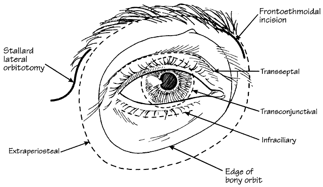
ANTERIOR Transseptal Anteriorly located tumors that are not subconjunctival but are visible within the substance of the eyelid or palpable within the anterior orbital space can be biopsied or removed completely through the transseptal approach, even with local anesthesia. The transseptal access may be performed in the upper eyelid or the lower eyelid (Fig. 22). This approach is most appropriate for well-demarcated lesions (e.g., dermoid cyst or cavernous hemangioma) situated in the anterior orbit that tend to bulge the septum forward, drainage of an abscess, or for incisional biopsy of a diffuse, infiltrative lesion, such as lymphoma, pseudolymphoma, or metastasis. The transseptal anterior orbitotomy may be performed under local or general anesthesia.
In the upper eyelid, the transseptal orbitotomy incision is best camouflaged within the upper eyelid crease. A 4-0 black silk traction suture is passed through the tarsal plate in a lamellar fashion, avoiding the marginal vascular arcade. When secured to the surgical drape inferiorly, this traction suture puts all lid structures posterior to the orbicularis on stretch, while allowing the overlying skin and orbicularis to be mobilized. The skin is incised along the lid crease initially marked with a scalpel. Hemostasis is achieved with a bipolar cautery. The skin and orbicularis muscles are tented anteriorly and a vertical cut is made in the avascular postorbicular fascial plane. A Westcott scissors is used to open this plane medially and laterally along the length of the wound. Gentle pressure applied to the globe will prolapse the orbital fat forward, bulging the shiny orbital septum. The septum is incised across the entire width of the upper eyelid to fully expose the preaponeurotic fat pad. Gentle palpation and blunt dissection of the orbital fat, coupled with careful placement of retractors, will facilitate exposure of the orbital lesion. After biopsy, excision, or drainage, the wound is closed in layers. The orbital septum is not closed as a separate layer. The reformation of the crease is accomplished by suturing the pretarsal skin-muscle edge to the levator aponeurosis. Four to five sutures are usually adequate to form a good eyelid crease. The remainder of the incision is closed with a running suture. In patients with unsuccessful transarterial embolization of cavernous-dural fistulas, a medial lid crease or a sub-brow incision can be used to locate the dilated anterior superior ophthalmic vein for transvenous embolization (Fig. 23; see also Volume 2, Chapter 17, Fig. 21C). Sharp dissection is carried superiorly in the preseptal plane until the arcus marginalis is reached. The orbital septum is incised along the width of the skin incision. The trochlea and superior oblique tendon are identified and insulated with neurosurgical cottonoids. The orbital fat adjacent to the trochlea is moved laterally with a pair of malleable retractors. Orbital fat septa are bluntly dissected apart to prolapse the superior ophthalmic vein forward. Once identified, the vessel is insulated with ½-inch neurosurgical cottonoids to prevent herniating fat from obscuring the surgical field. Two 2-0 silk ligatures are placed around the vein for traction. A guidewire is introduced through a venipuncture between the two ligatures. An angiographic catheter is threaded over the guidewire and secured to the vessel with a silk suture. With successful closure of the shunt, the vessel is no longer arterialized. The silk ligatures are then loosened and the venipuncture site cauterized with a bipolar cautery.
Occasionally, hemangiomas, pseudolymphomas, or lymphomas will appear through the lower lid. The lower eyelid transseptal anterior orbitotomy is similar in principle to the upper eyelid approach, with an incision made within 1 to 2 mm below the lashes, identical to the standard transcutaneous blepharoplasty technique. A myocutaneous flap is fashioned using sharp and blunt dissection. Dissection within the postorbicular fascial plane is carried inferiorly until the inferior orbital rim is reached. The orbital septum is opened to prolapse forward the preaponeurotic fat pad. Surgical retractors are used to expose the lesion. After either excision or biopsy of the lesion, the subciliary incision is closed with a simple running suture. In patients with lower lid laxity, suspending the lid with either a Steri-strip or traction suture may help avert postoperative ectropion. Transconjunctival The transconjunctival approach provides direct access to subconjunctival lesions and for some anterior orbital lesions located outside the muscle cone. The more common subconjunctival masses include congenital dermoids, dermolipomas, epidermoids, pseudolymphomas, lymphoid tumors, prolapsed palpebral lobe of the lacrimal gland, and orbital fat. This anterior orbitotomy technique can be used to drain a localized abscess or to gain access to the intraconal space by disinserting the appropriate rectus muscle. Entrance to the intraconal space is most commonly performed medially and will be described in relation to papilledema in pseudotumor cerebri. Indications for the intraconal transconjunctival orbitotomy with disinsertion of the medial rectus muscle include excision of localized cavernous hemangioma or neurilemoma, excision or biopsy of primary optic nerve tumors such as glioma or meningioma, and optic nerve sheath fenestration. In general, an incision through conjunctiva and Tenon's capsule is made near the lesion and extended radially to expose the deep tissue. Rectus muscles in the quadrant of the tumor are isolated and tacked with 4-0 black silk sutures to facilitate traction. In some cases, disinsertion of the appropriate rectus muscle is necessary for better exposure. With the eye gently retracted in the direction opposite the lesion, careful blunt dissection with malleable retractors aided by neurosurgical cottonoids, is undertaken. A retinal cryoprobe can be applied to the lesion for traction and to facilitate extraction. The closure of the transconjunctival approach includes reattachment of the rectus muscle, if applicable, and suturing of the conjunctiva. Another technique of exposing the inferior orbit is an inferior fornix approach utilizing a lateral canthotomy and cantholysis of the inferior limb of the lateral canthal tendon. Both the transconjunctival approach and the transcutaneous lower lid approach can give excellent exposure for the purpose of removing inferior orbital tumors, biopsy of tumors arising from the maxillary sinus, repair of blowout fractures of the orbital floor, and decompression of the bony orbit for thyroid-related orbitopathy (antral-ethmoidal decompression). Extraperiosteal Lesions located adjacent to the orbital roof, adjacent to the medial orbital wall, on the floor of the orbit, beneath the nasolacrimal sac, or within the frontal or ethmoid sinuses, including mucoceles of the frontal sinus, dermoids, and hemangiomas, are ideally suited for approach through extraperiosteal incisions. This incision is also useful for drainage of a subperiosteal hematoma or abscess. For exposure of the superonasal and medial quadrants of the orbit, an outline is made in the brow beginning at the supraorbital notch and carried medially into the medial canthus, and further inferiorly into the alar region of the nose (Lynch incision). The area of the incision is infiltrated with 2% lidocaine with 1:100,000 dilution of epinephrine. Skin incision is made with a no. 15 Bard-Parker blade and carried through the subcutaneous tissue and orbicularis muscle layers. The periosteum is incised along the orbital rim, avoiding the supraorbital neurovascular bundle that exits the supraorbital foramen. The incised periosteum is reflected off the bone with a Freer periosteal elevator. After entering the extraperiosteal space, dissection is carried as far posteriorly as necessary to expose the lesion. Care must be taken to avoid unnecessary damage to the superior oblique muscle and trochlea. The trochlea may be elevated from its bony fossa with the attached periosteum reflected inward. Once the lesion is palpated, it is exposed by a longitudinal incision of periosteum. A biopsy is taken or the tumor is completely removed as described for the subconjunctival approach. If the mass is encapsulated, it should be extracted with blunt dissection. If the tumor is cystic and ruptures, the lining should be removed by careful dissection. After removal of the lesion, the periosteal edges are reapproximated with 5-0 polygalactin 910 (Vicryl) suture. The muscle layer is closed with 5-0 chromic suture. The skin is closed with 6-0 nylon suture in a vertical mattress fashion. Mucoceles of the frontal or ethmoid sinuses are benign cystic lesions that may expand to displace the orbital bones and secondarily invade the orbit. If erosion of the posterior wall of the frontal sinus is present radiographically, neurosurgical consultation should be obtained. Most mucoceles are caused by obstruction of the sinus ducts and ostia through which fluid normally drains. Trauma or tumors that invade sinuses, or polyps obstructing the ostia, may lead to mucocele formation. Therefore, otolaryngologic consultation is advised. Surgical treatment of paranasal sinus mucoceles usually involves the evacuation of the mucoid contents with removal of the mucosal lining. Furthermore, since the formation of mucoceles is due to an obstructive sinus process, adequate drainage of the sinus into the intranasal cavity must be established. Surgical passage communicating the frontal sinus with the intranasal cavity can be made either through the ethmoids or the nasofrontal duct itself. Recurrence of frontal mucoceles is common if they are inadequately treated by intranasal drainage alone. For recurrent or bilateral mucoceles, effective surgery involves an osteoplastic midforehead flap approach, with removal of all sinus mucosa, followed by obliteration of the sinus cavity with abdominal fat transplantation. To avoid a conspicuous facial incision, a bicoronal forehead flap may be used to achieve exposure for superior, superior lateral, and superior nasal extraperiosteal orbitotomies. Inferior extraperiosteal orbitotomy is similar to the superior approach, and is useful for masses on the orbital floor, blowout fracture, and decompression for Graves' ophthalmopathy. We describe the transconjunctival approach to the inferior extraperiosteal space, though some surgeons prefer a transcutaneous incision. Initially, a lateral canthotomy is performed, with an incision directed horizontally, extending 3 to 5 mm in length. The scissors blades are then rotated 90° inferiorly, and insinuated between skin and conjunctiva, cutting all the tissues of the lateral retinaculum in between. Several snips are required to cut through the lateral canthal tendon and the overlying orbicularis muscle until the lateral edge of the lid can be completely distracted from the orbital rim. The lid margin is then retracted, while the fused edge of conjunctiva and lid retractor muscle is severed from the inferior margin of tarsal plate. This incision is begun at the lateral canthotomy and carried medially to the inferior lacrimal punctum. A silk suture is passed through the cut edge of conjunctiva and retractors, drawn superiorly, and clamped to the drapes. This maneuver facilitates the plane of dissection between conjunctiva and retractors posteriorly and orbicularis anteriorly, and also provides protection to the cornea. The plane of dissection is the same whether the percutaneous or transconjunctival approach is used. The only difference is that the plane is entered from the posterior surface if the transconjunctival approach is used. A scissors is used to dissect inferiorly between the orbicularis muscle and septum, down to the level of the orbital rim. During dissection, it is important to maintain traction on the lid margin to prevent folding of the skin and orbicularis. Otherwise, the plane of dissection may be obscured, which can result in full-thickness “button-holing” through the anterior lamella. Dissection performed within this plane also maintains an intact septum and prevents orbital fat from prolapsing into the surgical field. Dissection down to the inferior orbital rim is aided by frequent finger palpation to identify the position of the bony rim. After dissection to the rim, a Des-marres retractor can be used to retract the skin-muscle flap inferiorly. Soft tissue overlying the rim can be bluntly dissected with a Stevens scissors until the periosteum is exposed. While palpating the rim, the periosteum is incised with a scalpel blade. Incision is begun medially at the level of the punctum and carried across the rim, ending just below the lateral orbital tubercle. The incision is placed just below the rim on the anterior face of the maxilla, but care must be taken to avoid the infraorbital nerve. A periosteal elevator is used to reflect the posterior edge of the periorbita across the width of the rim, and the dissection continued posteriorly. Once the lesion is localized, an opening is made in the periorbita through which the tumor is biopsied or excised. The conjunctiva and lower eyelid retractors are reapproximated with a 6-0 plain suture. The lateral canthal tendon is sutured to the inner aspect of the lateral orbital rim with 4-0 Vicryl sutures. The lateral canthal skin incision is closed with interrupted 6-0 nylon sutures. This same approach may be used for floor fracture (blowout) repair or decompression for Graves' ophthalmopathy. LATERAL The lateral approach provides the best access to the retrobulbar compartments inside and outside the muscle cone. It is especially useful for lesions in the lacrimal fossa, but may be inadequate for lesions at the orbital apex. General endotracheal anesthesia is preferred when the retrobulbar area is explored. Induced intraoperative hypotension, though not usually required, may be considered to control hemostasis that facilitates tissue dissection. A fiberoptic headlight and magnifying loupes are essential to provide illumination and magnification of fine orbital structures during lateral orbitotomy. The patient is placed in a slight reverse Trendelenburg position to reduce orbital venous pressure. A suture tarsorrhaphy of the lids is not advisable, as it will prevent forward displacement of the globe and preclude monitoring of pupillary reaction during orbital manipulation. An S-shaped Stallard skin incision (see Fig. 22) is preferred because it gives excellent exposure and eliminates the necessity to reconstruct the lateral canthal angle. The lateral canthal region and the temporalis muscle are infiltrated with lidocaine 2% with 1:100,000 dilution of epinephrine prior to draping and scrubbing of hands. This allows the vasoconstrictive effect of epinephrine to work before an incision is made. After the surgical site has been prepped, the S-shaped skin outline is inscribed with a marking pen. The marking begins beneath the lateral one-third of the eyebrow, extending inferolaterally along the superior and lateral bony orbital rim, past the level of the lateral commissure to terminate over the zygomatic arch (see Fig. 22). The initial incision is carried down to, but not through, the periosteum. Subcutaneous tissues and orbicularis oculi muscles are bluntly dissected with a Freer periosteal elevator to expose the periosteum and fascia of the temporalis muscle. Bleeding from orbicularis muscle is controlled by bipolar cautery. Traction sutures of 4-0 black silk are positioned in both sides of the skin-muscle flaps and secured to the surgical drapes with hemostats. The periosteum is then incised with a no. 15 Bard-Parker parallel to and approximately 2 mm lateral to the orbital rim. The periosteal incision is carried superiorly above the zygomaticofrontal suture line and inferiorly past the superior aspect of the zygomatic arch. Periosteal relaxing incisions are made at the superior and inferior ends of the incision. The periosteum and temporalis muscles are reflected from the zygomatic process of the frontal bone and the frontal process of the zygomatic bone. This maneuver is accomplished by using either a Woodson or Freer periosteal elevator. Once the dissection is carried into the temporalis fossa, the separation of the periosteum and temporalis muscle is facilitated by forcing an opened 4 × 4 gauze into the dissection plane with a Freer elevator to just behind the sphenozygomatic suture line. By bluntly dissecting the temporalis muscle off its bony attachment with a gauze, shredding of the muscle by the sharp tip of the periosteal elevator can be avoided. Brisk bleeding may be encountered in the bed of the temporalis muscle or from avulsion of the zygomaticotemporal artery. This can be controlled either by pressure or with bipolar cautery. Oozing from the bony surface can be stopped with bone wax. After the periosteum and temporalis muscle have been separated from the external aspect of the lateral orbital wall, the periorbita is gently reflected away from the inner aspect of the lateral wall with a Freer periosteal elevator. Within the orbit, the periorbita is loosely adherent and can be easily separated from the bone. Care should be taken to maintain the integrity of the periorbita. The zygomaticotemporal or the zygomaticofacial artery may be seen as it penetrates the lateral wall from within the orbit. This vessel should be cauterized before further posterior dissection. It is unnecessary to dissect beyond the sphenofrontal suture line since removal of the lateral wall often stops short of this landmark. Once the full dimension of the lateral orbital wall has been delineated and hemostasis assured, the anticipated bony incisions are then determined. The superior bone incision is positioned about 5 mm above the zygomaticofrontal suture line, and the inferior bone cut is made above the upper margin of the zygomatic arch. If the inferior bone cut is made too low, there is a potential hazard of fracturing into the inferior orbital fissure during bone removal. Bone incisions are made with an oscillating saw, the vertical distance between the two cuts usually being 3 to 3.5 cm. To protect the periorbita and globe during the entire bone cutting process, a broad metal malleable retractor is positioned within the lateral wall of the orbit. Another malleable retractor is placed in the temporalis fossa to reflect and protect the temporalis muscle. The superior and inferior bone incisions should be parallel, and the posterior depth of incision usually does not exceed 1.5 cm. For the superior incision, the depth of the cut should terminate anterior to the sphenofrontal suture line. Before bone removal, holes are made with a pneumatic drill above and below the bone incision to allow fixation with sutures when the bone fragment is returned to its original position at the conclusion of the procedure. The lateral orbital rim is then grasped with a large double-action bone rongeur and gently rocked laterally until posterior fracture is induced and the section of lateral wall disinserted. This bone fragment is then wrapped in saline-soaked gauze and preserved for later replacement. Further resection of the lateral wall in the depth of the temporalis fossa may be accomplished in a piecemeal fashion with a bone rongeur, until the thick cancellous bone of the spheroid is reached, marking the most posterior limit before entering the middle cranial fossa. Bleeding from the cancellous bone can be controlled with bone wax. Hemostasis must be assured before opening the periorbita. The intact periorbita is now visualized and a T-shaped periorbital incision is made. The anteroposterior limb is made just above or below one side of the lateral rectus muscle and carried posteriorly as far as possible. The vertical incision is made beginning at the level of the lacrimal gland and extending inferiorly to below the lateral rectus. The incision can be started with a Bard-Parker blade, but is completed with a Westcott scissors to prevent injury to orbital tissues. The edges of the periorbital incisions are grasped with a forceps and gently reflected away from the orbital contents with a Freer elevator. With the periorbita opened, the perimuscular fascial sheaths are bluntly dissected with periosteal elevators to locate the lateral rectus muscle. It is preferable to displace the lateral rectus medially, with the globe, rather than dissecting the surrounding fat or encircling the muscle with a suture or a vascular band. Overmanipulation of the lateral rectus muscle may result in transient postoperative motility dysfunction. At this point, gentle finger palpation can usually locate the position of the orbital mass. For deeper exploration within the central surgical compartment (muscle cone), two malleable retractors are used to spread the orbital fat in a hand-over-hand fashion. Perhaps the most difficult feature of the dissection is the tendency of orbital fat to obscure normal anatomic landmarks. The fat is divided into lobules by fine connective-tissue septa; these lobules often billow over the edges of the retractors, screening the plane of dissection. Moistened ½-inch neurosurgical cottonoids, to which the orbital fat will adhere slightly, will prevent fat from billowing over the retractor edges, minimizing this problem. By gradually removing and reinserting the orbital retractor blades over the cottonoids, orbital fat can be kept away from the plane of dissection. Once the lesion is located, a Freer periosteal elevator is used to bluntly dissect the orbital tissues away from the surface of the tumor. If the lesion is encapsulated, it may be engaged with a retinal cryoprobe while blunt dissection is continued around the capsule with the periosteal elevator. Dissection should be blunt and tangential to the plane of the tumor or capsule. Under no circumstances should there be blind cutting with a scissors. If the lesion is infiltrative and adherent to vital structures, frozen section examination of the tissue will determine whether intraconal dissection should continue. If tissue planes are not clearly defined, it may be wise to leave portions of the lesion rather than risk ocular dysfunction by overly aggressive extirpation. After lesion removal, attention should be directed to meticulous hemostasis accomplished by pinpoint bipolar cautery with fine-tipped forceps. The use of unipolar Bovie cautery within the orbit is hazardous. Suction within the periorbita is to be avoided, as undue traction on orbital fat may cause bleeding from avulsed small blood vessels. Closure of an orbitotomy should not commence until complete hemostasis is assured. The use of microfibrillar collagen hemostat (Avitene) to stop bleeding within the orbit may cause tissue scarring. A regenerated oxidized cellulose hemostat (Surgicel) may be helpful in controlling slow ooze, but should be removed completely from within the orbit prior to closure after hemostasis. This material induces hemostasis, but it subsequently swells on contact with blood and may potentially exert considerable compressive force within the enclosed orbit, especially at the apex. Following removal or biopsy of an intraorbital tumor, the periorbita is closed with interrupted 5-0 chromic catgut or 5-0 polygalactin 910 (Vicryl) sutures. The lateral orbital wall bone fragment is returned to its original position and anchored with a 4-0 Prolene suture through the preplaced drill holes. The suture knots are then rotated and buried into a drill hole on the external surface of the bony rim. The periosteum and anterior temporalis fascia are reapproximated with 5-0 chromic sutures. Traction sutures are removed. Orbicularis muscle and subcutaneous tissues are also reapproximated with 5-0 chromic sutures in an interrupted fashion. The skin incision is closed with multiple interrupted vertical mattress sutures of 6-0 nylon. LATERAL COMBINED An inferior-lateral approach combines a modified blepharoplasty incision with a lateral orbitotomy. This technique is useful for large lesions occupying both the muscle cone and the inferior lateral portion of the orbit. Also, a lateral orbitotomy with an inferior orbitotomy through a fornix incision may be combined (Reese-Burke). The lateral orbital wall is removed as described. Medial-lateral orbitotomy is used for lesions occurring in the nasal apex of the orbit, or medial to the optic nerve in the deep orbit, which prove most difficult to remove. Examples include tumors such as gliomas or meningiomas at the nasal apex. Other common lesions located in the posterior nasal orbit are peripheral nerve tumors and hemangiomas. To obtain an adequate exposure, a combination of lateral orbitotomy and medial fornix approach allows the eye to be rotated laterally, exposing the medial retrobulbar space. After lateral orbitotomy, a lid speculum is placed in the fornices, and a nasal 180° limbal conjunctival peritomy is performed, followed by radial conjunctival relaxing incisions to expose the medial rectus muscle. The medial rectus is tacked with a double-armed 5-0 Vicryl suture and disinserted. With the medial rectus and conjunctival flap retracted medially and the globe laterally, retractors are inserted between the globe and the muscle to push billowing orbital fat away from the retrobulbar space. By carefully replacing and repositioning the retractors, the orbital lesion can usually be brought into view. Decompression of the optic nerve sheath or controlled excision or biopsy of a mass lesion is facilitated in this manner. Closure of transconjunctival medial orbitotomy and lateral orbitotomy incisions are as described previously. SUPERIOR-CRANIOTOMY Superior-craniotomy combines orbitotomy with neurosurgical craniotomy and provides excellent exposure of cranio-orbital lesions located in the superior-posterior orbit and otherwise inaccessible masses at the orbital apex. Optic nerve tumors with and without intracranial extension, trauma (fractures, foreign bodies), and complex lesions simultaneously affecting the orbit, middle cranial fossa, temporalis, and pterygopalatine fossae are best served by this combined approach. In most instances, a bicoronal skin incision is used and the scalp of the forehead, including the periosteum, is reflected anteriorly. The coronal scalp incision may be extended to just below the tragus for tumors extending into the pterygopalatine or inferotemporal fossa. At the superior orbital ridge, the periorbita, which is continuous with pericranium, is dissected off the surface of the orbital roof. The continuity of the periorbita and pericranium should be maintained. The supraorbital neurovascular bundle is carefully separated from the bony notch. Dissection must be done with particular care to prevent dehiscence of the periorbita and subsequent orbital fat herniation into the operative field. The orbital surgeon dissects the periorbita off the superior and lateral walls of the orbit. The dissection process extends toward the superior orbital fissure and lateral aspect of the inferior orbital fissure, or to the anterior limit of bone and soft tissue invaded by tumor. This allows the orbital surgeon to protect the periorbita during craniotomy by the neurosurgeon. The extradural component of the procedure begins with placement of two burr holes, one placed in the midline at the level of the orbital ridge and the other just behind the arch of the zygomatic process.76 The midline burr hole will often involve the frontal sinus. The two burr holes are then connected using a Gigli saw. This cut includes the superior orbital rim and part of the orbital roof. Using a craniotome, a bone flap is fashioned that incorporates the superior orbital rim and part of the orbital roof, ultimately permitting an excellent cosmetic closure. The remaining roof of the orbit and the lesser and greater wing of spheroid can be removed with rongeurs. While maintaining an intact dura, and with minimal retraction of the frontal lobe, a panoramic view of the superior orbit is achieved. The optic canal may be opened with a burr in those cases requiring exploration or extirpation of tumor or optic nerve. Opening the periorbita provides a large field for intraorbital exploration. The orbital soft tissues are dissected using blunt malleable retractors and neurosurgical cottonoids. Following removal of the cranio-orbital tumor, a pericranial flap is then developed from the skin flap and is turned down to cover the opening in the frontal sinus that may have been created by the initial craniotomy. The cranial bone flap is then secured with sutures or microfixation osteosynthesis plates. The skin and galea are then closed in the usual manner. |
POSTOPERATIVE MANAGEMENT |
RECOVERY ROOM After the patient awakens from anesthesia, visual acuity and pupillary reaction are assessed and the skin incisions inspected for evidence of bleeding. The head of the bed is elevated to a 45° position, and the patient is cautioned to report any deep orbital pain that might signal orbital hemorrhage. An ice pack is applied to the periorbital region, but an eye bandage is avoided since it obscures lid and conjunctival signs of retrobulbar bleeding. In the immediate postoperative period, an analgesic is given only for incisional pain. Any complaints of deep orbital pain should be investigated. A stool softener is used and the patient cautioned to avoid straining or Valsalva efforts. Systemic corticosteroids are given if any inflammatory lesion has been biopsied, or if the optic nerve was manipulated during surgery. The dressing over the incision is removed in 24 hours. An antibiotic ointment is applied onto the incision t.i.d. for 1 week. Sutures are removed after 7 days and the wound reinforced with a Steri-strip. POSTOPERATIVE COMPLICATIONS One of the most dreaded complications of orbital surgery is postoperative hemorrhage with visual loss, most likely due to compression of the optic nerve or interruption of ocular perfusion and resultant ischemia of the eye. Older patients with hypertension are especially at risk. As orbital pressure approaches the systolic blood pressure, central retinal artery or posterior ciliary artery flow may be compromised, resulting in decreased retinal and choroidal perfusion. Concomitantly, elevated intraocular pressure (IOP) from direct transmission of high orbital tension, or from acute angle closure glaucoma, may contribute to retinal and optic nerve ischemia. Recovery of vision is unlikely if retinal ischemia persists for more than a few minutes. For these reasons, any compressive bandage compounds the situation, and masks physical monitoring of the pupil, vision, and lid ecchymoses. In the worst case, therefore, maximum effort should be directed at detecting and restoring retinal and optic nerve perfusion within this vulnerable period. The main sources of bleeding are the temporalis muscle, bone incisions, and small vessels that traverse the orbital fat lobules. Treatment of an expanding orbital hematoma must be initiated without delay by surgical wound exploration. Blood clots are evacuated and the source of bleeding must be identified and cauterized. Intraocular pressure and retinal perfusion status should also be monitored and, in the presence of elevated IOP, medical therapy should commence. Topical timolol (Timoptic, 0.5%), intravenous acetazolamide (500 mg), and mannitol (1–2 g/kg of a 20% solution infused over 20 minutes) may be administered to decrease IOP and enhance retinal perfusion. Intravenous corticosteroids should also be given to reduce orbital swelling, decrease vascular permeability and to protect the optic nerve from ischemic damage. |
GRAVES' OPHTHALMOPATHY |
CLINICAL FEATURES The highly variable clinical course of thyroid ophthalmopathy ranges from no signs and symptoms to complete loss of vision and disfiguring exophthalmos. As discussed previously, ophthalmic evaluation includes standard assessment of visual function, with special attention to measurement of proptosis, eyelid morphology (edema), movement and position, corneal evaluation, and determination of motility. In situations of visual changes, optic nerve functions are included (see Chapter 5). Signs and symptoms of Graves' ophthalmopathy arise from a discrepancy between the expanded volume of the retrobulbar tissues and the fixed volume of the bony orbit. Treatment strategy is directed at either shrinking the volume of the hypertrophied orbital tissues or expanding the volume of the orbit. The overall approach is one of conservatism in the early stages until the systemic disease is evaluated, treated, and stabilized over a period of 6 to 12 months. More immediate and aggressive therapy may be instituted in cases of compressive optic neuropathy or serious corneal exposure complications. Management of the orbitopathy is mainly palliative to minimize ocular discomfort and prevent loss of vision and disfigurement. Nonsurgical measures used to reduce the expanded soft tissue volume include corticosteroids, cyclosporine, combinations of corticosteroids and cyclosporine,77 or orbital radiation therapy. Extraconal and intraconal fat extirpation without bone removal is a surgical attempt to ease this volume disparity. Mild to moderate corneal involvement can be treated with lubricants, moist chamber, and/or taping the eyelids at night. Severe corneal involvement may necessitate tarsorrhaphy or decompression surgery. During sleep, head elevation on more than one pillow or with other devices may reduce periorbital edema in the morning. Prisms in spectacles are effective in controlling double vision in primary gaze when the motility restriction is mild to moderate. Botox (botulinum toxin) injections have also been used to control strabismus in patients with restrictive strabismus. Corticosteroids are reserved for patients showing aggressive inflammatory signs (e.g., rapidly progressive proptosis, corneal exposure, and/or compressive optic neuropathy).31 Strabismus, lid retraction, and modest proptosis usually do not improve much with steroid therapy. The potential risks and side effects of corticosteroids must be considered. Radiation therapy is used to halt the progressive inflammatory component of the condition, rapidly progressive congestive orbitopathy, optic neuropathy, or where complications of corticosteroid therapy are intolerable, but is usually unproductive in chronic stable orbitopathy.31–33 External beam radiation dosage of 1500 to 2000 cGy is given, 180 to 200 cGy per sessions. Corticosteroid therapy may be continued during irradiation, then gradually tapered. A response is usually seen over a 6-week period, with maximum improvement observed over the course of 4 months. In the case of compressive optic neuropathy, if the optic neuropathy is severe or rapidly progressive, orbital decompression should be considered. Soft tissue inflammatory signs and proptosis are usually reduced; however, strabismus is infrequently improved with radiotherapy. Orbital radiation has been effective in about two-thirds of patients, although approximately 30% of these patients will require some surgical procedure at a later date. Orbital radiation is expensive to perform, and its potential complications include eyelid and facial erythema and dry eye; cataract, radiation retinopathy or optic neuropathy, and secondary malignancy are associated with doses greater than 5000 cGy, but are remote possibilities with low dosage used for Graves' or other inflammatory orbitopathies. Surgical rehabilitation needs to be individualized and performed in stages. Corrective surgical measures should not be undertaken until the inflammatory process has abated and the condition has remained stable for at least 6 months. If more than one type of surgery is indicated, the order in which it should be undertaken is as follows: orbital decompression, strabismus surgery, eyelid retraction repair, and blepharoplasty.78 This logical sequence is important because the first procedure could influence the indications and findings to be addressed by the following procedure. For example, orbital decompression may actually cause and worsen a pre-existing strabismus condition, and the subsequent muscle surgery would need to correct these abnormalities. If orbital decompression is not required prior to strabismus or eyelid surgery, strabismus surgery should be delayed until the orbits are no longer inflamed. ORBITAL DECOMPRESSION Orbital decompression is indicated for the following conditions: apical compressive optic neuropathy that fails to reverse with high-dose corticosteroids and/or radiation therapy, severe corneal exposure refractory to medical treatment, disfiguring proptosis, recurrent spontaneous globe subluxation, and severe orbital inflammation inadequately treated by corticosteroids or radiation therapy. Decompression has been described for all four walls of the orbit.79–82 Orbital decompression for the treatment of compressive optic neuropathy must emphasize decompression of the orbital apex and optic nerve. The medial and superior walls are the only walls that extend posteriorly to the optic nerve canal; thus at least one of these walls should be decompressed in the treatment of compressive optic neuropathy. Laterally, decompression of the apex is limited by the temporal lobe of the brain. Inferior orbitotomy does not reach the orbital apex, as decompression is limited to the posterior wall of the maxillary sinus. The most common method of orbital expansion to relieve apical compression is a two-wall decompression into the ethmoidal sinus and the maxillary antrum. This is typically performed through a subciliary or transconjunctival approach. Removal of the lateral wall permits additional expansion into the temporalis fossa. Decompression of the orbital roof is associated with greater risks and is typically performed by neurosurgeons. Four-wall decompression is reserved for patients with extreme exophthalmos. An endoscopic transnasal approach has been described particularly appropriate for decompression of the posterior portion of the medial wall, perhaps best suited for relief of traumatic optic neuropathy.83 Evaluation of patients for orbital decompression should include CT scan to assess the paranasal sinuses and bony anatomy. Thyroid function and medical status should be stabilized, and anticoagulants or platelet aggregation inhibitors such as aspirin, Motrin, or ticlid should be discontinued 2 weeks prior to surgery. Orbital decompression is performed under general anesthesia. The subciliary or transconjunctival approaches can be used to expose the bony orbit (the transconjunctival approach is described above). The subciliary incision is outlined with a marking pen and infiltrated with 2% lidocaine containing 1:100,000 epinephrine. A 4-0 silk traction suture is placed through the posterior lamella of the lower eyelid margin and anchored to the drape superiorly. The subciliary skin incision extends from the lateral canthus to just temporal to the punctum. Sharp dissection is then made inferiorly in the postorbicularis tissue plane anterior to the orbital septum. The dissection is continued until the periosteum of the orbital rim is identified. The periosteum is incised 2 mm from the rim using a no. 15 blade. With a periosteal elevator, periosteum is elevated to expose the bones of the orbital floor. Care is taken to avoid opening the periorbita and exposing the orbital fat. The orbital floor may be fractured medial to the infraorbital neurovascular bundle by using a hemostat or a periosteal elevator.79 The bone resection of the orbital floor extends laterally to the inferior orbital fissure, posteriorly to the posterior wall of the maxillary sinus, and medially to the inferior-medial strut. A Takahashi rongeur is used for bone removal. Care should be taken to identify and preserve the infraorbital neurovascular bundle and the lacrimal sac fossa. Along the medial wall the anterior and posterior ethmoidal arteries are identified, cauterized, and incised. Cauterization of the perforating ethmoidal vessels early in the procedure will substantially reduce bleeding during ethmoid sinus exenteration. The bone resection of the medial wall extends superiorly to the frontoethmoidal suture line, and inferiorly to the inferior-medial strut. The ethmoid air cells and mucosa are removed using the Takahashi rongeur. Posteriorly, the bone removal extends to the posterior ethmoidal foramen, which is approximately 5 mm anterior to the optic foramen. The lateral wall may be decompressed independently or in addition to an inferomedial, medial wall, or floor decompression.84 Through the lower lid incision, the lateral orbital wall can be removed with a burr. The periorbita is then opened in an anterior-posterior direction using a Wescott scissors. The surgical field is inspected for hemostasis prior to closing the incision. The periosteum is reapproximated with 5-0 polygalactin sutures and the subciliary incision closed with a running 7-0 nylon suture. Potential complications of orbital decompression include infraorbital nerve hypesthesia, lid abnormalities, strabismus, hypoglobus, nasolacrimal duct obstruction, cerebrospinal fluid leaks, infection, bleeding, decreased vision, and loss of vision.80–82,86,87 A review of 428 patients who underwent transantral decompressions at the Mayo Clinic82 showed an improvement in vision in 65% and a decrease in 12%, new diplopia in 64%, entropion in 9%, cerebrospinal fluid (CSF) leak in 3.5%, and significant blood loss requiring transfusion in 2.5%; only 3% required additional decompressive surgery. The natural history of compressive optic neuropathy is unclear; however, a number of eyes may spontaneously improve34 (see also Volume 2, Chapter 5). In combined series of 26 eyes with untreated thyroid optic neuropathy, 19 (73%) spontaneously improved to 20/50 or better; however, 6 (23%) had significant visual loss with visual acuity of counting fingers or worse.86 In a study by Hurwitz and Birt,78 orbital decompression for optic neuropathy resulted in a significant improvement in visual acuity in 22 of 27 (81%) eyes; the five patients who did not improve had been treated with lateral orbital decompressions, and three of the five subsequently improved following subsequent medial wall decompressions. In this study significant improvement was defined as at least three lines from the preoperative visual acuity. This study supports the efficacy of orbital decompression and the anatomic theory that lateral orbital decompression is not the most effective way to decompress the orbital apex. Mourits and associates79 described 25 patients who had inferomedial or inferomedial plus lateral wall decompressions for optic neuropathy; visual acuity and visual fields improved in 19 (76%); four of six patients who did not improve had diabetes mellitus. The authors noted that proptosis improved up to 6 months following decompression, with a mean reduction in proptosis of 2.0 mm (range 2–5 mm) for two-wall decompression and 4.3 mm (range 3–6 mm) for three-wall decompression. Warren and colleagues88 reviewed 305 cases of transantral inferomedial orbital decompression; 95% of patients had stable or improved visual acuity with a 4.0 mm mean reduction of proptosis (range 1–12 mm); strabismus surgery was subsequently performed in 69 of the patients for persistent diplopia following decompression. Carter et al86 evaluated 13 patients with compressive optic neuropathy treated with transantral inferomedial wall decompressions; all patients with preoperative visual acuity of 20/40 or better retained vision within this range after surgery; 8 of 10 patients with preoperative visual acuity of 20/50 to 20/100 improved to 20/40 or better; four of seven patients with acuity of less than 20/200 improved to better than 20/40; visual field defects improved in all but one case. Acquired postoperative diplopia in primary gaze position follows inferomedial decompression in 5% to 25% with the transorbital approach, and 30% to 65% with the transantral approach. Orbital decompression is an effective treatment for optic neuropathy and proptosis; however, there are other theories on how to effectively decompress the orbit. Harvey89 examined the role of inferomedial decompressions, but with leaving periosteum intact in four eyes with compressive optic neuropathy; all had significant improvement in visual acuity. Olivari87 advocated orbital decompression with removal of intraorbital fat only. He excised an average of 6 cm3 of orbital fat in 10 patients with reduced visual acuity, with acuity increase in six patients and no change in the remaining four. Such series involve small numbers of patients, with somewhat limited documentation of ocular examinations, and therefore these surgical procedures are not widely accepted. Other immune-modifying treatments are being assessed, including the use of cyclosporine, plasmaphoresis, and immunoglobulin therapy.76,90 Orbital decompression establishes a more posterior globe position and alters the muscle cone position, inducing or worsening strabismus. The patient needs to understand that restoration of single vision in all fields of gaze may be extremely difficult, and the goal of strabismus surgery is to achieve greatest range of fusion in both the primary and reading positions. Before contemplating muscle alignment surgery, it is imperative that the strabismus condition be stable for at least 6 months. Symptomatic diplopia may be treated with Fresnel prisms and patching, while ocular misalignment is still in evolution. Once ocular misalignment has reached a stable status, recession of the involved muscles can be performed using adjustable sutures. Superior oblique tenectomies or tenotomies may be necessary to correct torsional diplopia. LID MALPOSITION Eyelid retraction is one of the most common ophthalmic manifestations of Graves' ophthalmopathy. Eyelid malposition may occur with or without exophthalmos and is responsible for functional and cosmetic problems in many patients with thyroid-related eye disease. The etiology of eyelid retraction is not clear, but several factors seem to be contributory. In the upper lid, these factors include (1) Mueller's muscle overaction from sympathetic stimulation, (2) levator contraction from degeneration and thickening of the levator muscle or aponeurosis, (3) levator adhesions to the orbicularis muscle and orbital septum, and (4) overaction of the levator-superior rectus complex in response to a hypophoria produced by fibrosis and retraction of the inferior rectus. In the lower eyelid, adrenergic stimulation of the Mueller's muscle may play some role, but fibrosis of the inferior rectus exerting a retraction action on the lower eyelid through its capsulopalpebral head appears to be more influential. Recession of a tight inferior rectus muscle to correct ocular misalignment will accentuate lower lid retraction. Surgical treatment of eyelid retraction is usually reserved for patients whose endocrine status and eyelid height have been stable for at least 6 months to 1 year, and in whom retraction causes significant exposure to keratopathy, lagophthalmos, chronic conjunctival injection, and cosmetic imperfection. Muellerectomy along with recession of the fibrosed levator aponeurosis can be performed with or without the use of a spacer. In contrast to the upper eyelid, the lower eyelid often needs an interpositional graft, such as an ear cartilage or a hard palate, to achieve lid elevation. Normal lid function and closure is imperative to minimize exposure keratopathy. In view of the remarkable variations in physical presentation and unpredictable clinical course, Graves' ophthalmopathy poses a genuine challenge in both diagnosis and management, for which the treating ophthalmologist must be prepared. Responsibility begins with accuracy of diagnosis, maximal support of emotions and discomfort, prevention of visual loss, and, once stabilized, restoration in function and appearance. |
IDIOPATHIC INTRACRANIAL HYPERTENSION | |
The clinical constellation of signs and symptoms of raised intracranial
pressure, including idiopathic intracranial hypertension (IIH), is discussed
in detail in Volume 2, Chapter 5, Part II. As noted, the natural
history of patients with IIH followed over a long period demonstrates
that, in some older series, some 50% will have some permanent visual
deficit, with severe visual field defects in 25%. It is essential that
visual function be carefully monitored in all patients with papilledema; funduscopic
appearance alone may be misleading. Surgical decompression of the optic nerve should be considered sooner rather than later, and certainly before major field or acuity defects occur. To reiterate, because early damage to the optic disc manifests as peripheral visual field constriction, with normal visual acuity, progressive constriction of visual field is a consideration for surgical intervention, even if central visual acuity is not affected. Progression of visual loss despite maximum medical therapy is sufficient reason for optic nerve sheath decompression. Neurosurgical craniectomy for subtemporal bone resection is now rarely performed, and various ventricular shunt procedures are frought with risks of infection or shunt blockage, with variable long-term results. Indeed, the use of lumboperitoneal shunts has decreased, as optic nerve sheath fenestration has proven to be more effective, with fewer complications and with less morbidity. Optic nerve sheath fenestration is a well-accepted surgical technique employed to preserve vision in patients with papilledema that threatens vision. Indeed, DeWecker91 first described optic nerve decompression in 1872. Preoperative screening for metabolic or electrolyte abnormalities must be performed in patients who have been receiving long-term medical therapy, especially carbonic anhydrase inhibitors or diuretics. Systemic hypertension may contribute to optic disc damage and visual morbidity in these patients. Although systemic hypertension should be treated, abrupt or drastic reduction of blood pressure must be avoided since it may result in hypoperfusion to the optic disc in the presence of marked disc edema. Access to the optic nerve sheath can be achieved either through the medial transconjunctival approach, via a lateral orbitotomy, or by a combination of these two methods. The optimal optic nerve exposure is achieved by performing a lateral orbitotomy with bone removal.92 This approach is useful in patients with failed medial approach, or for patients with a previous scleral buckle that limits optic nerve exposure. The medial approach involves disinsertion of the medial rectus muscle to access the intraconal space and the optic nerve. A lid speculum is used to retract the eyelids, and a protective shield is placed over the other eye. A 180° conjunctival peritomy is performed along the medial limbus from the 12 o'clock to 6 o'clock positions. A tenotomy scissors is used to bluntly dissect along the sclera in the superonasal and inferonasal quadrants. The medial rectus muscle is isolated with a muscle hook and the intramuscular attachments are severed. A double-armed 5-0 polygalactin suture is passed partial thickness through the medial rectus muscle just behind the insertion, with locking full-thickness suture bites at the upper and lower muscle margins, as is standard in muscle displacement strabismus surgery. The medial rectus muscle is disinserted and retracted medially using the aforementioned suture. While good exposure is key for all orbital surgery, it is especially important for medial exploration of the intraconal space. It is critically important to recognize important anatomic landmarks. The vortex veins number between four and eight and lie 5 to 8 mm posterior to the equator of the globe. They are close to the vertical meridian and are most commonly seen in the temporal quadrants. The short posterior ciliary arteries branch from the ophthalmic artery to encircle the optic nerve. The paired long posterior ciliary arteries also course on either side of the optic nerve. Violation of any of these vascular structures can lead to serious hemorrhaging, which could jeopardize the surgical procedure or visual function of the eye. The intraconal space is now accessible by following the space between the sclera and the medial rectus. Two malleable ribbon retractors (1 cm in width) are bent 90° to form a right angle. The two retractors are used to enter the medial orbit oriented and in alignment with the medial rectus. Once the malleable retractors are in the intraconal space just behind the globe they are rotated 90° so that the retractors are oriented superiorly and inferiorly. The retractors are then separated to expose the intraconal space with the retraction away from the optic nerve and medial rectus muscle (Fig. 24). Neurosurgical cottonoids are used to assist in retracting the orbital fat superiorly and inferiorly. The optic nerve sheath may still be obscured by surrounding orbital fat, which is separated just behind the globe using two periosteal elevators. The malleable retractors and cottonoids are repositioned to retract the separated fat. The fat dissection is performed carefully until the optic nerve sheath is exposed. Throughout the procedure all retraction is directed superiorly and inferiorly to eliminate traction on the globe or optic nerve. The pupil is monitored for dilation during the procedure, which would indicate manipulation of the posterior ciliary nerves. If this occurs, dissection is stopped and the malleable retractors are repositioned to reduce traction against the ciliary nerve fibers. Once the retrobulbar portion of the optic nerve is exposed, a binocular-operating microscope mounted on a Contraves (Contraves, Zurich) stand is moved into position. This stand provides a unique balancing system, allowing rapid, stable adjustments for different viewing angles during surgery. Under microscopic visualization, adipose tissue surrounding the bulbous portion of the nerve immediately behind the globe is gently retracted. The short ciliary nerve, the posterior ciliary arteries, and the fine vascular plexus made up of collateral branches of the ophthalmic artery on the epidural surface should be identified (Fig. 25). Extreme care must be exercised to preserve the fine vasculature surrounding and supplying the nerve; these vessels should be neither manipulated nor cauterized. Blood vessels and ciliary nerves are brushed behind the cottonoids to delineate an area on the optic nerve sheath for incision.
The dura of the distal bulbous portion of the optic nerve sheath is best grasped with a fine neurosurgical microforceps and incised with a bayonet microsurgical scissors (Fig. 26). As the dura is incised, arachnoid bulges slightly through the incision. The arachnoid and the edge of the dura are then regrasped together with the forceps, and the subarachnoid space is entered by snipping the arachnoid with the microscissors. Because CSF escapes once the arachnoid is incised, it is important to prevent the arachnoid from collapsing onto the pia. The arachnoid and dura should be excised without touching the pia. One blade of the microscissors is inserted into the subarachnoid space, and a rectangular window at least 3 × 5 mm is excised from the sheath (Fig. 27). If an epidural vessel is inadvertently cut, hemostasis can be obtained by gentle pressure on the cut edge of the vessel with a cotton-tip applicator; bipolar cauterization is often not necessary. The dural edge of the window is examined with a microforceps; two distinct cut edges should be seen: the arachnoid and the overlying dura. The arachnoid layer within the sheath window must be excised since an intact arachnoid is an effective barrier to CSF egress. The malleable retractors and cottonoids are then removed from the wound. Some surgeons prefer to use a blade and make several slits on the nerve sheath instead of a window, but there are no data that show any advantage. Once hemostasis has been assured, the medial rectus is reattached at its insertion with the preplaced polygalactin muscle margin sutures. The conjunctiva is closed with interrupted 6-0 plain gut suture. The lateral orbitotomy is patterned after that described by Stallard, which was described earlier. After en bloc removal of the bone of the lateral orbital wall, a T-shaped periorbital incision is made. Dissection within the central surgical space to gain access to the optic nerve is performed with malleable orbital retractors. As with the medial approach, the most difficult feature of lateral access is the tendency of orbital fat to obscure normal anatomic landmarks. The fat lobules often billow over the edges of the retractors, obscuring the plane of dissection. Using ½-inch (13-mm) neurosurgical cottonoids to insulate the billowing orbital fat lobules can eliminate this problem. By gradually removing and reinserting the orbital retractor blades over the cottonoids, the fat can be displaced from the plane of dissection. The optic nerve can be reached easily if posterior dissection follows the surface of the scleral wall. The nerve sheath fenestration is performed as described above for the medial approach. After optic nerve fenestration is effected, the periorbita is reapproximated with interrupted 5-0 poly-galactin sutures. The lateral orbital wall bone fragment is replaced and fixed with a nonabsorbable suture in the preplaced drill holes. The periosteum, muscle, subcutaneous tissues, and skin are closed in separate layers. Kersten and Kulwin93 described a lateral approach without bone removal. This approach begins with a lateral canthotomy, and sharp dissection is carried down to the periosteum at the lateral orbital rim. A 4-0 silk suture is passed beneath the lateral rectus muscle and used to adduct the eye. The periosteum at the lateral rim is incised from the frontozygomatic suture to the zygomatic arch. A periosteal elevator is used to elevate the periosteum and the periorbita approximately 2-mm posterior to the rim. A T-shaped incision is made in the periorbita and the flaps are retracted with 4-0 silk sutures. The lacrimal gland is then identified immediately within the periorbita and retracted superiorly with a Sewell retractor. The lateral rectus muscle is then identified and retracted inferiorly. The plane of blunt dissection is between the lacrimal gland and the lateral rectus muscle. The retractors and cottonoids are repositioned as the dissection proceeds toward the nerve. Once the optic nerve is identified, the overlying ciliary nerves and blood vessels are cleared from the fenestration site; fenestration is performed in the same manner as described before. The lateral canthal angle is reapproximated with a double-armed 6-0 polygalactin suture and the skin is close with 7-0 nylon. The advantages of this technique are that bone does not need to be removed and the rectus muscle does not need to be disinserted. The potential complications of optic nerve sheath fenestration include diplopia, pupillary dilation, angle closure glaucoma, conjunctival bleb, microhyphema, infection, decreased vision, and loss of vision. The most common complication of the medial approach is transient adduction deficit. A disadvantage of the lateral approach is the location of the ciliary ganglion on the lateral aspect of the optic nerve. Damage to the ganglion or to the ciliary nerves can result in pupillary and accommodation abnormalities. Another potential concern is the location of the papillomacular bundle nerve fibers along the lateral side of the optic nerve, although there is no evidence of different visual outcomes based on the surgical approach. The most severe complication of this procedure is permanent visual loss. This may be due to direct trauma to the optic nerve or central retinal artery occlusion. The incidence of permanent visual loss from optic nerve decompression has been reported as low as 2%. However, results of several clinical studies clearly have validated the efficacy of optic nerve sheath fenestration as an effective and safe surgical treatment to limit visual loss in idiopathic intracranial hypertension.92–95 Optic sheath fenestration may also have a role in the treatment of optic nerve sheath hemorrhage, acute retinal necrosis, and papilledema secondary to a nonresectable central nervous system mass. There is no substantial evidence to indicate that nerve sheath decompression is useful in the treatment of the progressive form of nonarteritic anterior ischemic optic neuropathy (see Volume 2, Chapter 5, Part II). |
TRAUMATIC OPTIC NEUROPATHY |
Traumatic optic neuropathy may occur by direct trauma in association with
penetrating injury or by indirect closed-head orbitofacial trauma. An
indirect injury to the optic nerve is usually associated with an anterofrontal
impact with rapid deceleration of the head. Trauma can affect
any portion of the optic nerve, from the intraocular segment to the
intracranial.96,97 The sinusoidal shaped intraorbital segment is redundant and cushioned
by orbital fat. The surplus in its length allows the nerve to be displaced
away from penetrating objects and makes it less liable to indirect
injury. In contrast, the intracanalicular segment of the nerve is most
vulnerable and subject to shearing forces, as it is tethered firmly
to the bony optic canal entrance. Within the canal, the dural sheath
of the nerve fuses with the periosteum of the lesser wing of the sphenoid. At
the intracranial entrance of the optic foramen, the nerve is firmly
adherent to the falciform dural fold superiorly. In the posterior
indirect type of traumatic optic neuropathy, usually from a midfacial
blow with rapid deceleration of the head, the brain and orbital contents
continue to shift forward while the intracanalicular segment of the
nerve remains immobile.98 The shearing forces generated result in microscopic injuries to the optic
nerve. Postulated mechanisms of injury include fracture of the canal
with bone impingement of the optic nerve, edema or hemorrhage within
the tight confines of the bony canal, contusion necrosis of the nerve
fibers, and ischemia or infarction secondary to shearing of the nutrient
pial vessels.84,99–101 In assessing a patient with possible traumatic optic neuropathy, the systemic search for life-threatening injuries must take priority. All patients should receive a thorough ophthalmic examination, emphasizing the assessment of optic nerve function. Traumatic optic neuropathy is a diagnosis of exclusion, so other ocular injuries or causes of decreased vision should be evaluated. The visual acuity in traumatic optic neuropathy patients can range from 20/20 to no light perception. A relative afferent pupillary defect with other signs of optic nerve dysfunction in association with an otherwise normal ocular examination is suggestive of traumatic optic neuropathy. It is important to note that a relative afferent pupillary defect does not rule out the possibility of bilateral traumatic optic neuropathy, and no relative afferent pupillary defect also does not rule out bilateral traumatic optic neuropathy. In patients suspected of having traumatic optic neuropathy, orbital CT should be performed. The orbit, optic canal, and paranasal sinuses are assessed with axial and coronal projections, preferably with 1.5 mm or smaller sections. Fractures may be seen in up to 50% of indirect posterior traumatic optic neuropathies. The severity of the optic neuropathy is independent of the presence of an optic canal fracture.97,98,102 In cases of suspected anterior optic nerve injury including avulsion of the optic nerve and optic nerve sheath hemorrhage, ultrasonography may be helpful in evaluating the globe and the anterior aspect of the orbit. The management of traumatic optic neuropathy is controversial, with no clear consensus. Treatment options include observation, corticosteroids, and optic canal decompression. Spontaneous visual improvement has been reported in as high as 20% to 33% of patients with traumatic optic neuropathy.99,103,104 High-dose corticosteroids have shown a beneficial effect in prospective studies involving spinal cord injuries. Therapy is usually instituted immediately following injury. In the National Acute Spinal Cord Injury Study, patients treated within 8 hours of injury had fewer permanent neurologic deficits than those whose treatment was initiated after 8 hours.105 The corticosteroid therapy may be continued for up to 72 hours, although there is no clear evidence that treatment beyond 24 hours is beneficial. If clinical improvement is noted after 72 hours of intravenous corticosteroids, the patient is placed on oral prednisone and the prednisone is slowly tapered. If no improvement is noted after 72 hours, the corticosteroids may be discontinued and no prednisone taper is necessary. Patients are typically started on an H-2 blocking agent as gastric ulcer prophylaxis. It is important to note that it is unclear whether the conclusions of the spinal cord injury studies apply to traumatic optic neuropathy. The megadose corticosteroids treatment of traumatic optic neuropathy should only be considered empirical at this time. Once the diagnosis is made, the current suggested regimen is 30 mg/kg of methylprednisolone IV loading and 5.4 mg/kg/hour continuous IV infusion thereafter.96,97,99 Optic canal decompression has been shown in retrospective studies to be beneficial in cases of traumatic optic neuropathy.106–111 This is a consideration in a patient whose vision deteriorates or fails to improve on megadose of corticosteroids within the first 48 hours, or in whom vision deteriorates on oral taper. This procedure is usually reserved for those cases in which a fracture of the optic canal is documented on imaging; however, some have advocated that the absence of a canal fracture on imaging is not a contraindication for decompression, as small fractures can be identified at the time of surgery.97,98,106 Optic canal decompression may be more beneficial in patients younger than 40 years of age.100 Extracranial access to the optic canal can be achieved through transorbital, transethmoidal, and transnasal approaches.107–112 Optic canal decompression performed transcranially does not appear to be effective and is normally only performed if other intracranial procedures are necessary. An adequate canal decompression should fulfill the criteria of removing at least 50% of the circumference of the bony canal, bone removal along the entire length of the canal, and total longitudinal incision of the dural sheath including the annulus of Zinn.96,112 This procedure should be performed by an experienced surgeon, as potential risks include carotid artery injury, cavernous sinus injury, hemorrhage, cerebrospinal fluid leak, diplopia, decreased vision, loss of vision, and death. The literature regarding the management of traumatic optic neuropathy is limited to retrospective small series. Cook et al113 performed a meta-analysis on all case series and case reports of traumatic optic neuropathy in the English-language literature. The authors identified 244 cases of traumatic optic neuropathy of which 49 patients were treated only with observation. Visual acuity improved in 22% of patients who had no treatment. Patients were separated into three treatment groups: corticosteroids, 64 patients; extracranial decompression, 68 patients; and corticosteroids and decompression, 63 patients. Visual acuity improved 42% to 59% in the treatment groups; however, the data were insufficient to determine which treatment was most effective. The authors concluded that treatment with corticosteroids, extracranial decompression, or both was better than observation for traumatic optic neuropathy. The International Optic Nerve Trauma Study was supposed to prospectively examine the role of megadose corticosteroids in comparison to corticosteroids and extracranial canal decompression; however, the study was stopped due to limited enrollment. |