Chapter 28
Acute Proptosis in Adults
DAVID R. JORDAN, LOUISE MAWN and BERNARD HURLEY
Main Menu   Table Of Contents

Search

PATIENT EVALUATION
ORBITAL INFECTIONS
INFLAMMATORY DISEASE OF THE ORBIT: (NONINFECTIVE, NONTHYROID)
THYROID ORBITOPATHY
ORBITAL HEMORRHAGE
ORBITAL VASCULAR MALFORMATIONS
ACQUIRED ARTERIOVENOUS SHUNTS: DURAL CARTOID CAVERNOUS FISTULAS
NEOPLASMS
TRAUMA
REFERENCES

Proptosis that occurs over several minutes, several hours, or 1 to 2 days is considered acute. When the onset is over a period of weeks, proptosis is considered subacute, and proptosis with a more insidious onset over several months is defined as chronic. The globe can shift forward acutely with the accumulation of blood, air, infectious or inflammatory material, mass effects secondary to tumor, structural changes (orbital fractures, spontaneous globe luxation, etc.), or with a variety of other traumatic events.

This chapter highlights the clinical features of several common as well as uncommon disease processes that may cause acute proptosis in adults and provides a framework for diagnosis and management.

Back to Top
PATIENT EVALUATION
Evaluation of the patient with acute proptosis is similar to that performed for any orbital disease. The goal is to establish a diagnosis based on a thorough history and physical examination.This clinical analysis sets the stage for further investigations and is a valuable tool.1 There are several basic patterns of disease presentation that provide a framework for the study of each case. The history should elicit the following information: rapidity of onset, progression (intermittent or gradually progressive), accompanying symptoms (pain, diplopia, loss of vision), duration of symptoms, current medications, past disease (thyroid, sinus), trauma history, current or past systemic disease (cancer history), and family history. The temporal sequence of events preceeding the acute proptosis may act as a clue to the responsible disease process. Evidence of a temporal variation such as diurnal fluctuation or relapsing and remitting symptoms in the days, weeks, or months prior to presentation may be extracted from the patient's history. The rapidity of development of acute proptosis is also important. A catastrophic change occurring over a matter of minutes suggests a vascular event (e.g., a high-flow carotid-cavernous sinus fistula), hemorrhage into a preexisting lesion (e.g., lymphangioma) or air accumulation (e.g., after a nose blow or sneeze in patient with an orbital fracture). A change that is somewhat less rapid, occurring over several hours or 1 to 2 days, but progressive, suggests an inflammatory or infectious process. An intermittent nature of the proptosis, pulsation, or alteration with a Valsalva maneuver in the weeks or months prior to the acute event suggests either a bony defect in the orbit or a vascular lesion such as an orbital varix. It is important to determine where the disease process is located and how the process has affected the orbital structures.1 When a process shifts the orbital structures, the direction of shift may be a clue to the location of the disease. The effect can be viewed as positive if the lesion occupies space and pushes orbital structures away, negative if it draws structures toward, or both. The physical examination should provide information about the degree and direction of displacement of the affected orbital structures.1

The patterns of orbital involvement with various disease processes can be divided into four basic clinically discernable categories. These are not necessarily independent but provide a working framework for characterization of the orbital problem1:

  1. Inflammatory effect: inflammation may be either infectious or noninfectious in nature and is associated with signs and symptoms of redness, swelling, pain, heat, and loss of function. There is often a mass effect present.
  2. Mass effect: displacement with or without signs of involvement of sensory or neuromuscular structure. Displacement points to the location of the diseases and may help to characterize its nature.
  3. Vascular change: features suggesting vascular disease include venous dilation, pulsation, expansion with straining (Valsalva), and hemorrhage.
  4. Infiltrative change: infiltrative diseases are usually associated with evidence of destruction, entrapment, or both. These include effects on ocular movement or neurosensory function (e.g., optic neuropathy, pain, or paresthesia).

The history and physical examination should allow the clinician to discern whether the disease process is characterized by features of inflammation (infective or non-infective), mass effect, vascular change or infiltrative effect as well as the location of the disorder. Subsequent investigations are used to confirm clinical suspicion about the disease process and may include visual function assessment (e.g., color vision, visual fields), orbital imaging (computed tomography, magnetic resonance imaging, echography, arteriography, venography), systemic survey (complete blood cell count, white blood cell count, erythrocyte sedimentation rate, thyrotropin, etc.) and pathologic study (cytology, immunohistochemistry, electron microscopy, etc.) of a biopsy specimen. In the final analysis it is generally a combination of the historical features, clinical presentation, and investigative results that confirms a diagnosis and leads to the rationale management of the process affecting the orbit.

Back to Top
ORBITAL INFECTIONS

ORBITAL CELLULITIS

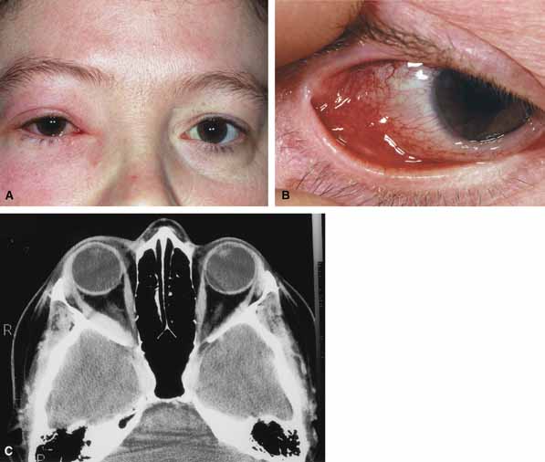
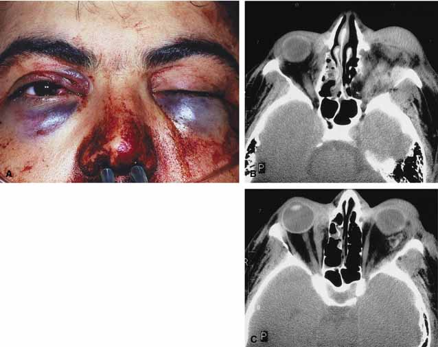
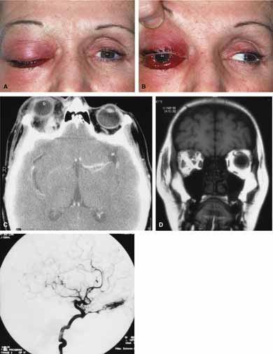
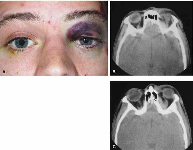
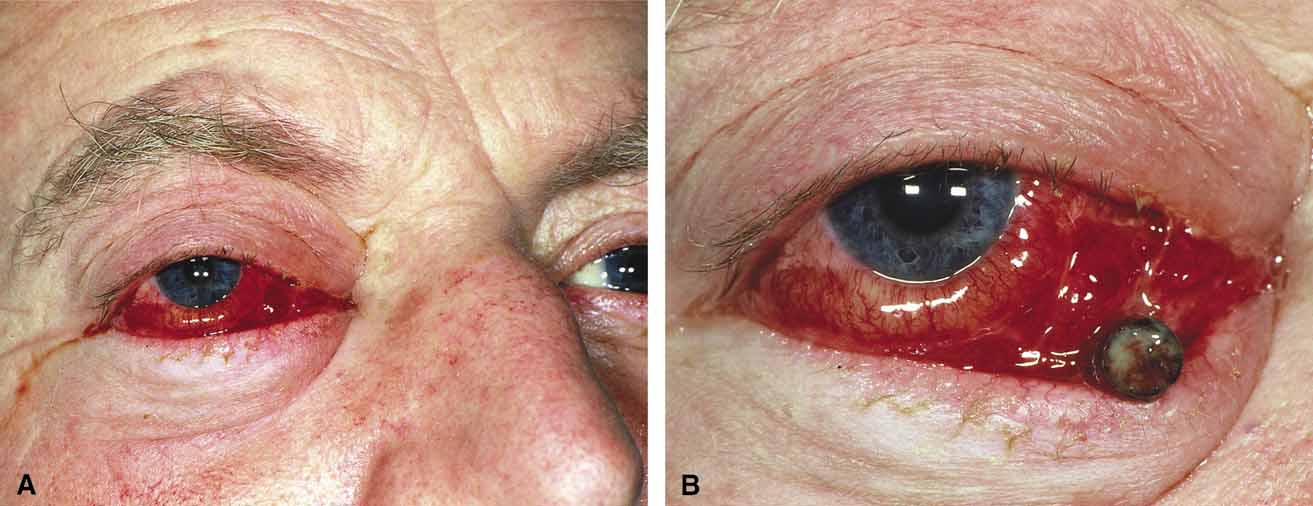
Orbital cellulitis is the model for acute inflammation and a major cause of orbital inflammation in adults.2 It is characterized by a rapid development (over 1 to 2 days) of inflammatory signs and symptoms. These include: eyelid swelling, redness, warmth, pain, conjunctival injection, chemosis, proptosis, and mobility impairment with or without reduced vision. These patients generally feel unwell (malaise) and are febrile (Fig. 1A and 1B). The malaise and fever are key features in differentiating cellulitis from a rapid-onset nonspecific orbital inflammation (pseudotumor) and should be obtained from the patient's history.

Fig. 1 A. A 25-year-old patient presented with proptosis, lid swelling, conjunctival chemosis/injection, pain, malaise, and fever. B. Conjunctival chemosis. C. Ethmoid sinus opacification is present with a superior subperiosteal abscess.

The majority of orbital cellulitis is secondary to extension from an adjacent sinus infection (Fig. 1C). Organisms gain access to the orbit directly through the thin ethmoidal bone, through congenital or acquired dehiscences in the thin orbital walls, pass through preexisting orbital foramina, pass retrogradely through the valveless venous orbital system, or along the veins as a periphlebitis. Orbital cellulitis may also be secondary to an endophthalmitis, systemic bacteremia (e.g., after dental work), infection of a nearby skin wound, dacryocystitis, or penetrating trauma.

The history and physical examination are crucial in distinguishing between preseptal and true orbital cellulitis. The orbital septum delineates the anterior eyelid soft tissue from the orbital soft tissue. Infections anterior to the septum are classified as preseptal cellulitis while those posterior to the septum are termed orbital cellulitis. Recognition of true orbital involvement is important not only because of the threatened visual loss associated with the orbital involvement but also because of the potential for central nervous system complications including cavernous sinus thrombosis, meningitis, and death.

Preseptal cellulitis is characterized by lid edema, erythma, and discomfort. Inflammation posterior to the orbital septum (orbital cellulitis) is heralded by the development of pain, chemosis, proptosis, motility disturbance, and visual deterioration. The extent of ocular involvement in preseptal and orbital cellulitis can be determined by assessing visual acuity, extraocular motility, pupillary reaction, color vision, confrontation visual fields, intraocular pressure, and optic nerve head appearance. Evidence of facial/head trauma or surgical wounds may be apparent. Constitutional signs such as fever, lethargy, and irritability should be sought. The examiner should assess for meningeal signs and neurologic defects. After the initial examination, the physician should follow patients with orbital cellulitis with at least daily assessments of visual acuity, motility, and pupil reaction.

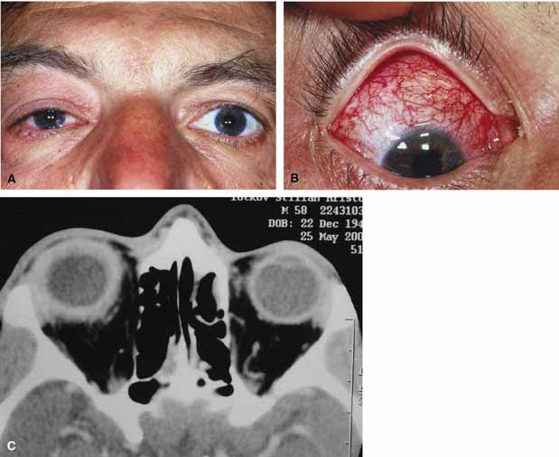
Progression of the disease process despite appropriate antibiotic therapy suggests abscess formation. Orbital abscesses may be either localized, diffuse, or subperiosteal (Fig. 1C). Subperiosteal abscesses most commonly occur along the medial wall and may expand rapidly, compromising optic nerve function even in the absence of many signs of infection.3–6 Several mechanisms may contribute to visual deterioration including direct optic nerve compression, elevation of the intraorbital pressure, and proptosis causing a “stretch” optic neuropathy. Clinically, the eye typically is displaced away from the subperiosteal abscess, and orbital imaging shows a convex mass adjacent to the involved sinus.

Orbital infection with potential orbital abscess formation occurs when bacteria break through the periorbita and gain access to the extraoconal or intraconal space. Diffuse or localized orbital abscess formation may also lead to visual loss through an increase in intraorbital pressure, posterior ischemic optic neuropathy, optic nerve inflammation, or vasculitis. Acute visual loss in the presence of an orbital infection is almost always a surgical emergency in which immediate drainage of the abscess is required to save vision. Continued posterior extension of the infection may result in an orbital apex syndrome, decreased function of cranial nerves,2–6 thrombosis of the cavernous sinus, and even death. Hallmarks of cavernous sinus thrombosis include cranial neuropathy and central neurologic impairment.7

Organisms responsible for orbital cellulitis vary widely and may include Staphylococcous aureus, Streptococcus species, as well as a mixture of aerobes and anaerobic organisms. Recent studies indicate that streptococcus is the most common cause of orbital cellulitis associated with sinusitis in children.8 With increasing age the pathogens increase in complexity. In patients older than 15 with subperiosteal abscesses, polymicrobial infections are typical with anaerobes cultured in every case.5

Orbital imaging in the axial and coronal plane should be obtained in all patients suspected of having orbital cellulitis. Computed tomography (CT) is preferred to magnetic resonance imaging (MRI) because the orbital tissues have higher contrast and bone is well visualized. Axial CT views allow evaluation of the medial orbit and ethmoid sinuses, whereas coronal scans image the orbital roof, floor, frontal, and maxillary sinus. A subperiosteal abscess appears as a homogeneous opacification between the orbital wall and the displaced periorbita.6 Contrast agents are not necessary to visualize a subperiosteal abscess.

Management of orbital cellulitis is dictated by the rapidity of onset. Oral antibiotics may be appropriate for mild cases whereas intravenous antibiotics are required for more fulminant cases. Antibiotic coverage should be broad spectrum and include coverage for gram-positive organisms and anaerobes. Examples include cloxicillan and clindamycin or a late-generation cephalosporin and clindamycin. In those patients allergic to penicillin agents, erythromycin and clindamycin or vancomycin and clindamycin are considerations.

Most patients with orbital cellulitis also benefit from a nasal decongestant as well as an oral decongestant and warm compresses to the affected site (10 minutes out of every hour). Daily or more frequent reexaminations are required depending on the fulminancy of the disease. Once antibiotics are initiated, a 24-hour wait-and-watch period is generally observed (unless the process is aggressive and rapidly developing). If there is no progression at 24 hours and the vision is stable, continued observation is appropriate. If the orbital cellulitis is rapidly developing and fulminant, frequent assessments (every 2 to 4 hours) are required. If the vision, motility, or neurologic status is deteriorating, immediate surgical intervention may be required.

The indication for surgery in a patient with orbital cellulitis has been controversial over the years. The simple presence of a subperiosteal abscess (SPA) was at one time an indication to drain but this is no longer always the case. Providing the vision is normal, the SPA may resolve with medical management. The clinical course, and not the radiographic appearance should dictate management.5,6,9 Surgical therapy may be influenced by many factors including the visual status, size, and location of the SPA; intracranial complications; the sinus involved; the presumed pathogenesis, and the anticipated bacterial response to antibiotic treatment.10 Immediate drainage of SPAs and sinuses is recommended for patients of any age whose vision is compromised. Urgent drainage (as soon as practical) should be considered for large SPAs, extensive superior or inferior abscesses, intracranial complications at the time of presentation, frontal sinusitis where the risk of intracranial extension is increased, and in those suspected of having anaerobes (postdental procedures). However, cookbook approaches should never take the place of good clinical judgement and therapeutic decisions regarding early drainage versus medical management with observation are ultimately up to the managing physician.10 Older children (older than 9 years of age) and adults benefit from early surgical drainage. Although surgical treatment does not guarantee rapid resolution, a complicated course is more likely without it.10

Abscesses may also develop within the intraconal space. The prescence of an abscess within the orbital tissue coupled with any signs of visual loss, afferent pupillary defect, or a significant motility defect should prompt emergent surgery. If visual acuity is stable and extraocular motility essentially full, initial treatment may consist of empiric antibiotic therapy and close observation with serial CT scans.

The prescence of neurologic symptoms such as altered mental status or seizures implies intracranial extension with potential intracranial abscess. Infection may spread from the sinuses to the intracranial cavity via retrograde thrombophlebitis, directly through osteitic bone or from congenital or acquired bony defects. In the past, intracranial abscess formation had a poor prognosis with a high mortality rate. Successful management of suppurative abscesses requires early recognition of the disease process, intravenous antibiotics, serial neuroimaging, and surgical management of at least the orbit and sinus disease and often the intracranial disease.11

PHYCOMYCOSIS (RHINO-ORBITAL MUCORMYCOSIS AND ORBITAL ASPERGILLOSIS)

Rhino-orbital mucormycosis, a fungal infection of class Phycomycetes and order mucorales, is notable for its high morbidity and mortality.12,13 Orbital involvement is an acute, aggressive, and often lethal infection if not recognized early. Phycomycetes (common bread molds) are ubiquitous fungi occurring in soil, air, skin, body orifices, manure, and food including fruit.13 Inoculation occurs by inhalation reaching the nasopharynx and oropharynx. At this stage most patients are able to contain the disease. However, individuals whose cellular and humoral defense mechanisms have been compromised by disease or immunosuppressive treatment may not be able to generate an adequate response. The fungus may then spread to the paranasal sinus, orbit, meninges, and brain by direct extension.13 Mucormycosis preferentially involves blood vessel walls resulting in vascular occlusion, thrombosis, and infarction.14 This frequently affects the ophthalmic artery and in more serious cases may involve the internal carotid artery and cavernous sinus.

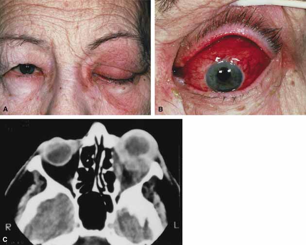
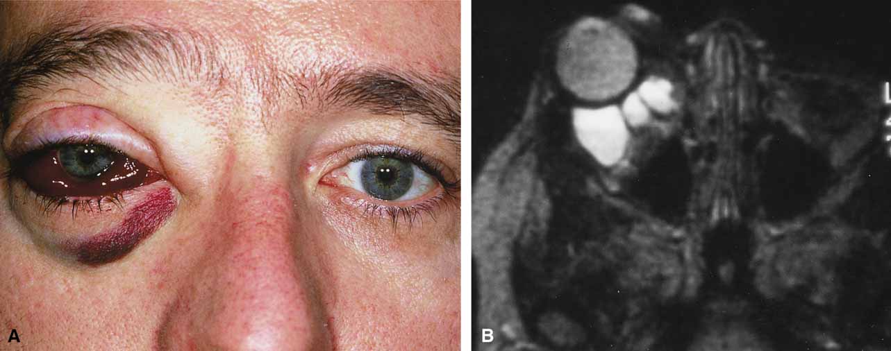
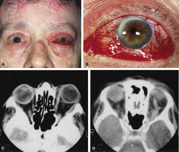
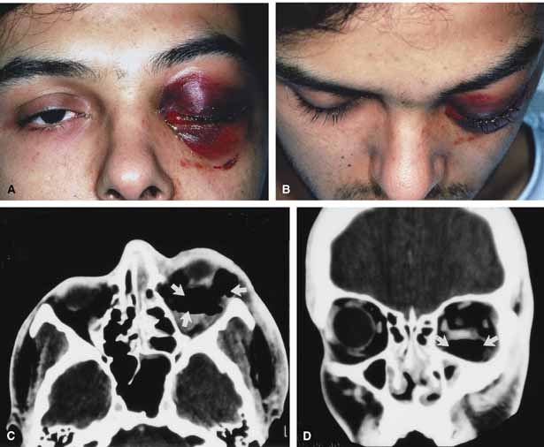
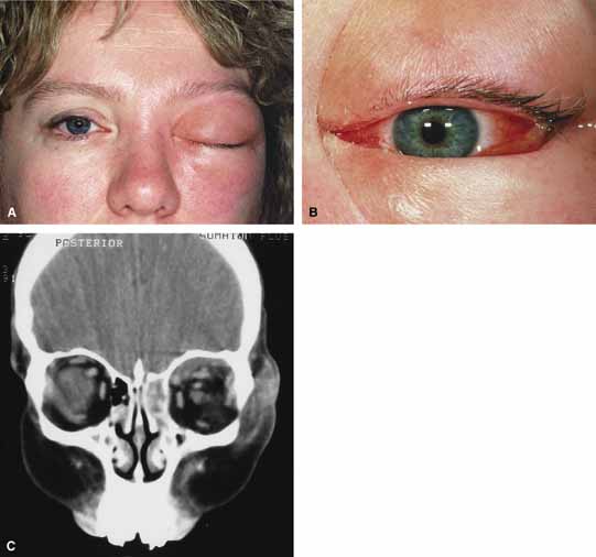
Although there have been a few reports of mucormycosis occurring in healthy individuals, virtually all other patients have had previous severe underlying disease. The patient most vulnerable to this infection is an one with uncontrolled diabetes with ketoacidosis. A host of other conditions also predispose patients to the disease including: multiple myeloma, lymphoma, organ transplantation with immunosuppresion, chemotherapy, corticosteroid treatment, acquired immunodeficiency syndrome, etc.(Fig. 2A).12–14 Mortality is extremely high for patients with phycomycosis infection, and successful treatment is contingent on early recognition and prompt treatment.

Fig. 2 A. A 65-year-old man with a myelodysplastic disorder and pancytopenia developed a pimple on the side of his nose followed by rapidly developing cellulitis and skin necrosis. The patient developed a frozen globe and loss of vision over a matter hours. Despite aggressive surgical debridement of the necrotic areas and intravenous, amphotericin, the patient died. B. Black eschar (necrosis) along right side of nose, ala, upper lip, and hard palate. C. Right orbital infiltration is seen along with opacification of the right ethmoid sinus and nasal passage. D. The optic nerve is on stretch with tethering of the globe, extensive soft tissue swelling anteriorly, ethmoidal opacification, and enlargement of the right medial rectus muscle.

A characteristic pattern of clinical symptoms and signs occurs, the recognition of which should lead to the immediate institution of antifungal treatment and possible surgical intervention to increase the patient's chances of survival. Early diagnosis while the disease is still somewhat anatomically confined is essential for a more favorable outcome.12–14

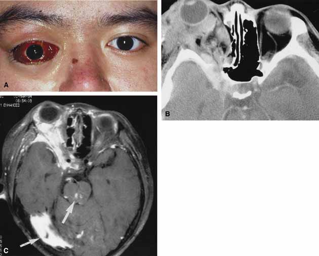
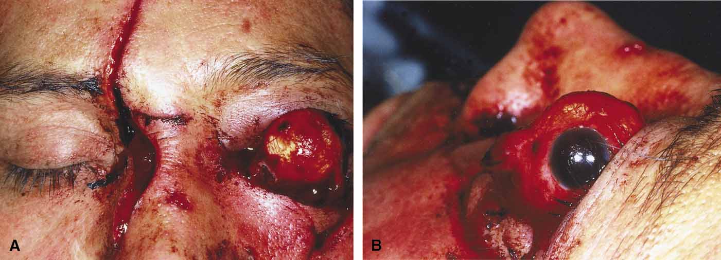
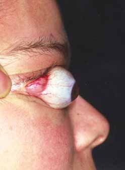
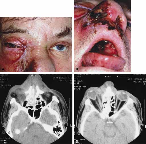
Characteristic features of orbital mucormycosis include an immunocompromised patient with sinusitis, pharyngitis or nasal discharge who develops cellulitis of the face or lid. Signs and symptoms include orbital/periorbital pain, acute proptosis, abrupt visual loss, orbital apex syndrome with acute motility changes (external ophthalmoplegia), pupillary changes (internal ophthalmoplegia), ptosis, and decreased corneal sensation. Infarction of tissue results in black eschar formation of the skin, nasal mucosa and hard palate (Fig. 2A and 2B).12,13 With intracranial extension, the patient generally become obtunded, develops convulsions, contralateral hemianaesthesia or hemiplegia, and lapses into coma.

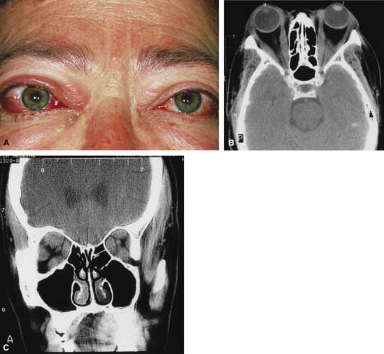
CT scanning demonstrates an orbital mass often with bone destruction and sinus involvement (Fig. 2C and 2D). The diagnosis is confirmed by biopsy of involved tissue with demonstration of characteristic nonseptate, large, branching hyphae, which can be seen on routine hematoxylin and eosin stains. Material should be submitted for both frozen and conventional paraffin-embedded sections. Frozen sections are not always definitive and the surgeon must have considerable confidence in the skill of the pathologist. 13 Management includes: (1) early definitive diagnosis; (2) correction of any underlying metabolic disturbance; (3) wide local excision with debridement of all involved and devitalized oral, nasal, sinus and orbital tissue; (4) establishment of adequate sinus and orbital drainage; (5) daily irrigation and packing of the involved orbital and paranasal areas with amphotericin B; and (6) intravenous amphotericin B.12,13,15

The extent of surgical excision should balance the degree of morbidity and mutilation against the life-threatening risk this organism represents. In limited cases, surgical excision may be confined to those tissues clearly infarcted. Should infection be extensive as demonstrated by widespread necrosis, then aggressive surgery, including exenteration of the orbit and any involved paranasal sinuses, may prove necessary and lifesaving.13

ORBITAL ASPERGILLOSIS

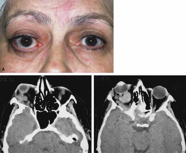
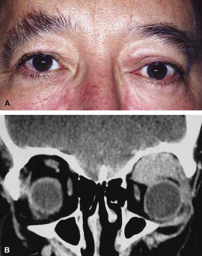
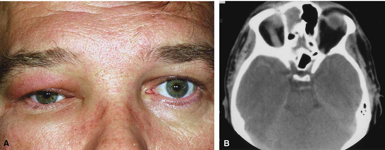
Aspergillus, a fungus of the Ascomycetes class, is a common environmental organism that may colonize the aerodigestive tract. Although widespread, the fungus has a low intrinsic virulence and clinically apparent aspergillosis is rare in the immunocompetant host. Invasive aspergillosis, similar to mucormycosis, occurs more often in the immunocompromised host, including patients undergoing transplantation or steroid therapy, neutropenic patients undergoing chemotherapy, alcoholics, patients with diabetes, and patients with acquired immune deficiency syndrome (AIDS).16–20 Orbital involvement may be slowly progressive or abrupt in onset with orbital inflammation, proptosis, pain, ophthalmoplegia, and sudden loss of vision.16,19 Fulminant aspergillus infection of the nose, paranasal sinus, and orbit often associated with intracranial extension has been reported with increasing frequency in immunocompromised individuals.17,18 In these patients, local invasion of the sinus mucoperiosteum produces a rapidly progressive gangrenous necrosis.18 With bone destruction, infection may extend into the orbit and intracranial cavity; the latter has a poor prognosis. The fungus may also spread by hematogenous routes.17 Imaging studies reveal sinus involvement, heterogenous soft tissue masses with bony erosion, and calcification.16 A definitive diagnosis of aspergillosis is based on tissue biopsy and fungal cultures.

Treatment of invasive sino-orbital aspergillosis involves aggressive surgical debridement combined with a systemic antifungal agent. Intravenous amphoticin B has been the mainstay of medical therapy but toxic side effects, especially renal, require discontinuing the medication in some patients. Newer systemic antifungal agents include liposomal amphotericin B (fewer renal toxic effects) and oral intraconazole.21–24 Adjuvant local irrigation of amphotencin B has also been recommended.24 For patients unable or unwilling to undergo surgery, intralesional injection of amphotericin B has been used successfully as palliative treatment.25 Despite aggressive therapy, the mortality in those with invasive sino-orbital aspergillosis remains high.17

Back to Top
INFLAMMATORY DISEASE OF THE ORBIT: (NONINFECTIVE, NONTHYROID)
Orbital inflammatory processes can be viewed as either nonspecific or specific.26 The nonspecific orbital inflammatory diseases were previously known as inflammatory orbital pseudotumor. The frequency of diagnosis of nonspecific orbital inflammation appears to be decreasing as we improve our understanding of the various types of clinical specific orbital inflammation.26

The definition of nonspecific inflammation remains clinical and consists of processes that are acute and subacute and have characteristic anatomic localization within the orbit. Although the exact mechanism of the inflammation is unknown, an immunologic disorder of some sort is most likely the cause.26,27 Histopathologically, the spectrum of disease is characterized by a nonspecific polymorphous infiltrate of inflammatory cells.26,27 In contrast to nonspecific inflammations, specific inflammations represents three possible types of processes:

  1. Those identified on the basis of a specific pathogen (i.e., infections) and infestations (e.g., orbital cellulitis).
  2. Those that have specific local and/or systemic constellations of findings that identify them as distinct, e.g., Wegener's granulomatosis.
  3. Those diseases that have a specific histopathology that identifies them such as some of the granulomatous diseases, e.g., sarcoidosis.

This section examines the nonspecific orbital inflammations (noninfective and nonthyroid) as well as some of the specific types of inflammations (vasculitis, collagen vascular disorders).

NONSPECIFIC INFLAMMATION OF THE ORBIT

The common feature of nonspecific inflammation of the orbit (previously known as orbital pseudotumor) is the acute or subacute presentation of orbital inflammation. The etiology undetermined. There are several clinical categories of disease, defined by the location of the inflammation. The nonspecific inflammatory syndromes can be divided into a myositic, lacrimal, anterior, diffuse or apical pattern. Imaging reveals an irregular margin adjacent to the primary focus of disease with evidence of tissue swelling and contrast enhancement. Each pattern may present acutely over 24 to 48 hours with orbital pain, tenderness, lid swelling, conjunctival injection, chemosis, proptosis, and decreased extraocular motility. In contrast to patients with orbital cellulitis, these patients are afebrile and do not have malaise. Myositis is the most common pattern of presentation. Patients are usually young (mid 30s) but the age of presentation may range from 9 to 84 years.27 They characteristically present with the acute onset of periorbital pain aggravated by extraocular movement. Signs include eyelid edema, redness, chemosis, conjunctival injection, and motility restriction (Fig. 3A and 3B). The areas over the involved muscles are generally tender to the touch. Most (68%) present with one muscle involvement (horizontal recti > vertical recti) but some present with two or three muscles involved.27,28 Bilateral involvement is rare.27 CT shows muscle enlargement associated with tendon involvement (in contrast to thyroid associated enlargement) and a fuzzy border to the contrast-enhancing muscle (Fig. 3C). Echography is usually pathognomic showing a thickened tendon and a low reflective muscle belly.27

Fig. 3 A. A 22-year-old female presented with a 24-hour history of lid swelling, conjunctival chemosis/injection, pain with abduction, and double vision in horizontal fields of gaze. B. Conjunctival injection was most noticeable over the insertion site of the lateral rectus muscle. C. An axial computed tomography (CT) scan revealed an enlarged lateral rectus muscle on the right.

Lacrimal gland involvement (dacryoadenitis) presents with pain, tenderness, injection of the temporal portion of the upper lid and conjunctival fornix with an associated tender palpable gland and an s-shaped contour deformity of the upper lid. There may be mild proptosis and a slight shift of the globe inferonasally. Imaging shows an enlarged gland with irregular margins. Other causes of dacryoadenitis, such as viral/ bacterial infections, Sjögren's syndrome, Wegener's granulomatosis, or sarcoidosis must be ruled out.29 A significant number of patients have the potential for some type of associated systemic disorder.29

In those with anterior nonspecific orbital inflammation the main focus of inflammation is the globe and adjacent orbital tissue (Fig. 4A and 4B). Features include pain, proptosis, lid swelling, injection, and occasionally decreased vision. Scleritis and uveitis may also be present. In those with diffuse involvement there are extraocular and neurosensory structures involved as well. Imaging shows an irregular orbital infiltrate adjacent to the globe with scleral thickening in the anterior group (Fig. 4C) and more extensive involvement with the diffuse group (Fig. 5A, 5B and 5C).

Fig. 4 A. A 45-year-old male presented with a 24-hour history of increasing eye pain and conjunctival injection. B. Diffuse scleral and episcleral injection is present. C. An axial computed tomography (CT) scan revealed diffuse enhancement of the sclera and adjacent orbital tissues.

Fig. 5 A. A 72-year-old female presented with a 36-hour history of ptosis, conjunctival injection, pain, double vision, and proptosis. B. Diffuse conjunctival injection and chemosis is seen. C. Diffuse orbital inflammation is present with a mass effect anteriorly, adjacent to the globe.

Apical involvement with nonspecific orbital inflammation is uncommon but may present with pain, decreased vision, restricted motility, mild proptosis, and chemosis. The functional abnormality is typically more than the degree of inflammation would suggest.26 These patients require careful consideration as a wide variety of disorders can present with these findings (including neoplastic processes, cavernous sinus thrombosis, etc.).

Treatment for the acute nonspecific orbital inflammatory syndrome include nonspecific anti-inflammatory medication. In mild cases nonsteroidal anti-inflammatory agents (NSAIDs) such as ibuprofen21 may help but most cases require steroids.26,27 A rapid steroid response, especially for the associated pain, is typical and most patients notice a significant improvement of symptoms over 24 hours. If the presentation is typical, as is generally the case with the myositic and anterior varieties of nonspecific orbital inflammation, a course of steroids without biopsy is common. Because a large number of the lacrimal inflammations are associated with a systemic disease, a biopsy is recommended prior to steroid use. For diffuse and apical inflammations, and those that fail to respond to steroids or have recurrent disease, a biopsy is wise to confirm the disease process.

SPECIFIC INFLAMMATIONS OF THE ORBIT

In addition to the nonspecific orbital inflammatory syndromes there are a number of more specific inflammations that may present with acute proptosis. These can be divided into those with specific local and/or systemic findings and those with specific histopathology that identifies them.26

Vasculitis

Vasculitis includes a wide range of inflammatory angiodestructive processes that involve varying calibers of vessels and diffuse types of infiltrates ranging from polymorphonuclear to necrotizing granulomatous.26 The unifying feature is evidence of vessel inflammation with varying degrees of destruction. Clinically, patients may present with acute inflammatory changes similar to the nonspecific orbital inflammation patients. The various entities are classified on the basis of symptoms related to the organs or tissue affected, as well as their principle histopathologic features.26

Although many of these disorders have relatively distinct systemic symptoms and signs, it may be difficult to diagnose them and differentiate them from nonspecific inflammatory disease if the process is restricted to the orbit during the early stages.

Wegener's granulomatosis

Wegener's granulomatosis (WG) is a disease of presumed autoimmune origin that in its pure form is characterized by necrotizing granulomatous inflammation of the upper and lower respiratory tract, vasculitis involving small to medium vessels, and a focal glomerulonephitis.30 It has a broad range of presentations and may occur as a systemic or localized (limited) disease. When systemic, it is characterized by widespread inflammatory effects with multisystemic damage, upper and lower respiratory tract involvement followed by renal involvement.26,30–33 In the more localized form involving the orbit, the patient may present with proptosis, periorbital inflammation, an orbital mass, and associated ocular inflammation (iritis, episcleritis, scleritis, retinal vasculitis). Bilateral orbital involvement is not uncommon.26,31

The vasculitis is necrotizing and the process is progressive and associated with destruction. Imaging may show a unilateral or bilateral poorly outlined orbital mass, infiltration and destruction of fat planes, involvement of sinuses, and bone erosion. Cytoplasmic antineutrophil cytoplasmic antibody (C-ANCA) laboratory testing is useful even in limited disease where the results are said to be positive in 60% to 70% of cases,34,35 however, it may initially be negative.31,34 Biopsy of involved tissue is essential for a diagnosis. The three classic histopathologic features of WG are vasculitis, granulomatous inflammation, and tissue necrosis. The diagnosis should be based on a combination of clinical (orbital involvement, scleritis, respiratory tract involvement, renal and sinus disease) laboratory, radiologic, and pathologic features. A specific diagnosis is essential before initiating the necessary aggressive treatment (corticosteroids and cyclophosphamide).

Polyarteritis nodosa

Polyarteritis nodosa is also a vasculitis of the medium to small arteries. Orbital involvement, although unusual, presents with nonspecific orbital inflammatory features. This entity is distinguished as a specific orbital inflammatory disorder by the pattern of involvement and histopathology. Sites of predilection are kidney, heart, liver, and gastrointestinal tract.36 It may also involve the skin, peripheral nerves, pancreas, testicles, and skeletal muscles. Clinically, the patients often have fever, malaise, leukocytosis, and evidence of multisystem involvement. Other ophthalmologic features include retinal and choroidal infarcts, corneal and scleral necrosis. Definitive diagnosis requires a biopsy of involved tissue. Treatment, similar to that for WG, involves corticosteroids and cyclophosphamide.

Collagen Vascular or Connective Tissue Disorders

Any of the connective tissue disorders may also be associated with a systemic vasculitis including systemic lupus, rheumatoid arthritis, and dermatomyositis.26,37,38 Orbital involvement is uncommon. The clinical picture may be identical to the nonspecific orbital inflammations previously discussed and may be treated as such. A definitive diagnosis is based on the pattern of systemic disease, laboratory testing and/or radiologic and pathologic features.

Back to Top
THYROID ORBITOPATHY
Thyroid-related eye disease (Grave's disease) has been recognized by the medical community for more than two centuries. The eye signs of Grave's disease are a symptom complex that describes the response of the eyes to an inflammatory disorder within the orbital tissues. It is therefore best thought of as an infiltrative orbitopathy rather than an ophthalmopathy.38,40

Thyroid orbitopathy occurs in a genetically preselected population, affecting females four to five times more frequently than males, except in the older age groups where the female to male ratio decreases.39 A specific cause remains unknown but evidence links the orbitopathy, the immunogenic thyroid disorder, and pretibial myxedema to immune mechanisms of both cellular and humoral mediation.39,41–43 Recent insights into the pathogenesis of thyroid associated orbitopathy suggests the orbital fibroblast may play a central role.41 Some individuals think that the previously overlooked fibroblast may orchestrate a complex immune cascade that initiates, perpetuates, and eventually attenuates the clinical disease.41 Pathologically, the extraocular muscles may be infiltrated with lymphocytes, macrophages, plasma cells, and mast cells.39 In addition, there is deposition of hydrophilic mucopolysaccharides. The pathology varies depending on the intensity and stage of the disease when the tissues are examined. The various mechanisms at play result in the clinical manifestations of increased mass, inflammation, swelling, muscle restriction, and secondary compressive features. Varying degrees of symmetric soft tissue swelling, chemosis, proptosis, lid retraction, temporal lid flare, and extraocular motility restriction are seen.

The clinical presentation of Grave's orbitopathy varies from mild symptoms of exposure to a more rapidly progressive course resulting in compromised visual function. In most cases, the disease has an insidious onset and the patients may have symptoms of dry eye secondary to proptosis, lid retraction, and lagophthalmos. Conjunctival injection and chemosis are commonly seen.

In some patients the orbitopathy is more aggressive and rapidly evolving with acute proptosis over 1 to 2 days, swelling of the lids and conjunctiva, diplopia, corneal exposure, and optic neuropathy (Fig. 6A). This presentation may be confused with disorders such as nonspecific orbital inflammation or carotid-cavernous fistula.44 The differential diagnosis of acute proptosis with enlarged extraocular muscles includes thyroid orbitopathy, vascular causes (arteriovenous fistulas), inflammation (orbital myositis, sarcoid), infection, primary invasive tumor, metastasis to the extraocular muscles.45,46

Fig. 6 A. A 56-year-old female presented with a 2-day history of proptosis, lid swelling, conjunctival chemosis/injection, and double vision. Thyrotropin (TSH) level was extremely low and a computed tomography (CT) scan revealed grossly enlarged extraocular muscles. A diagnosis of acute thyroid eye disease was made. B. Axial computed tomographt (CT) scan illustrating diffuse enlarged extraocular muscles characteristic of thyroid eye disease. C. Coronal computed tomography (CT) scan showing enlargement of extraocular muscles.

Thyroid orbitopathy usually occurs close to or within 18 months of the hyperthyroid state.39 Some patients will be hyperthyroid at the time of diagnosis, others euythyroid, and some might even be hypothyroid. The hyperthyroid state is characterized by elevation of the plasma thyroid hormones triiodothyronine (T3) and thyroxine (T4). For patients presenting primarily with features of thyroid orbitopathy and no clinical thyroid abnormality, the laboratory investigations should include sensitive thyrotropin (TSH); free T4 or total T4 (if TSH is low or altered); T3 (if TSH is low); and normal T4 testing. Additional tests that may be of benefit include thyroid antibody titer and TSH receptor antibody test.

CT classically reveals enlarged extraocular muscles (Fig. 6B and 6C). One or all of the muscles may be involved and there is bilateral involvement in the majority. The most frequently involved muscle is the inferior rectus followed by the medial, superior, and lateral rectus (opposite to the spiral of Tillaux).40 The tendons of the affected recti muscles are typically not thickened, resulting in a characteristic fusiform enlargement of the muscle, whereas in myositis secondary to nonspecific orbital inflammation the tendons are generally thickened. Other signs are also useful to distinguish between the two. Bilaterally is unusual in myositis but common with thyroid. Thyroid usually has several muscles involved while myositis more often only has one muscle involved. In myositis the muscle border is more likely to be irregular or fuzzy with or without involvement of adjacent fat whereas in thyroid disease the muscle border is regular and there is no fat involved. Clinically there are also several distinguishing features. Myositis classically presents more rapidly and is associated with pain on extraocular movement. This is rare with thyroid eye disease. The eyelid signs (retraction, flare) are common with thyroid but absent in myositis.

Management of thyroid orbitopathy is dependant on the phase of disease. Severe disease with a rapid onset of inflammatory and infiltrative features causing early mechanical and significant soft tissue involvement requires aggressive treatment to avoid compromise of visual function. Conservative measurements for corneal protection include topical artificial tears and lubricating ointment with or without a the use of a moisture chamber at night. Elevating the head of the bed and using cool compresses over each eye three to four times per day for 45 to 60 minutes may be helpful. The role of corticosteroid treatment of the acute congestive phase of Grave's orbitopathy is well established. Steroids appear to affect not only the inflammatory component of the disorder but also retrobulbar fibroblastic proliferation and function, which are thought to play an integral rate in the overall pathogenesis.39,41 When visual function is being compromised, high dose steroids can be invaluable. Doses of 80 to 120 mg of oral prednisone are commonly used initially and then tapered over 6 to 8 weeks. Pulsed intravenous methylprednisolone may also be used.39 Radiotherapy is another possibility for treatment of those with severe congestive orbitopathy.41 However, the role of radiotherapy has become increasingly controversial.47–50 If the visual function (visual acuity, color vision, visual fields) are not improving despite high-dose steroids, surgical intervention (orbital decompression) may be required. There are various techniques to decompress the orbit either through an orbital sinus or combined approach.51–54 The goal is to expand the apical region of the orbit by removing bone from the adjacent sinuses. In recent years fat removal has also been beneficial to decrease the orbital volume but only rarely is used on its own in those with compromised vision secondary to compressive optic neuropathy.55,56

Back to Top
ORBITAL HEMORRHAGE
Orbital hemorrhage develops most often as a result of accidental or surgical trauma (Fig. 7A, 7B, and 7C). It has been known to occur during or after a variety of periocular procedures including blepharoplasty, retrobulbar injection, fine-needle biopsy of the orbit, repair of orbital fractures, sinus surgery, dacryocystorhinostomy, lacrimal gland surgery and others.57–61 Spontaneous orbital hemorrhage is uncommon but may occur in patients with lymphangioma (Fig. 8A), orbital varices, leukemia, hemophilia, scurvy, hypertension, aneurysm of the ophthalmic artery, use of thrombolytic agents, labor and delivery etc.62–66 Orbital hemorrhage may also occur spontaneously after a Valsalva maneuver or other straining effort.62,67,68 Another important but more rare cause of spontaneous orbital hemorrhage is a postoperative hemorrhage that occurs approximately 3 to 4 days after orbital surgery. The hemorrhage may be caused by dissolution of intravascular clots. In part it may be related to postoperative inflammation or the release of proteolytic enzymes that results from damage to the orbital fat.59

Fig. 7 A. A 20-year-old male presented to the emergency department after a knife attack with bilateral periocular hemorrhages, ptosis, and proptosis of the left eye; normal vision on the right; and a vision of 20/200 on the left. Both globes were intact. B. Axial computed tomography (CT) scan with signs of orbital hemorrhage bilaterally (Lt > Rt). C. Axial computed tomography (CT) scan with signs of orbital hemorrhage bilaterally (Lt > Rt).

Fig. 8 A. A 45-year-old male with a known past history of orbital lymphangioma developed sudden proptosis, subconjunctival hemorrhsage and eyelid ecchymosis. B. Magnetic resonance imaging (MRI) scan showing multiple cystic lesions with a fluid level, characteristic of lymphangioma.

Orbital hemorrhage may be dramatic in presentation because it can develop over a matter of minutes. However, the signs and symptoms may be variable and are dependent on the location of the hemorrhage. Blood may accumulate within the intraconal or extraconal space, subperiosteally, within the optic nerve sheath, preseptally, or in multiple locations. Rapid accumulation of a significant amount of blood within the orbital tissues, developing over 30 to 60 seconds, is associated with acute proptosis, pain, nausea, vomiting and decreased ocular motility. A marked elevation of intraorbital pressure occurs with secondary elevation of intraocular pressure. The rise in intraorbital pressure often produces a posterior ischemic optic neuropathy.69

Treatment of orbital hemorrhage with visual compromise is a matter of utmost urgency. After injecting local anesthetic into the lateral canthal area a lateral canthotomy, inferior cantholysis and/or superior cantholysis is performed.70 If a surgical wound is present (e.g., post blepharoplasty incision), the sutures should be removed and the wound opened. These measures often decrease the intraorbital pressure with resultant return of vision. Intravenous mannitol (20 to 40 mL of 20%) and high-dose intravenous steroid (decadron or solumedrol) is also beneficial. If the intraorbital pressure fails to decrease, a CT scan should be performed and plans made for orbital exploration with possible decompression. Subperiosteal hemorrhage especially those along the posterior medial wall can compress the optic nerve causing visual loss but may not cause a dramatic rise in intraocular pressure or significant proptosis. The motility may only be mild restricted in horizontal gaze.

In a patient presenting post trauma with decreased vision, no evidence of ocular injury but evidence of a posterior medial subperiosteal hemorrhage (that may not be dramatic) three possible explanations exist for the decrease in acuity: traumatic optic neuropathy, compression of the optic nerve from the subperiosteal blood, or a combination of the two. In these instances it is best to treat what is treatable and drain the hemorrhage. High-dose steroids may also be worthwhile for the possible traumatic optic neuropathy.

Hemorrhage into the optic nerve sheath is accompanied by marked visual loss and requires direct opening of the optic nerve sheath in addition to high-dose steroids.

Back to Top
ORBITAL VASCULAR MALFORMATIONS
Spontaneous hemorrhage with sudden proptosis also can occur in all types of vascular malformations. The nomenclature for orbital vascular malformations has been hampered over the years by conflicting definitions and terminology particularly as it applies to lymphangiomas and varices. Some prefer to consider them in the same family of disorders while others prefer to consider them as separate entities.71,72 Terminology for these entities may therefore be used and interpreted differently by various authors and readers.71 Members of the Orbital Society, in an attempt to reconcile the various controversies, proposed a classification system of orbital vascular malformations based on hemodynamic factors (Table 1).71 The system does not apply to capillary and cavernous hemangiomas. Pertinent clinical and imaging features should be considered before a given case is assigned to one of the categories. Universally accepted terminology is essential to compare therapeutic protocols and advance management of these often problematic lesions. Several of the vascular malformations in Table 1 may present suddenly (over minutes) with acute proptosis, pain, nausea and/or vomiting, reduction of ocular motility and decreased visual function.


TABLE 1. Orbital Vascular Malformations


1No Flow Malformationshemodynamically isolated, applicable to the so-called lymphangiomas
2Venous Flow Malformationsapplicable to so-called primary varices including distensible and non distensible varieties as well as mixed forms with venous and no flow components
3Arterial Flow Malformationsapplicable to arteriovenous malformations or arteriolized veins distal to them and also applicable to so-called secondary varices


NO-FLOW MALFORMATIONS

No-flow malformations (so-called lymphangiomas) have nonfunctional vascular systems that arborize through variable portions of the orbit.73 They may appear as superficial, deep, combined, or complex lesions.74 Clinically they are characterized by syndromes consisting of episodes of acute exacerbation and remission, often related to an upper respiratory tract infection. Although they are hemodynamically isolated from the systemic circulation, nutrient vessels within their fragile walls may be the source of sudden hemorrhage into their lumens, which transforms microscopic channels and cysts into blood-filled macrocysts (so-called chocolate cysts). Hemorrhage within a retrobulbar lesion may be dramatic, causing sudden proptosis (Fig. 8A and 8B). The patient has pain, decreased ocular motility, and decreased vision. Involvement of superficial components will lead to disfigurement of the lid, as well as swelling and ecchymosis of the conjunctiva. Management is similar to other orbital hemorrhages with canthotomy, cantholysis, and/or orbital decompression. Surgical decompression if required is aimed at evacuating the blood-filled cysts. Combined and complex lesions are frequently massive in size involving the intraconal, extraconal, preseptal, and postseptal spaces (Fig. 9A and 9B).75,76 They produce significant cosmetic disfigurement and may extend beyond the confines of the orbit into the intracranial space. In addition, isolated intracranial vascular anomalies may be noted (Fig. 9C). These combined lesions often require orbital surgery either on an acute basis, when there is an acute retrobulbar hemorrhage with optic nerve compression, or on an elective basis to deal with cosmetic disfigurement or chronic compression. Such surgery must be approached cautiously because of the risk of iatrogenic hemorrhage. Oral or intravenous steroids are also beneficial in the treatment of acute flare-ups.

Fig. 9 A. A 20-year-old male with extensive right facial and orbital lymphangioma presented acutely with proptosis, subconjunctival hemorrhage, double vision, and orbital pain. B. Axial computed tomography (CT) scan reveals extensive involvement of the orbit. The abnormal tissue also extends through the superior orbital fissure to the cavernous sinus area. C. Magnetic resonance imaging (MRI) scan reveals not only the extensive orbital involvement but also vascular anomalies in the midbrain and cerebellum (arrows).

VENOUS FLOW MALFORMATIONS

Venous flow malformations (so-called varices) generally refer to those vascular malformations with weakened segments of the orbital venous system, of variable length and complexity.76,77 They may appear as superficial, deep, or combined lesions and may be of a distensible or nondistensible variety. Because they are intrinsic to the systemic circulation, distensible primary varices enlarge with increased venous pressure, and their distensibility varies with the residual thickness and strength of their wall. The lesions often cause intermittent proptosis, pain, and/or bruising because of expansion brought about by physical effort (straining, Valsalva maneuver) or bending (Fig. 10A, 10B and 10C).78 Sudden proptosis may result from hemorrhage into one of the varices or the creation of a one-way valve effect trapping blood within the lesion. High-dose steroids, canthotomy, and cantholysis may be of benefit to decrease the orbital pressure. Direct surgical excision is difficult because of the tortuous tangles of fragile, thin-walled malformed vessels and their tendency to rupture and bleed excessively.59 Nondistensible varices may also appear as superficial, deep, or combined lesions. Clinically they are similar to lymphangiomas and characterized by episodes of acute exacerbation and remission related to hemorrhage or thrombosis within the lesion. Hemorrhage into deeper lesions causes sudden proptosis, pain, decreased motility, and reduced vision. Profound orbital hemorrhage with visual deterioration and pain is treated in a manner similar to that of lymphangioma and orbital hemorrhage (canthotomy, cantholysis, and/or orbital exploration with evacuation of clotted blood and excision of the associated lesion).

Fig. 10 A. A 68-year-old female presented with intermittant proptosis occurring on bending over. B. Multiple, oval shaped, contrast-enhancing masses are present in the right orbit. The masses enlarged with valsalva and were consistant with a diagnosis of orbital varix. C. Multiple, oval shaped, contrast-enhancing masses are present in the right orbit. The masses enlarged with valsalva and were consistant with a diagnosis of orbital varix.

ARTERIAL FLOW MALFORMATIONS

Arterial flow malformations are distinguished by evidence of arterial flow. The primary examples are arteriovenous malformations that have a direct flow from the arterial side, through a malformation, and then to the venous side of the circulation. Arteriovenous malformations are characterized by high arterial flow directly into the anomaly that exits through the normal venous channels, which may arterialize. Thus, they bypass the orbital system and more commonly act to shunt blood away from rather than into normal vascular channels.59 Clinically they are associated with pulsating exophthalmos or recurrent episodes of hemorrhage or thrombosis (Fig. 11A and 11B). They are often associated with a bruit (which may be heard by the patient) and may cause pain when engorged with straining or Valsalva maneuver. With imaging, orbital arteriovenous malformations are characterized by irregular, rapidly enhancing masses that may have high flow characteristics on Doppler studies and flow voids on MRI scanning (Fig. 11C, 11D, and 11E). Direct selective angiography will demonstrate the engorged, rapidly filling proximal arterial system, the malformation and the distal venous outflow. Management may involve selective gluing followed by excision. For those lesions with feeding vessels that are outside the orbit, a direct cut-down followed by embolization and surgery is also a safe and direct approach.59

Fig. 11 A. A 48-year-old female presented with sudden proptosis (developing over several minutes), pain, ptosis, decreased ocular motility, decreased visual acuity, and conjunctival chemosis. B. Conjunctival chemosis and restricted motility is seen. C. On axial computed tomography (CT) scanning a contrast enhancing mass is seen superiorly. D. Coronal magnetic resonance imaging (MRI) scanning revealed the superior anomaly. E. Angiography demonstrated an arteriovenous malformation of the right orbit. The vascular malformation was successfully removed surgically.

Back to Top
ACQUIRED ARTERIOVENOUS SHUNTS: DURAL CARTOID CAVERNOUS FISTULAS
Cartoid cavernous sinus fistula (CCSF) is the most common arteriovenous malformation affecting the orbit and involves abnormal communication between a branch of the carotid artery and the cavernous sinus.79 The resulting high-flow fistula can produce proptosis (may be pulsatile), pain, engorgement of epibulbar veins (corkscrew configurations), limited extraocular motility, raised intraocular pressure and loss of vision (Fig. 12A and 12B). In many cases a bruit may be heard by the patient or examiner. The clinical features may mimic acute thyroid disease, but in contrast to thyroid eye disease, fistulas are usually unilateral. Seventy-five percent of fistulas occur as a result of head trauma; the rest occur spontaneously.79,80 Traumatic causes include head injury (penetrating or nonpenetrating) as well as iatrogenic causes including complications of carotid endarterectomy surgery. The spontaneous fistulas often occur in post-menopausal women. They may be the result of a congenital malformation (e.g., aneurysm or arteriovenous malformation) or associated with atherosclerotic changes, hypertension, collagen vascular disease, or childbirth. Some cases remain idiopathic. The spontaneous fistulas tend to be low-flow shunts in contrast to high-flow traumatic fistulas. Carotid cavernous fistulas can be subdivided as: (1) etiologic (spontaneous or traumatic); (2) hemodynamic (high or low flow), and 3) anatomic (direct or dural).79,80 CT and MRI are useful screening tests and may show dilation of the superior ophthalmic vein, increased extraocular muscle size and enlargement of the cavernous sinus (Fig. 12C and 12D). The definitive diagnostic test is bilateral selective internal and external carotid angiography.

Fig. 12 A. A 68-year-old female developed sudden proptosis (over 2–3 minutes), decreased vision, conjuntival injection, chemosis, and restricted motility. B. Multiple corkscrew vessels are seen secondary to engorgement of the epibulbar vessels. C. Axial computed tomography (CT) scan revealed proptosis and enlargement of the extraocular muscles. D. Axial computed tomography (CT) scan also revealed an enlarged superior ophthalmic vein.

Treatment may not always be required because 10% to 60% of spontaneous CCSFs will spontaneously close.79,80 The indication for intervention include visual deterioration, diplopia, intolerable bruit, headache, and proptosis causing untreatable corneal exposure.79,80 The goal is to cure the patient's symptoms without causing cranial nerve palsies or compromising carotid circulation. Methods of treatment involve various procedures to obliterate either the fistula, its afferent vessel, or its efferent vessel. High-flow fistulas are best treated with a detachable balloon because most will not close spontaneously. Closure may be facilitated by carotid compression applied by the patient. Fistulas between meningeal branches of the external carotid and cavernous sinus are supplied by single feeders and can be obliterated by selective embolization. Fistulas with shunts between the meningeal branches of both the internal and external carotid artery and the cavernous sinus require embolization of all feeding branches, which will eradicate fistulas in approximately 50% of cases. Alternatively, retrograde obliteration of the cavernous sinus may be required through a superior ophthalmic vein approach or internal jugular vein.59,81,82

Back to Top
NEOPLASMS
A number of neoplasms (primary, metastatic, secondary) may present rather acutely over 1 to 2 days with symptoms of proptosis, lid edema, chemosis, ptosis, motility disturbance, and/or some degree of visual compromise. The diagnosis may be suggested by history, ocular/orbital evaluation, systemic evaluation with orbital imaging studies and confirmed by fine-needle aspiration or open biopsy.

LYMPHOPROLIFERATIVE DISORDERS

Lymphoproliferative disorders represent a group of diseases with a basic unifying histopathologic substrate of a densely cellular infiltrate comprised mostly of small lymphocytes. Over the past several years the classification of orbital lymphoproliferative lesions has undergone profound changes, primarily as a result of increasingly sophisticated and specific immunodiagnostic and molecular techniques in tissue pathology. This has resulted in numerous updates to the various classificatation schemes.83

The lymphoproliferative disorders involving the orbit primarily involve reactive and lymphomatous lesions of the non-Hodgkin's variety. In contrast, Hodgkin's lymphoma rarely involves the orbit.84

Orbital involvement with lymphoid tumors is generally characterized by insidious development of painless proptosis, lid swelling, and diplopia. A rapidly developing and fulminant course may occasionally occur with poorly differentiated high-grade lymphomas (Fig. 13A).83,85–88 Lymphoid tumors may occur anywhere in the orbit but have a predilection for superior and anterior locations often resulting in a downward displacement of the globe. A firm or rubbery mass not fixed to bone is frequently palpable. Subconjunctival extension, presenting as a fleshy, salmon-colored mass of the conjunctival fornix, may occur. On CT scan, lymphoid tumors typically appear as an enhancing soft tissue mass, usually oblong in shape, with well-defined margins and putty-like molding to the contours of adjacent orbital structure (Fig. 13B). Orbital bone erosion is rare in lymphoid tumors.89

Fig. 13 A. A 52-year-old male awoke one morning with swelling of the left upper eyelid and a pressure feeling behind the right eye. B. Coronal computed tomography (CT) scan revealed a large orbital mass that was biopsied, revealing a poorly differentiated B-cell Lymphoma.

Patients with orbital lesions require an incisional biopsy to confirm the diagnosis followed by a thorough medical examination to investigate for the presence of systemic disease. The patient's disease is staged and appropriate treatment is organized. For lesions confined to the orbit, radiation is required. Systemic involvement requires chemotherapy.

LEUKEMIAS

Leukemias that involve the orbital tissues are always a part of a systemic disease. In some cases, however, the orbital manifestations of leukemia may occur prior to the onset of systemic manifestations.90 There are a host of leukemic disorders, both acute and chronic, which fall mainly into the lymphoid and myeloid groups that may involve the orbital tissue (usually as a complication of late stage disease). Generally, soft tissue involvement of the orbit is more frequent in acute (especially lymphoblastic) rather than in chronic leukemia. Unilaterality is the rule with only 2% having bilateral disease.83 Involvement may be the result of soft tissue infiltration or hemorrhage and may occur acutely over 1 to 2 days. Orbital disease usually implies a rapid demise but local irradiation and both intrathecal and systemic chemotherapy may significantly prolong survival. This emphasizes the importance of prompt recognition and treatment.83

Granulocytic sarcoma (chloroma or extramedullary myeloid cell tumor) represents a localized, usually extramedullary form of acute myeloblastic leukemia or of a chronic granulocytic leukemia entering a blast crisis.83,90,91 This tumor has also been called a chloroma because the myeloperoxidase within the tumor imparts a green hue to its surface that can be seen on gross examination.90 Granulocytic sarcomas are known for their rapid development.83,91,92 Because the clinical presentation may be fulminant, confusion with local inflammation and other malignant disease may occur. The sites of orbital involvement include soft tissue, lacrimal gland, or bones.83 CT may demonstrate diffuse infiltration or an irregular circumscribed mass with or without bone erosion. A diagnosis is established by biopsy. The prognosis for patients with this aggressive tumor is poor. Treatment consists of local irradiation and intensive chemotherapy.83,91

METASTATIC DISEASE

Metastatic tumors to the orbit are a rare but important cause of orbital disease. Although the prognosis for patients with metastatic cancer is poor, new regimes of radiotherapy and chemotherapy have substantially improved the duration and quality of life for many cancer patients.93

A number of malignant neoplasms are associated with metastatic disease to the orbit, most of which are carcinoma.93,94 Breast, prostate, melanoma, lung and gastrointestinal primaries are typical sources of orbital metastasis.93–95 Metastatic disease to the orbit may present many years after discovery of the primary neoplasm. In other cases, the primary neoplasm may be unknown (10% to 25%) at the time of orbital presentation.93,94 Metastatic disease is not always easy to diagnose. The most common misdiagnosis in one series were related to orbital inflammation and to bizarre motility disturbances.94

Orbital metastasis may occur anywhere within the orbit. Clinical features are secondary to unrestricted growth with local infiltration and entrapment of structures. Patients often display rapidly progressive proptosis which may be axial or non axial. More than 90% of orbital metastasis occur unilaterally.94 Diplopia, pain, blepharoptosis, proptosis or enophthalmos, conjunctival chemosis/injection, and some degree of visual loss may be present. A hard mass firmly fixed to underlying bone may be palpable along the superior orbital rim. These lesions are often tender to touch as a result of periosteal or bone invasion. On CT scan, a metastatic tumor usually appears as an infiltrating, contrast-enhancing, soft tissue intraconal or diffuse mass and/or bone, and/or muscle involvement. Bone destruction or hyperostosis (especially with prostatic carcinoma) may be present.93,94 A diagnosis is established by history (including a careful systems review and past history) and biopsy (incisional or needle). Management involves a search for a primary if there is cancer history known followed by chemotherapy, hormonal therapy, irradiation, surgical excision or observation depending on the clinical circumstances.93 Radiation is the mainstay of treatment for control of the orbital tumor and often helpful for those experiencing significant orbital pain. Local orbital radiotherapy can often dramatically improve vision and ocular function in patients with space occupying metastatic lesions. Such symptomatic treatment can have a profound effect on a patient with a terminal disease.

Secondary orbital tumors such as malignant tumors of the sinus (e.g., squamous cell carcinoma) may abruptly involve the orbit producing acute proptosis over 1 to 2 days with lid swelling, chemosis, motility disturbance, and pain (Fig. 14A and 14B). Diagnosis is confirmed by CT imaging and biopsy of the tissue.

Fig. 14 A. A 43-year-old male presented with a 2-day history of right upper eyelid swelling. B. Axial computed tomography (CT) scan revealed a mass in the right frontal sinus with extension into the right orbit. A biopsy disclosed poorly differentiated squamous cell carcinoma.

Back to Top
TRAUMA
Acute traumatic proptosis may be secondary to a variety of processes including orbital hemorrhage, air accumulation, or, reduction in orbital volume. At times, more than one entity is responsible for the globe displacement.

ORBITAL FRACTURES

Acute proptosis may occur with any orbital fracture secondary to orbital hemorrhage (diffuse or subperiosteal), orbital emphysema, by a reduction in orbital volume secondary to inward displacement of the orbital walls, or the presence of an encephalocele.96 Traumatic encephaloceles may be seen as a possible complication of orbital roof fractures when significant bone fragments are displaced upward (blow-out fracture) or downward (blow-in fracture). Ocular findings may include proptosis, globe ptosis, ecchymosis, lid edema, restriction of ocular motility and/or visual loss. Orbital pulsations may also be noted.97,98 Imaging studies (CT, MRI) usually demonstrate the presence of the fracture(s), blood, and air (released from sinuses). Short-term management is based on the extent of the injuries and the threat to vision. Orbital hemorrhage and orbital emphysema are discussed below. Traumatic encephaloceles are generally repaired via a subfrontal route with resection of herniated contused brain tissue, direct closure, and orbital roof reconstruction. The timing of surgery depends on the status of the patient. When the patient's clinical condition is stable, it is advisable to wait until the other brain contusions are better defined to avoid unnecessary resection of normal brain tissue.96 Acute proptosis may also occur years after an orbital fracture repair secondary to hemorrhage within the pseudocapsule that forms around the floor implant.99

ORBITAL HEMATOMA

Trauma is one of the numerous causes of orbital hemorrhage. Hemorrhage may be localized and of little consequence, diffuse or subperiosteal and compromise optic nerve function.100–102 Management is dependent on the extent of visual compromise. Orbital hemorrhage without visual compromise will resolve without treatment. Accumulation of blood within the confined orbital space may also form a hematoma. Orbital hematomas can be classified as intraorbital or subperiosteal. Intraorbital hematomas are more common and include the characteristic findings of lid ecchymosis, lid edema, axial globe displacement, diffuse subconjunctival hemorrhage, restricted ocular motility, and varying degrees of visual dysfunction (Fig. 7A, 7B, and 7C). Subperiosteal hematomas are less common and the characteristic findings include proptosis, downward globe displacement, lid ecchymosis, motility impairment, and varying degrees of visual dysfunction. Subperiosteal hematomas most often occur acutely after orbital trauma but may also present days later (Fig. 15A, 15B, and 15C). They more commonly involve the roof and are secondary to rupture of the subperiosteal blood vessels or extrusion of a subgaleal hematoma with subsequent accumulation of blood between the underlying bone and periosteum.104 Nontraumatic subperiosteal orbital hemorrhages (i.e., spontaneous, after straining, vomiting, childbirth, scuba diving, etc.) may also occur, although uncommon.62,63 Similar to the traumatic ones they more commonly occur superiorly.

Fig. 15 A. A 19-year-old university student fell 10–15 feet from a parking garage. While in the emergency department he developed eyelid ecchymosis and reduced visual acuity of the left eye. B. Axial computed tomography (CT) scans revealed a subperiosteal hematoma posteriorly along the medial orbital wall. The blood was drained and postoperatively the acuity returned to 20/20. C. Axial computed tomography (CT) scans revealed a subperiosteal hematoma posteriorly along the medial orbital wall. The blood was drained and postoperatively the acuity returned to 20/20.

The diagnosis of intraorbital or subperiorbital hematoma is made with imaging. CT typically shows a well-defined nonenhancing mass. The presence or absence of orbital wall fracture can be visualized easily. Subperiosteal and orbital hematomas can regress without treatment. When visual compromise is present, rapid evacuation of the hematoma is recommended. Both orbital exploration with drainage of the hematomas and needle aspiration have been described.104–108 Advantages of orbital exploration and drainage include the ability to remove coagulated blood, cauterize any active bleeding, place a drain at the wound site, and repair associated fractures. Needle aspiration is technically easier to perform and does not require operating room time. However, its limitations include rebleeding and the inability to remove clotted blood.

ORBITAL EMPHYSEMA

Orbital emphysema is commonly seen after a trauma with an associated floor or medial wall fracture with air entering from the sinus space into the orbital space.109,110 Fifty percent of orbital fractures have been noted to have some degree of orbital emphysema. Orbital emphysema has also been documented with compressed air injuries without fractures,111,112 recreational use of an inner tube,113 tire explosion,114 as well as occurring spontaneously with violent nose blowing or sneezing.115

The diagnosis of orbital emphysema is generally made by palpation of the upper or lower lids with crackling and crepitation of the tissue on light touch (Fig. 16A and 16B). Generally air within the orbit is benign and self-limited causing only transient proptosis and diplopia. The air can generally escape through the fracture sites as readily as it enters.116 However, if the air cannot escape because of a ball-valve mechanism preventing the exit, intraorbital pressure can increase. This may result in loss of vision secondary to the optic nerve or retinal ischemia.109,110,115,117 The small pial vessels supplying the optic nerve, the central retinal artery and/or the posterior ciliary artery are all at risk of compression from the entrapped air.

Fig. 16 A. An 18-year-old male with a floor fracture, blew his nose and experienced sudden proptosis, ptosis' and orbital pain. The vision decreased to 20/200 in this eye. B. Proptosis is obvious on the right side. C. Axial computed tomography (CT) scan showing air within the orbit (arrows). D. Coronal computed tomography (CT) scan showing air within the orbit (arrows).

Orbital emphysema with visual compromise is an ocular emergency. Needle aspiration, canthotomy and cantholysis, or orbital exploration with drainage of air are available to decompress the orbit rapidly.109,110,117 Needle aspiration is simple, has minimal risk to the patient, and dramatically reduces intraorbital pressure. The technique involves localizing the main accumulation of the air by imaging (preferably CT but plain roentgenogram may suffice) Fig. 16C and 16D). With the patient seated semiupright or upright, a sterile 25-gauge (1- or 1½-inch needle) attached to a 10-mL syringe is introduced inside the orbital rim toward the air using caution to avoid the globe. Once the air pocket is entered, air generally rushes into the attached syringe under sufficient pressure to push the plunger out the barrel.

BAROTRAUMA

Barotrauma refers to the tissue injury caused by extreme changes in atmospheric pressure. A marked decrease in local atmospheric pressure may result in diffuse edema and hemorrhage of the involved tissues of the orbit and face.118 Evidence suggests that sudden decreased extravascular pressure, much like increased intravascular pressure, can result in rupture of capillary walls with subsequent hemorrhage and edema.118 Management should consist of initial evaluation to determine the extent of orbital hemorrhage and ocular compromise. CT is helpful to delineate the extent of orbital hemorrhage.

EVULSION OF THE GLOBE

Complete or partial evulsion of the globe as a result of trauma is uncommon but has a dramatic presentation (Fig. 17A and 17B). Autoenucleation by psychiatric patients, while also rare, appears to be more commonly reported than traumatic removal of the eye as a result of an accident or assault.119–121 Traumatic evulsion of the globe may cause the optic nerve and its sheath to be disrupted at varying distances from the eye and may involve the optic nerve and its sheath together or separately. One or more extraocular muscles may remain attached to the evulsed globe. Proposed mechanisms for globe evuslion include the forceful entry of an object into the orbit with a lever effect, a wedge effect or a direct lacerating effect by the incoming object.120 Treatment involves severing any remaining attachment to the orbital tissue followed by placement of an orbital implant.

Fig. 17 A. A 35–year-old mechanic was working under a car when one of the jacks slipped, pinning him beneath the car. The patient presented to the emergency department with an evulsed globe. B. Globe evulsion.

GLOBE SUBLUXATION

Globe subluxation is defined as an acute event of anterior displacement of the globe with displacement of the equator anterior to the orbit rim and eyelid retraction posterior to the equator of the globe.122 Globe subluxation may occur with trauma, advanced thyroid orbitopathy, floppy eyelid syndrome, space occupying lesions (orbital tumors, cysts, hemorrhage) congenital malformations or with an extremely shallow orbit (Fig. 18).122–124 Acute globe subluxation, when it occurs, is quite alarming for the patient as well as hospital staff and requires urgent medical attention to avoid potentially devastating complications of the eye (severe corneal exposure with ulceration and possible infection as well as optic nerve compromise). The length of the intraorbital segment of the optic nerve is approximately 25 mm while the distance of the posterior globe from the orbital apex is 18 mm. This allows stretching of the optic nerve up to 7 mm if proptosis occurs. In subluxation, axial displacement of the globe is often dramatic with subsequent stretching of the optic nerve.122 Vision may be acutely threatened by both optic neuropathy and severe exposure keratopathy. The acute episode of subluxation can be treated effectively with normal repositioning of the globe with axial pressure applied by the fingers or palm of the hand. Moisture drops, gels, or lubricating ointment may be used prior to or after globe repositioning. Adjunctive use of steroids (orally or intravenously) may be beneficial if there is any sign of optic neuropathy.120

Fig. 18 With lateral traction on the eyelids in a 26-year-old female thyroid patient (Hertel readings 28 mm, 28 mm) globe subluxation is recreated.

HYDRAULIC ORBITAL INJECTION INJURIES

Hydraulic lines or fuel injections may release streams of high-pressure gas or liquid during service or malfunction.125,126 Such a stream may be very fine and nearly invisible. The injection of noxious and toxic petrochemicals or other liquids may result in acute proptosis with severe orbital/ocular injury. Because of the velocity of liquid released under such pressures, direct contact with the point of release is not required for the skin or mucous membrane to be breached with the implantation of a foreign substance.125,126 The retained material (e.g., hydrocarbon material) frequently causes a chronic granulomatous inflammatory response. Hydraulic injection injuries require prompt evaluation and treatment dictated by the nature of the material, point of entry, and pathophysiologic alteration. The interplay of inflammatory stimulus and host response determine acute and chronic sequelae of injury. High-dose steroids to limit the acute inflammatory response and antibacterial coverage are worthwhile.125 Urgent orbital exploration and/or decompression with removal of the material may be required if a significant amount was injected. Careful observation is essential to rule out a developing compartment syndrome and prevent adverse outcome.

FOREIGN BODIES

Intraorbital foreign bodies usually occur after a high-velocity injury but may also occur after relatively trivial trauma. A high index of suspicion is required in projectile injuries or in orbital inflammation after a history of periocular trauma, no matter how trivial it seems, because retained foreign bodies may give rise to severe orbital implications.127–130 Inorganic foreign bodies usually cause visual loss or orbital complication from direct trauma whereas organic foreign bodies have a higher incidence of developing severe orbital infection.128 Retained organic foreign bodies are capable of causing purulent inflammation, abscess formation, gangrene, tetanus, as well as granulomatous tissue reaction, fistula formation and osteomyelitis (Fig. 19A and 19B).127

Fig. 19 A. A 70-year-old man presented to the eye clinic with a 2-day history of increasing proptosis, lid swelling conjunctival chemosis, and injection. One day earlier, while walking in a field, he bent over to tie his shoe and thought something might have hit him in the eye area. A computed tomography (CT) scan was performed and reported as normal. The patient was admitted and treated as an orbital cellulitis. B. Forty-eight hours later while examining the patient using the slit lamp, the lower eyelid was retracted downward. A foreign body was seen in the inferior fornix. With gentle pressure on the lower eyelid a 1 inch piece of tree branch spontaneously extruded.

Numerous mechanisms of injury are responsible for intraorbital foreign bodies including shootings (air-gun pellets, shotgun pellets, rifle bullets, shrapnel), falling, industrial accidents, assault, motor vehicle accidents, gardening, etc. The foreign bodies may be metallic or nonmetallic. Associated ocular and orbital injuries are numerous and may be severe leading to loss of vision. Visual loss is usually caused by the initial trauma. Acute proptosis may be secondary to the physical presence of the foreign body, or secondary effects such as hemorrhage, infection, fracture of the orbital walls, orbital emphysema, etc. The clinical findings are numerous and variable depending on the structures injured. Projectile injuries to the orbit may also be associated with trauma to other parts of the body including the central nervous system. A team approach to the patient may be necessary.125 Imaging studies such as CT and MRI scanning are extremely useful in determining the presence of a foreign body as well as the injuries they have caused. CT scanning is the standard because it demonstrates most intraocular foreign bodies and it is safe in the presence of metallic foreign bodies and is more readily accessible. MRI is contraindicated as the primary imaging modality when there is a possibility of a retained metallic foreign body. Wooden foreign bodies may be missed on CT scanning or misdiagnosed as intraorbital air.129–132 MRI scans are better at demonstrating wooden foreign bodies and should be performed after a negative CT scan if there is a possibility of a wooden foreign body. Ultrasound may also be useful when CT and MRI are negative.

Management of intraorbital foreign bodies may include broad-spectrum antibiotics, steroids, and surgical exploration with removal of the foreign body depending on the clinical situation. Certainly not all foreign bodies require removal. The decision regarding surgical removal depends on the location and type of foreign body. Anteriorly located localized foreign bodies are more easily removed than posterior ones. Small metallic foreign bodies located posteriorly causing few problems are best left alone.125,128 The removal of anterior metallic foreign bodies allows patients to undergo further MRI studies without concern of dislodgement. Usually organic foreign bodies are removed because they have a much higher incidence of potentially sight-threatening complications than inert, nonorganic foreign bodies. If surgery is contemplated, the approach should be tailored to the individual situation. Usually the foreign body is approached through the original wound entry. In all patients with orbital foreign bodies (organic or nonorganic) consideration should be given to antitetanus prophylaxis, broad-spectrum antibacterial and at times steroids depending on the nature of the injury.125,128

Back to Top
REFERENCES

1. Rootman J. An approach to diagnosis. In Rootman J, ed. Disease of the Orbit: A Multidisciplinary Approach, 2nd ed., Philadelphia: Lippincott Williams & Wilkins, Philadelphia, 2003:85

2. Mawn L, Jordan DR: Prespetal and orbital cellulitis. Ophthal Clin North Am 13:4:633, 2000

3. Harris GJ: Subperiosteal abscess of the orbit. Arch Ophthalmol 101:751, 1983

4. Garcia GH, Harris GJ: Criteria for nonsurgical management of subperiosteal abscess of the orbit: analysis of outcomes 1988–1998. Ophthalmology 107;1454, 2000

5. Harris GJ: Subperiosteal abscess of the orbit. Age as a factor in the bacteriology and response to treatment. Ophthalmology 101:585, 1994

6. Harris GJ: Subperiosteal abscess of the orbit: computerized tomography and clinical course. Ophthal Plast Reconstr Surg 12:1, 1996

7. Watkins LM, Pasternack MS, Banks M, et al: Bilateral cavernous sinus thrombosis and intra orbital abscess secondary to Streptococci miller. Ophthalmology 110:569, 2003

8. Donahue SP, Schwartz G: Preseptal and orbital cellulites in childhood: A changing microbiologic spectrum. Ophthalmology 105;1902, 1998

9. Dudin A, Othman A: Acute periorbital swelling: evaluation of management protocol. Pediatr Emerg Care 12:16, 1996

10. Harris GJ: Subperiosteal abscess of the orbit older children and adults require aggressive treatment. Ophthal Plas Reconstr Surg 17:395, 2001

11. Harstein ME, Steinvurzel S, Cohen CP: Intracranial abscess as a complication of subperiosteal abscess of the orbit. Ophthal Plas Reconstr Surg 17:398, 2001

12. Ferry AP, Abedi S: Diagnosis and management of rhino-orbitocerebral mucomycosis (phycomycosis). Ophthalmology 90:1096, 1983

13. Kohn R, Hepler R: Management of limited rhino-orbital mucormycosis without exenteration. Ophthalmology 92:1440, 1985

14. Hejny C, Kerrison JB, Newman NJ, et al: Rhino-orbital mucormycosis in a patient with acquired immunodeficiency syndrome (AIDS) and neutropenia. Am J Ophthalmol 132:111, 2001

15. Seiff SR, Choo PH, Carter SR: Role of Local Amphotericin B Therapy for sino-orbital Fungal Infections. Ophthal Plast Reconstr Surg 15:28, 1999

16. Johnsen TE, Casiano RR, Kronish JW, et al: Sino-orbital aspergillosis in acquired immunodeficiency syndrome. Arch Ophthalmol 117:57, 1999

17. Harris G, Will BR: Orbital aspergillosis-conservative debridment and local amphotericin irrigation. Ophthal Plast Reconstr Surg 5:207, 1989

18. McGill TJ, Simpsen G, Healey G: Fulminant aspergillosis of the nose and paranasal sinus: a new clinical entity. Laryngoscope 90:748, 1980

19. Mauriello JA, Yepez N, Mostafovi R: Invasive rhinosino-orbital aspergillosis with precipitous visual loss. Can J Ophthalmol 30:124, 1995

20. Kronish JW, Johnson TE, Gilberg SM, et al: Orbital nfections in patients with human immunodefieciency virus infection. Ophthalmol 103:1483, 1996

21. Hay RJ: Liposomal amphotericin B, Ambisone. J Infect 28(Suppl 1):35, 1994

22. Denning DW, Lee JY, Hostetle JS: NIAID mycosis study group multicentre trial of oral intraconazole therapy for invasive aspergillosis. Am J Med 97:135, 1994

23. Denning DW, Tucker RM, Hanson LH, et al: Treatment of invasive aspergillosis with intraconazole. Am J Med 86:791, 1989

24. Harris GJ, Will BR: Orbital aspergillosis: conservative debridement and local amphotericin irrigation. Ophthal Plast Reconstr Surg 5:207 1989

25. Cahill KV, Hagan CD, Koletar SL, et al: Intraorbital injection of amphotericin B for palliative treatment of aspergillosis orbital abscess. Ophthal Plast Reconstr Surg 10:276, 1994

26. Rootman J: Orbital inflammatory disease. In Rootman J, ed. Disease of the Orbit: A Multidisciplinary Approach, 2nd ed. Philadelphia: Lippincott Williams & Wilkins, 2003:455

27. Siatkowski RM, Capo H, Byrne SF, et al: Clinical and echographic findings in idiopathic orbital myositis. Am J Ophthalmol 118:343, 1994

28. Mannor GE, Rose GE, Mosley IF, et al: Outcome of orbital myositis. Clinical features associated with recurrence. Ophthalmology 104:409, 1997

29. Aburn NS, Sullivan TJ: Infectious mononucleosis presenting with dacryoadenitis. Ophthalmology 103:776, 1996

30. Wou TL, Francis IC, Wilcsek GA, et al: Australian orbital and adnexal Wegeners granulomatosis. Ophthalmology 108:1535, 2001

31. Perry SR, Rootman J, White V: The clinical and pathologic constellation of Wegener granulomatosis of the orbit. Ophthalmology 104:683, 1997

32. Power WJ, Rodriquez A, Neves RA, et al: Disease relapse in patients with ocular manifestation of Wegeners granulomatosis 102:54, 1995

33. Kopstein AB, Kristopaitis T, Gujrati M, et al: Orbital Wegeners granulomatosis without systemic findings. Ophthal Plast Reconstr Surg 15:6:467, 1999

34. Nolle B, Specks U, Ludeman J: Anticytoplasmic autoantibodies: their immunodiagnostic value in Wegener's granulomatosis. Ann Intern Med 111:28, 1989

35. Puilido JS, Goeken JA, Nerad JA: Ocular manifestations of patients with circulating antineutrophil cytoplasmic antibodies. Arch Ophthalmol 108:845, 1990

36. Koike R, Yamada M, Matsunago T, et al: Polyarteritis Nodosa (PN) Complicated with unilateral exophthalmos. Intern Med 32:232, 1993

37. Stavrow P, Murray PI, Batta K, et al: Acute ocular ischemia and orbital inflammation associated with systemic lupus erythematosis. Br J Ophthalmol 86:474, 2002

38. Jordan DR, McDonald H, Olberg B, et al: Orbital panniculitis as the initial manifeststion of systemic lupus. Ophthal Plast Reconstr Surg 9:71, 1993

39. Rootman J, Dolman PJ: Thyroid Ophthalmopathy. In Rootman J, ed. Disease of the Orbit: A Multidisciplinary Approach, 2nd edition, ed. J. Rootman. Philadelphia: Lippincott Williams and Wilkins, 2003:169

40. Kazim M, Trokel S, Moore S: Treatment of acute Graves orbitopathy. Ophthalmology 98:1443, 1991

41. Kazim M, Goldberg RA, Smith TJ: Insights into the pathogenesis of thyroid-associated orbitopathy. Evolving rational for therapy. Arch Ophthalmol 120:380, 2002

42. Weetman AP: Recent progress in thyroid autoimmunity: an overview for the clinician. Thyroid Today 14:1, 1996

43. Yamada M, Li AW, Wall JR: Thyroid associated ophthalmopathy: clinical feature, pathogenesis and management. Crit Rev Clin Lab Sci 37:523, 2000

44. Sergott RC, Glaser JS: Graves ophthalmopathy: a clinical and immunological review. Surv Ophthalmol 26:1, 1981

45. Patrinely JR, Osborn AG, Anderson RL: Computerized tomographic features of nonthyroid extraocular muscle enlargement. Ophthalmology 96:1038, 1989

46. Standard K, Spalton DJ: Sarcoidosis with infiltration of extraocular muscles. Br J Ophthalmol 69:562, 1985

47. Gorman CA, Garrity JA, Fetourechi V, et al: A prospective, randomized, double blind, placebo controlled study of orbital radiotherapy for Graves ophthalmopathy. Ophthalmology 108:1523, 2001

48. Bartley GB, Gorman CA: The Mayo orbital radiotherapy for Graves (ORGO) study: lessons learned. Ophthal Plast Reconstr Surg 18:170, 2002

49. Kazim M: Radiotherapy for Graves ophthalmopathy: the Columbia University experience. Ophthal Plast Reconstr Surg 18:173, 2002

50. Wiersinga WM: Retrobulbar irradiation in Graves orbitopathy: the Dutch experience. Ophthal Plast Reconstr Surg 18:175, 2002

51. Garrity JA, Fatourechi V, Bergstralh EJ, et al: Results of transanted orbital decompression in 428 patients with severe Graves' ophthalmopathy. Am J Ophthal 116:533, 1993

52. Sieff SR, Tovilla JL, Carter SR, et al: Modified orbital decompression for dysthyroid orbitopathy. Ophthal Plast Reconstr Surg 16:62, 2000

53. Goldberg RA, Perry JD, Hostaleza V, et al: Strabismus after balanced medial plus lateral wall versus later wall only orbital decompression for dysthyroid orbitopathy. Ophthal Plast Reconstr Surg 16:271, 2000

54. Kikkawa, DO, Pornpanich K, Cruz RC Jr, et al: Graded orbital decompression based on severity of proptosis. Ophthalmopathy 109:1219, 2002

55. Trokel S, Kazim M, Moore S: Orbital fat removal decompression for Graves orbitopathy. Ophthalmology 100:674, 1993

56. Kazim M, Trokel SL, Acaroglu G, et al: Reversal of dysthyroid optic neuropathy following orbital fat decompression. Br J Ophthalmol 84:600, 2000

57. Krohel GB, Wright JE: Orbital hemorrhage. Am J Ophthalmol 88:254, 1979

58. Lowry J, Bartley GB: Complications of blepharoplasty. Surv Ophthalmol 38:327, 1994

59. Rootman JR, Marotta TR, Graeb DA, Vascular Lesions. In Diseases of the Orbit, A Multidisciplinary Approach, 2nd edition, J.B. Lippincott, Williams & Wilkins, Philadelphia, 2003, page 385–416

60. Neuhaus R. Orbital complications secondary to endoscopic sinus surgery. Ophthalmology 97: 1512–1518; 1990.

61. Buus DR, Tse DT, Farris BK. Ophthalmic complications of sinus surgery. Ophthalmol 97:612, 1990

62. Atalla ML, McNab AA, Sullivan TJ, et al: Nontraumatic subperiosteal orbital hemorrhage. Ophthalmology 108:183, 2001

63. Griffith MT, Neb O, Dailey RA, et al: Bilateral spontaneous subperiosteal hematomas of the orbits; a case report. Arch Ophthalmol 115:679, 1997

64. Sloan B, Kulwin DR, Kersten RC: Scurvey causing bilateral orbital hemorrhage. Arch Ophthalmol 117:842, 1999

65. Kim JW, Stewart WB, Spencer WH, et al: Acute expansion of an orbital vascular malformation. Arch Ophthalmol 117:844, 1999

66. Chorich LJ, Derick RJ, Chambers RB, et al: Hemorrhagic complications associated with the use of systemic thrombolytic agents. Ophthalmology 105:428, 1998

67. Boyer MM, Lucarelli MJ: Valsalva-induced subperiosteal hemorrhage during migraine. Arch Ophthalmol 116:106, 1998

68. Hunt KE: Orbital hemoohage in the non-operated eye as a complication of general endotracheal anesthesia. Arch Ophthalmol 116:105, 1998

69. Petrelli RL, Petrelli A, Allen W: Orbital hemorrhage with loss of vision. Am J Ophthalmology 89:593, 1980

70. Yung C, Moorthy RS, Lindley D, et al: Efficacy of lateral canthotomy and cantholysis in orbital hemorrhage. Ophthal Plast Reconstr Surg 10:137, 1994

71. Harris GJ: Orbital vascular malformation: a consensus statement on terminology and its clinical implications. Am J Ophthalmol 127:453, 1999

72. Wright JE, Sullivan TJ, Garner A, et al: Orbital venous anomalies. Ophthalmology 104:905, 1997

73. Harris GJ, Sakol PJ, Bonavolanta G, et al: An analysis of thirty cases of orbital lymphangioma: pathophysiologic considerations and management recommendations. Ophthalmology 97:1583, 1990

74. Rootman J, Hay E, Graeb D, et al: Orbital adnexal lymphangiomas: a spectrum of hemodynamically isolated vascular hamaetoma. Ophthalmology 93:1558, 1986

75. Katz SE, Rootman J, Vangveeravong S, et al: Combined venous-lymphatic malformation of the orbit (so-called lymphangiomas). Ophthalmology 105:176, 1998

76. Harris GJ: Orbital lymphangioma vs. primary orbital varices: the clinicalcal importance of pathophysiological differentiation. Scientific exhibit 8, American Academy of Ophthalmology Annual Meeting, San Francisco, California, 1985

77. Lacey B, Rootman J, Marotta TR: Distensible venous malformations of the orbit: clinical and hemodynamic features and a new technique of management. Ophthalmology 106:1197, 1999

78. Bullock JD, Goldberg SH, Connelly PJ: Orbital varix thrombosis. Ophthalmology 97:251, 1990

79. Keltner JL, Satterfield D, Dublin AB, et al: Dural and carotid cavernous sinus fistulas. Diagnosis management and complications. Ophthalmology 94:1585, 1987

80. Barrow DL, Spector RH, Braun IF, et al: Classifiation and treatment of spontaneous carotid-cavernous sinus fistulas. J Neurosurg 62:248, 1985

81. Debrun GM, Vinuela F, Fox AJ: Indications of treatment from classifications of 132 carotid-cavernous fistulas. Neurosurgery 22:285, 1988

82. Goldberg RA, Goldey SH, Duckwiler G, et al: Management of cavernous sinus-dural fistulas. Indications and techniques for primary embolization via the superior ophthalmic vein. Arch Ophthalmol 114:707, 1996

83. Rootman J, White VA, Connors JM, et al: Lymphoproliferative, leukemic and histiocytic lesions of the orbit. In Rootman J, ed. Diseases of the Orbit: A Multidisciplinary Approach, 2nd ed. Philadelphia: Lippincott Williams & Wilkins, 2003:385–416

84. Klapper SR, Jordan DR, McLeish W, et al: Unilateral proptosis in an immunocompetent man as the initial clinical manifestation of systemic Hodgins disease. Ophthalmol 106:338, 1999

85. Coupland SE, Krause L, Delecluse HJ, et al: Lymphoproliferative lesions of the ocular adnexa: analysis of 112 cases. Ophthalmology 105:1430, 1998

86. Jenkins C, Rose GE, Bunce C, et al: Histological features of ocular adnexal lymphoma (REAL classification) and their association with patient morbidity and survival. Br J Ophthalmol 84:907, 2000

87. Johnson TE, Tse DT, Byrne GE Jr, et al: Ocular-adnexal lymphoid tumors: a clinicopathologic and molecular genetic study of 77 patients. Ophthal Plast Reconstr Surg 15:171, 1999

88. Au Eong KG, Choo CT: Burkitt lymphoma manifesting as acute proptosis. Am J Ophthalmol 123:856, 1997

89. Yeo JH, Jakobiec FA, Abbott FG, et al: Combined clinical and computed tomographic diagnosis of orbital lymphoid tumors. Am J Ophthalmol 94:235, 1982

90. Shields JA: Lymphoid tumors and leukemia. In Shields JA, ed: Diagnostic and Management of Orbital Tumors. Philadelphia: WB Saunders, 1989:316–340

91. Bulas RB, Laine FJ, Das Narla L: Bilateral orbital granulocytic sarcoma (chloroma) proceeding the blast phase of acute myelogenous leukemia: CT findings. Pediatr Radiol 25:488, 1995

92. Watkins LM, Remulla HD, Rubin PA: Orbital granulocytic sarcoma in an elderly patient. Am J Ophthalmol 126:854, 1997

93. Goldberg RA, Rootman J: Clinical characteristics of metastatic orbital tumors. Ophthalmol 97:620, 1990

94. Shields JA, Shields CL, Brotman HK, et al: Cancer metastatic to the orbit, the 2000 Robert M Curtis lecture. Ophthal Plast Reconstr Surg 17:5:346, 2001

95. Boldt HC, Nerad JA: Orbital metastasis from prostate carcinoma. Arch Ophthalmol 106:1403, 1988

96. Antonelli V, Cremonini AM, Campobassi A, et al: Traumatic encepholocele related to orbital roof fracture: report of six cases and literature review. Surg Neurol 57;117, 2002

97. Flanagan JC, McLachlan DL, Shannon GM: Orbital roof fractures. Ophthalmology 87:325, 1980

98. Smith RR, Blount RL: Blow out fracture of the orbital roof with pulsating exophthalmos blepharoptosis and superior gaze paresis. Am J Ophthalmol 71:1052, 1971

99. Jordan DR, St. Onge P, Anderson RL, et al: Complications associated with Alloplastic implants used in orbital fracture repair. Ophthalmology 99:1600, 1992

100. Larsen M, Weislander S: Acute orbital compartment syndrome after lateral blow-out fracture effectively relieved by lateral cantholysis. Acta Ophthalmol Scand 77:232, 1999

101. Mootha VV, Cowden TP, Sires BS, et al: Subperiosteal orbital hemorrhage from retrobulbar injection resulting in blindness. Arch Ophthalmol 115:123, 1997

102. Guirgis MF, Segal WA, Lueder GT: Subperiosteal orbital hemorrhage as the initial manifestation of Christmas disease (factor IX defiency). Am J Ophthalmol 133:584, 2002

103. Pope-Pegram LD, Hamill MB: Post traumatic subgaleal hematoma with subperiosteal orbital extension. Surv Ophthalmol 30:258, 1986

104. Carrison LT, Edwards WC, Perry LD: Spontaneous subperiosteal orbital hematoma. Ann Ophthalmol 11:1753, 1979

105. Kersten RC, Kersten JL, Bloom HR, et al: Chronic hematic cyst of the orbit. Ophthalmology 95:1549, 1988

106. Wolter JR, Leenhouts JA, Coulthart SW: Clinical picture and management of subperiosteal hematoma of the orbit. J Pediatr Ophthalmol 13:136, 1976

107. Wolter JR: Subperiosteal hematomas of the orbit in young males: a serious complication of trauma or surgery in the eye region. Trans Am Ophthalmol Soc 77:104, 1979

108. Landa MS, Landu EH, Levine MR: Subperiosteal hematoma of the orbit: case presentation. Ophthal Plast Reconstr Surg 14:189, 1998

109. Jordan DR, White G, Anderson RL: Compressive optic neuropathy following a floor fracture. Ann Emerg Med 17:853, 1988

110. Fleshman JA, Beck RW, Hoffman RO: Orbital emphysema as an ophthalmic emergency. Ophthalmology 91:1389, 1984

111. Teller MD: A rare mechanism of orbital emphysema. Ann Ophthalmol 17:532, 1985

112. Li T, Mafee M, Edwards DP: Bilateral orbital emphysema from compressed air injury. Am J Ophthalmol 128:103, 1999

113. Gross JG, Doxanos MJ: Traumatic optic atrophy caused by compressed air. Ann Ophthalmol 19:69, 1987

114. Li T, Mafee MF, Edward DP: Bilateral orbital emphysema from compressed air injury. Am J Ophthalmol 128:103, 1999

115. Duke-Elder S, MacFaul PA: Part 1: Mechanical Injuries. In System of Ophthalmology, Vol. 14, St. Louis: CV Mosby Co, 1972:191–194

116. Carter K, Nerad J: Fluctuating visual field loss secondary to orbital emphysema. Am J Ophthalmol 104:604, 1987

117. Lindberg JV: Orbital emphysema complicated by acute central retinal artery occlusion: a case report on treatment. Ann Ophthalmol 14:747, 1982

118. Stern JH, Meyer DR: Barotrauma. Ophthal Plast Reconstr Surg 11:49, 1995

119. Krauss HR, Yee RD, Foos RY: Autoenucleation. Surv Ophthalmol 29:179, 1984

120. Morris WR, Osborn D, Fleming JC: Traumatic evulsion of the globe. Ophthal Plast Reconstr Surg 18:4:261, 2002

121. Schneck M, Oshry T, Marcus M, et al: Attempted bilateral manual enucleation (gouging) during a physical assault. Ophthalmology 110:575, 2003

122. Rubin PAD, Watkins LM, Rumelt S, et al: Orbital computerized tomographic characteristics of globe subluxation in thyroid orbitopathy. Ophthalmology 105:2061, 1998

123. Jordan DR: Voluntary globe subluxation. Can J Ophthalmol 35:3:146, 2000

124. Alexandrakis G, Tse DT, Chang WJ: Spontaneous globe luxation with floppy eyelid syndrome and shallow orbits. Arch Ophthalmol 117:138, 1999

125. Holds JB, Patrinely JR, Zimmerman PL, et al: Hydraulic orbital injection injuries. Ophthalmology 100:1475, 1993

126. Goel N, Johnson R, Phillips M, et al: Grease gun injuries to the orbit and adnexa. Ophthal Plast Reconstr Surg 10:211, 1994

127. Finkelstein M, Legmann A, Rubin PAD: Projectile metallic foreign bodies in the orbit. Ophthalmology 104:96, 1997

128. Nasr AM, Haik BG, Fleming JC, et al: Penetrating orbital injury with organic foreign bodies. Ophthalmology 106:523, 1999

129. Fulcher TP, McNab A, Sullivan TJ: Clinical features and management of intraorbital foreign bodies. Ophthalmology 109:494, 2002

130. Liu D, Al Shail E: Retained orbital wooden foreign body: a surgical technique and rationale. Ophthalmology 109:393, 2002

131. Green BF, Kraft SP, Carter KD, et al: Intraorbital wood. Detection by magnetic resonance imaging. Ophthalmology 97:608, 1990

132. Cartwright MJ, Kurumety UR, Frueh BR: Intraorbital wood foreign body. Ophthal Plast Reconstr Surg 11:44, 1995

Back to Top