Chapter 34 Orbital Infections Allan E. Wulc and Brenda C. Edmonson Table Of Contents |
SYSTEMS OF CLASSIFICATION SYMPTOMS AND SIGNS LABORATORY AND IMAGING STUDIES MICROBIOLOGY OF ORBITAL INFECTIONS MANAGEMENT COMPLICATIONS FUNGAL INFECTIONS OF THE ORBIT REFERENCES |
Orbital and periorbital infections are the most common causes of acute
orbital inflammation and are distinguished clinically by anatomic location. Preseptal
cellulitis and periorbital cellulitis are synonymous terms
that refer to superficial spreading cellulitis without associated
spread to the postseptal tissues. Orbital cellulitis, in contrast, is
an infection of the postseptal orbital tissues sometimes associated with
superficial spreading cellulitis. Both conditions are more commonly
seen in children and young adults.1–7 In Schramm's series of 303 cases of orbital cellulitis, 68% of
patients were younger than 9 years of age, and only 17% were
older than 15 years of age.8 Interestingly, there seems to be an unexplained preponderance of left-sided
orbital infections as compared with right-sided infections.8–10 In the preantibiotic era, Gamble11 reported a mortality rate of 17% in patients with orbital cellulitis. An additional 20% of survivors had loss of vision. Even now, with the ubiquitous use of modern imaging techniques and new antibiotics, the relative ease and rapidity of spread of contiguous infection to the cavernous sinus and the brain renders complications associated with misdiagnosis or inappropriate therapy life threatening. Estimates of the case fatality rate in one series suggest an improvement from 9.4% to 2%, but this mortality rate is unacceptably high, particularly in newborns in which the mortality rate approaches 11%.11 Although these alarming figures are 10-fold less in patients with preseptal cellulitis rather than with orbital cellulitis, preseptal cellulitis can be associated with sepsis and bacterial meningitis and therefore is not necessarily a benign process.12,13 The discussion in this chapter focuses on the differential diagnosis, symptoms and signs, radiologic diagnosis, and microbiology of preseptal and orbital cellulitis. Over the past decade, the management of sinusitis has evolved. New vaccines, better imaging and new antibiotics along with refined methods of surgery all have improved the diagnosis and treatment of sinusitis and as a consequence, the diagnosis and treatment of periorbital and orbital cellulitis. A rational approach to the evaluation and management of these conditions, as well as their sequelae, is provided. |
SYSTEMS OF CLASSIFICATION | |||
Preseptal and orbital cellulitis are two distinct conditions that have
different anatomic problems at their source but often present with a similar
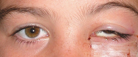
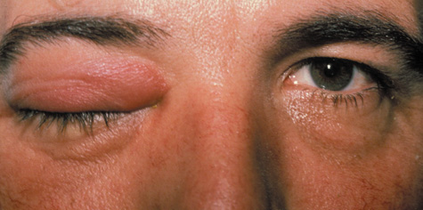
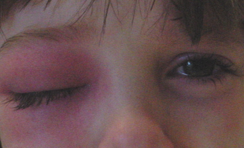
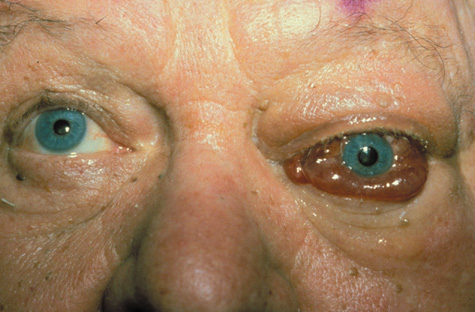
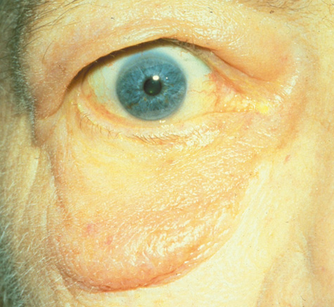
clinical picture. Preseptal cellulitis usually occurs in either of four clinical settings. These settings include: (a) sinusitis, (b) bacteremia, (c) upper respiratory infection, or (d) local cutaneous infection.14–16 In the latter setting, preseptal cellulitis can arise from spread of a contiguous anterior eyelid infection such as a chalazion, local trauma resulting in infection such as an insect bite, or a foreign body.(Fig. 1)
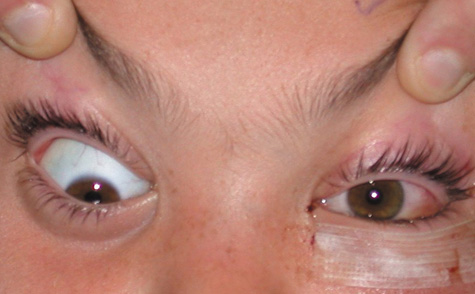
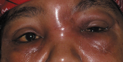
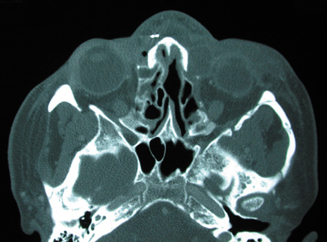
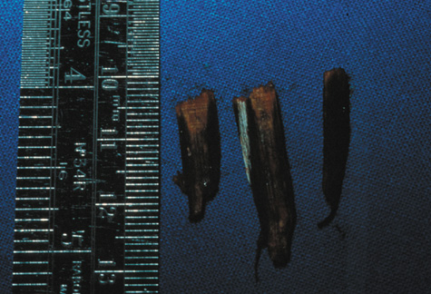
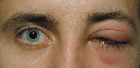
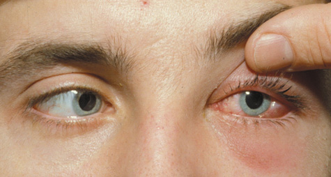
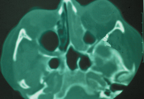
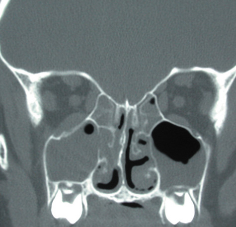
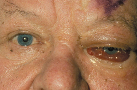
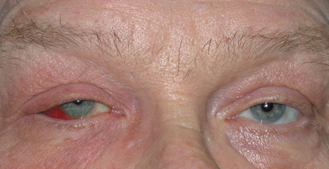
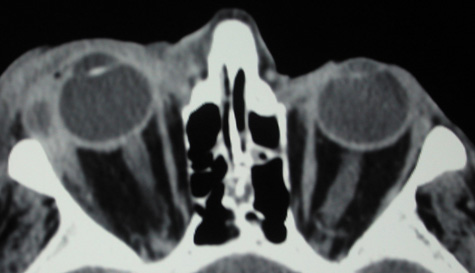
Prior to the Haemophilus influenzae type b (Hib) vaccine, the most common clinical presentation was of a child younger than the age of 36 months with a history of antecedent upper respiratory tract infection, otitis media, and bacteremia and usually resulted from infection with Hib.4 However, since the introduction of the Hib vaccine, the incidence of Hib induced preseptal cellulitis has declined dramatically with some studies reporting up to a 90% reduction.15 Orbital cellulitis, in contrast, usually arises from spread of infection from the paranasal sinuses. Orbital involvement occurs in 0.5% to 3% of patients with acute sinusitis.14 In a large series of patients with orbital cellulitis, sinusitis was responsible for orbital infection in 75% to 90% of cases.8,18,19 The ethmoid sinus is the most commonly implicated sinus in infections that spread to the orbit in children; the frontal and sphenoidal sinuses do not develop until age 7.20 In adults, pansinusitis often is associated with orbital cellulitis and spread is believed to occur through the ethmoid or frontal sinuses. In addition to anatomic proximity, several other factors predispose the orbit to the spread of contiguous sinus infection. Dehiscences often are present in the orbital walls, particularly in the thin-walled lamina papyracea.21–23 As a consequence, pus or transudate may rupture through the thin-walled lamina into the orbit and result in subperiosteal abscess formation. This theoretical mechanism is consistent with the observation that most abscesses occur in the medial orbit, adjacent to the ethmoidal sinuses.24–26 Posteriorly, the optic nerve within the optic canal is adjacent to the lateral wall of the sphenoidal sinus. Dehiscences can also be present in the lateral wall of the sphenoidal sinus along the optic canal, in approximately 6% of cases.27 Another factor predisposing the orbit to the spread of adjacent sinus infection is the free vascular communication between the orbit and the sinuses. The orbital veins are valveless, and flow occurs in either direction28 through the anterior and posterior ethmoidal foramina. Increased pressure within the sinuses caused by obstruction to mucus outflow in sinusitis may hamper venous drainage and manifest as the eyelid edema that accompanies acute sinus inflammation. Septic thrombophlebitis can also occur within these vessels and allow bacterial spread into the orbit.29–33 However, all orbital cellulitis resulting from sinus disease is not secondary to acute sinusitis. Orbital fracture can spread existing chronic sinus infection into the orbit (Fig. 2).25,31–33 Mucoceles, slowly enlarging mucus-filled cysts within the sinuses that enlarge by pressure erosion of adjacent orbital and intracranial structures, can become infected and result in mucopyoceles that cause both cellulitis and abscess formation.34–37 Approximately 15% of orbital cellulitis occurs from other sources.8 In particular, dacryocystitis is sometimes associated with a cellulitis that may spread posteriorly into the orbit (Figs. 3 and 4). 8 Infection can spread to the orbit from the eye, the teeth, the middle ear, or the face. Foreign bodies, such as wood, glass, or orbital floor implants, can cause orbital cellulitis refractory to treatment with antibiotics that requires removal of the nidus of infection (Fig. 5). Alternatively, a granulomatous reaction may develop around the foreign body or a cutaneous fistula may occur.25 Orbital infection has been described after uncomplicated eyelid, strabismus, and retinal surgery.38–40 Rarely, dental work or an infected dental cyst may cause orbital cellulitis through spread of infectious phlebitis across the pterygopalatine venous plexus.41,42 Endophthalmitis from a variety of endogenous or exogenous causes may extend extraocularly to involve the orbit. Finally, orbital cellulitis can occur secondarily from embolic spread in subacute bacterial endocarditis.2,3,12
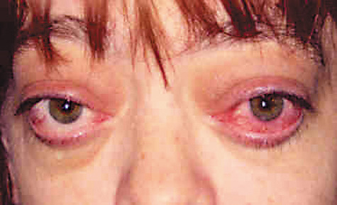
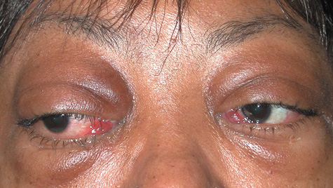
Regardless of its anatomic site of origin, edema develops as the orbital infection spreads within the connective tissues. Septic thrombophlebitis may ensue and increase both edema and orbital pressure as blood flow is impeded. Congestive signs such as chemosis, intraocular pressure, and proptosis increase. The infection may consolidate to form an abscess. The infection, in turn, may spread through the vascular emissaries into the cavernous sinus or intracranially, causing cavernous sinus thrombosis, meningitis, carotid occlusion, and subdural, epidural, or brain abscess. A spectrum of progressive infectious changes in orbital cellulitis initially was described by Hubert30 and modified by Smith and Spencer43 and later by Chandler,29 and is useful to the understanding of the nature of the evolution of orbital cellulitis, in classifying the severity of infection and defining progression and management. Stage 1 inflammation represents preseptal cellulitis. All changes that are seen on examination are confined to the anterior eyelid tissues. Occasionally, edema may spread secondarily posterior to the septum but does not represent the spread of cellulitis posteriorly. Chemosis may be present, and it may even cause minimal limitation of ocular movement. Stage 2 inflammation represents orbital cellulitis, with leukocytosis, and often fever, proptosis, extraocular movement limitation, and the congestive signs discussed earlier (Fig. 6).
With stage 3 inflammation, subperiosteal orbital abscess formation occurs. The globe is often displaced and limited in the field of gaze of the abscess, which occurs concomitant with the signs of orbital cellulitis seen in stage 2. In stage 4 inflammation, a true orbital abscess develops. These patients have severe proptosis, chemosis, ophthalmoplegia, and, often, visual loss. Stage 5 inflammation represents retro-orbital spread of infection into the cavernous sinus. In these instances, orbital signs may occur in the fellow eye and other central nervous system signs supervene.29 |
SYMPTOMS AND SIGNS | ||||||
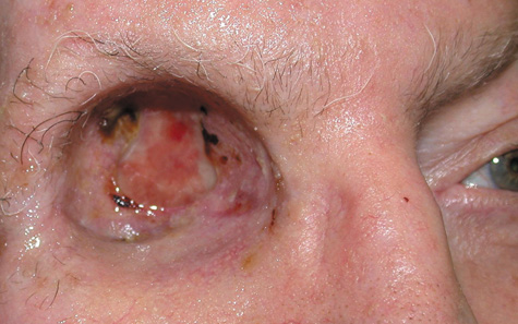
PRESEPTAL CELLULITIS Patients with preseptal cellulitis often present with a short history of painless swelling of the eyelids. A history of previous upper respiratory tract infection, trauma, insect or animal bite, conjunctivitis, or chalazion may be elicited. Fever is an inconstant feature but may be present in approximately 62% of cases.12 The eyelid characteristically is erythematous and edematous and may show signs of the trauma, bite, or chalazion that initiated the cellulitis (see Fig. 1). The eyelid may be painful to palpation. The superficial spreading cellulitis is confined to the eyelid and delimited by the attachments of the orbital septum to the arcus marginalis of the orbital rim so that infection will extend to this area and not beyond. Rarely, the etiology of the eyelid infection may be diagnosed clinically. Necrotizing eyelid cellulitis may be caused by erysipelas, an infection with a group A β-hemolytic Streptococcus, and can give rise to lid necrosis (Fig. 7).44,45 H. influenzae produces a characteristic nonsuppurative preseptal cellulitis with a purplish discoloration of the eyelids.46 Staphylococcal species may cause a suppurative cellulitis with abscess formation.
Chemosis and extraocular motility deficits are seen only occasionally and are presumed to result from anterior edema rather than spread of infection. Vision and pupillary examination are always unaffected by preseptal cellulitis, and proptosis and globe displacement never are present. Therefore, in the examination of patients with presumed preseptal cellulitis, vision, proptosis, and extraocular movements are key distinguishing points. ORBITAL CELLULITIS Orbital cellulitis presents as pain, proptosis, globe displacement, double vision, and/or vision loss (Figs. 6 and 8). Patients often have accompanying headache and malaise. In children fever occurs with equal incidence as in preseptal cellulitis (62%),12 whereas in adults it may be absent 66% of the time.47
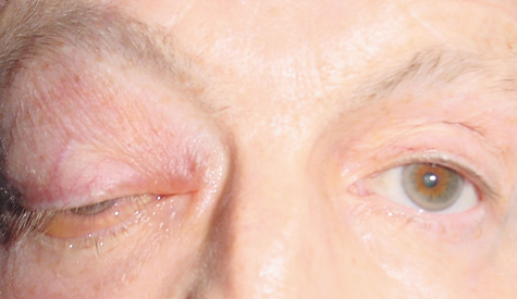
Antecedent and significant past medical history may include a history of headache, rhinitis, sinusitis, polyposis, nasal discharge, anosmia, or recent upper respiratory tract infection. The patient may give a history of prior orbital fracture or orbital fracture surgery with implantation of alloplastic materials. Scleral buckling procedures and strabismus surgery have been noted to cause orbital cellulitis; thus, the patient should be queried regarding prior ophthalmic surgery.38–40 The patient may also give a history of recent dental work, specifically dental extractions.42 The patient may have had prior surgery of the eyelids or of the facial skeleton with insertion of wires, rigid internal fixation (miniplates), alloplastic plates, or nondissolving suture material.34 Pain is not invariably present. It is notably absent in approximately 40% of cases, even those presenting with abscess.25 Fever is a nonspecific sign. In the presence of purulent sinusitis, pain may be present when the affected sinus is palpated; and with ethmoid sinusitis, pain may be elicited with pressure on the inner canthus. The sinus may not transilluminate. Mucopurulent rhinorrhea may be present, and the ophthalmologist when possible should look in the nose or seek the opinion of an otorhinolaryngologist. Proptosis, chemosis, and extraocular motility deficit are the clinical hallmarks of orbital cellulitis (see Fig. 6). Visual acuity decrease, pupillary signs, and vision loss may occur rapidly. Rarely, if sphenoid sinusitis occurs in isolation, the patient may present with atypical optic neuritis without proptosis or with little in the way of superficial cellulitis.48 Optic neuropathy, with the associated pupillary finding of a relative afferent pupillary defect and optic nerve head findings such as optic disc edema, may be observed. As orbital pressure increases, patients can show signs of retinal and choroidal arterial and venous stasis with congestion and a picture resembling chronic vein occlusion or central retinal artery occlusion. Hypesthesia of the nasociliary or frontal nerves is rarely seen. Hypotony, choroidal folds, and anterior segment inflammation with hypopyon can occur and imply intraocular spread. A dilated nonreactive pupil implies third nerve involvement. Patients may show signs of third, fourth, or sixth nerve involvement when the infection extends into the orbital apex. Subperiosteal abscess formation most often is diagnosed on neuroimaging and is indistinguishable clinically from orbital cellulitis. Signs of an abscess include displacement of the globe away from the affected sinus and limitation of abduction and adduction,24 but these signs can occur with orbital cellulitis as well. Intraorbital abscess formation can present in the patient who has been partially treated for an orbital infection and may present as proptosis or globe displacement without any infectious signs or as orbital cellulitis.25 If infection spreads posteriorly through the superior orbital fissure to involve the cavernous sinus, additional signs may supervene. Signs of cavernous sinus thrombosis include headache, ipsilateral hypesthesia from involvement of the ophthalmic and maxillary division of the trigeminal nerve; third, fourth, and sixth cranial nerve palsies; and mental status changes from confusion to obtundation. The contralateral side may develop cranial nerve signs, periorbital edema, or cellulitis (Fig. 9). DIFFERENTIAL DIAGNOSIS Several conditions can mimic orbital and periorbital cellulitis. Most of these present as the same constellation of symptoms and signs and often can have a similar appearance on neuroimaging. PRESEPTAL CELLULITIS Conditions that may masquerade as preseptal cellulitis include allergic edema of the eyelids, severe blepharoconjunctivitis, dacryoadenitis (Fig. 10), trauma, thyroid eye disease (Fig. 11), leukemic infiltrates,49 blepharochalasis syndrome, and autoimmune inflammatory disorders such as lupus.
ORBITAL CELLULITIS Conditions that may mimic orbital cellulitis include thyroid eye disease, idiopathic inflammatory orbital pseudotumor (Fig. 12), orbital myositis,50 orbital abscess,51 ruptured dermoid cyst, necrotic intraocular melanoma or retinoblastoma,52,53 orbital trauma, orbital foreign body, orbital vasculitis, Wegener's granulomatosis, herpes simplex or zoster (Fig. 13), and carotid cavernous fistula. Acute-onset proptosis with inflammatory signs in a child always should alert the clinician to the possibility of rhabdomyosarcoma (Fig. 14).
|
LABORATORY AND IMAGING STUDIES | |||||
As a routine, white blood cell counts and at least two sets of blood cultures
are obtained in all patients who present with orbital infections. However, since
the decline in Hib as an etiologic agent, blood cultures
in patients with preseptal cellulitis are less likely to yield positive
results.14,15 Cultures of the nasopharynx and conjunctiva may be obtained, although
this may not yield significant information unless a predominant organism
is present. Cultures of the leading edge of the cellulitis are performed
if there is an obvious sign of an entry wound responsible for the
cellulitis. Cultures of purulent discharge may also be helpful. Cerebrospinal
fluid is obtained for culture if the patient shows central nervous
system signs or bilateral disease. Neuroimaging via computed tomography (CT) usually is obtained in patients with orbital cellulitis
in Chandler stages 3 through 5.16,54,55 However, CT may be obtained in Chandler stages 1 and 2 based on the clinical
situation, and index of suspicion of posterior infection, sinus
disease, or abscess formation. Repeat CT is also indicated if there is
no improvement after 48 hours of appropriate antibiotic treatment or
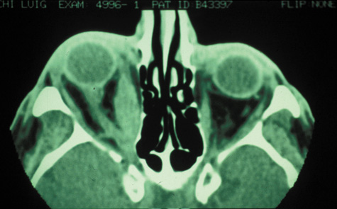
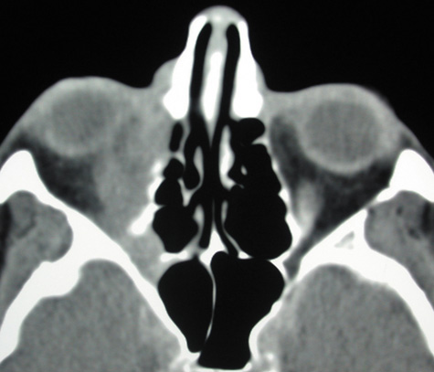
if there is worsening of the clinical signs and symptoms.16,54,55 The white blood cell count may be elevated with leukocytosis in both orbital and preseptal cellulitis. In one study, elevated absolute white blood cell counts were seen in children with periorbital cellulitis and bacteremia but not as frequently as in orbital cellulitis.12 The erythrocyte sedimentation rate may be elevated. If the infection is from α-hemolytic Streptococcus, the antistreptolysin O titer may be elevated. If meningitis is present, the cerebrospinal fluid may demonstrate pleocytosis, increased protein, and decreased sugar. ULTRASOUND Ultrasound has higher resolution (0.1 mm) when compared with CT (0.8 mm) but adds little clinically significant information because ultrasound misses the posterior third of the orbit and does not image the bone and sinuses.56 Ultrasound in orbital cellulitis, however, may be useful in ruling out orbital myositis, determining the location of orbital foreign bodies or abscesses, and following patients with drained orbital abscesses to rule out reaccumulation. Ultrasound findings in subperiosteal abscess can include a signal of low or medium reflectivity adjacent to the involved orbital wall. If the abscess is intraconal, a low reflective signal is encountered within the cone, the muscles may be thinned as they are placed on stretch, the sclera may be thickened, or a T sign (usually associated with orbital pseudotumor) may be seen. Ultrasound is not helpful in distinguishing inflammatory transudate from infectious exudate or hemorrhage. NEURORADIOGRAPHIC STUDIES Routine skull films and polytomography have been supplanted by CT in the evaluation of patients with orbital cellulitis.57 CT allows the clinician to differentiate a preseptal cellulitis from an orbital cellulitis.58 If orbital cellulitis has resulted from adjacent intercurrent sinus infection, the diagnosis can be made and the extent of the sinus disease estimated. Sinuses may show changes of osteomyelitis with blurring of the osseous margins of the sinuses, air–fluid levels, or inflammatory tissue within the normally aerated sinus.59 Central nervous system complications can be assessed by neuroimaging, and progression of disease can also be monitored.58 CT should be performed using thin-section (2–4 mm) high-resolution scanning with multiple views of both bone and soft tissue detail.53 Axial and coronal views should be obtained; in one-third of patients with subperiosteal abscesses, the abscess was seen in the coronal sections only.18 Helical CT is a fairly new technology that allows increased resolution with decreased imaging time.60 This type of scan may be especially beneficial in children because of the ability to obtain good imaging with a shorter imaging time.60 elica He HhIntravenous contrast material is not advocated at all centers because there is intrinsically high contrast between infectious changes and orbital fat. However, some authors believe that it is essential to the diagnosis, and it thus remains the preference of the individual clinician, as well as the neuroradiologist.22,59,62 With preseptal inflammation, CT demonstrates soft tissue swelling of the eyelids and tissue adjacent to the orbital septum (Fig. 15). The orbit is not involved, and usually the sinuses do not show evidence of inflammation. The distinction between inflammatory preseptal cellulitis and edema cannot be made.63
An extraconal or intraconal mass may be present in orbital cellulitis. Proptosis also may be visible. In particular, with intraconal involvement, proptosis is seen with obliteration of the normal soft tissue shadows. “Patchy enhancement” of the intraconal fat in orbital cellulitis has been described.59 The rectus muscles, particularly the medial rectus, and the optic nerve may be thickened.58 CT is particularly useful for imaging orbital and subperiosteal abscesses. Because the periorbit is not adherent to the orbital walls except at the suture lines, an abscess lifts the periorbit, creating a convexity in the orbital periosteum (Fig. 16). Usually subperiosteal abscess formation occurs adjacent to the involved sinus,25,64 but occasionally it occurs at a remote location such as the superolateral orbit.65 Gas may be found within a subperiosteal abscess or within the orbit, arising either from gas-forming bacilli or free communication with sinus air or from prior trauma (Fig. 17). 57,66 CT cannot accurately predict whether a subperiosteal mass represents exudate, inflammatory transudate, or hematoma.67,68
A subperiosteal abscess may rupture or invade the periorbit, resulting in an orbital abscess. This may or may not be contiguous with the subperiosteal collection on CT. There may be gas or air–fluid levels within the mass.51,56,58,59 An orbital abscess may present as an enhancing ringlike peripheral mass that can be either heterogeneous or homogeneous (Fig. 18). Finally, CT can demonstrate intracranial involvement such as epidural or cerebral abscess, which is better appreciated on coronal imaging.62,63 The importance of coronal sections on CT of abscesses has been emphasized; in one series, one-third of abscesses were seen only on coronal sections.62 However, CT also has limitations. CT of the orbits may not demonstrate the telltale signs of orbital involvement, because the increased water content of the orbital fat may cause only a small rise in the attenuation coefficient of the orbital fat by 5 to 10 Hounsfield units66 at the routine window settings. The normal fat shadows within the cone may be obliterated.67 In other series, abscesses drained at surgery were not demonstrable on high-resolution CT.68 CT may also miss wooden orbital foreign bodies.69,70 Recent publications have stated the relative risk of radiation-induced fatal cancer in pediatric CT imaging.60 Although the risk was small, it was not negligible. CT should be used judiciously in the management of pediatric orbital cellulitis, and every attempt to minimize radiation dose should be attempted.60 Magnetic resonance imaging (MRI) is purported to be more useful than CT in the diagnosis of preseptal cellulitis. It is less reliable at diagnosing the subtle signs of muscle enlargement and periscleritis and thus is not as useful in differentiating orbital cellulitis from other inflammatory orbital diseases.71 On MRI with gadolinium contrast, orbital cellulitis may show a smearing or linear streaking of the normal fat shadows on T2-weighted images. MRI is excellent for demonstrating localized fluid collections such as abscesses. It is not helpful in distinguishing a transudate from an exudate, because both appear liquid and are of low intensity on T1-weighted images and bright on T2-weighted images (Fig. 19).
MRI is superior to CT in the diagnosis of cavernous sinus thrombosis. T2- and proton-weighted images show high signal luminal narrowing as well as absent flow or localized parenchymal infarcts (Fig. 20).72 Absent flow can be demonstrated as well in the superior ophthalmic vein in cases of carotid or cavernous sinus thrombosis.72 MRI with gadolinium can help define these abnormalities and can detect dural invasion.
MRI may be helpful in distinguishing orbital inflammatory diseases such as orbital pseudotumor from orbital cellulitis.73 MRI is most helpful in distinguishing the diffuse orbital form of orbital pseudotumor as well as the localized inflammatory pseudotumors such as the myositis, periscleritis, and scleritis forms. The diffuse orbital form is most commonly seen on MRI as a reticular pattern with isointense signals in the orbital fat in both T1- and T2-weighted studies.73 Orbital cellulitis is best characterized by a diffuse pattern that is isointense in T1-weighted images and hyperintense on T2-weighted images.73 Both CT and MRI can also help in the planning of surgery and the evaluation of treatment efficacy. |
MICROBIOLOGY OF ORBITAL INFECTIONS |
The organisms responsible for orbital infections often are difficult to
determine, and all cultures must be interpreted cautiously. Information
can be gained from cultures of blood, conjunctivus, nasopharyngeal
tissues, an aspirate of the leading edge of the spreading cellulitis, sinus
aspirates, purulent discharge, abscess, and cerebrospinal fluid; however, mixed
infections often are seen, and normal conjunctival or
nasal contaminates can confound or obscure the diagnosis. Furthermore, patients
who were previously treated with antibiotics may have negative
cultures, despite demonstrably purulent sinus infection or abscess
formation. Aerobic and anaerobic cultures should be performed, because
unsuspected anaerobes are present in approximately 50% of cases
of presumed aerobic sinusitis and are the sole organisms in 30% of
cases.74 The organisms presumed responsible for orbital infections vary by patient age and whether the infection is preseptal or orbital. Prior to vaccination for Hib, the most common organism responsible for preseptal cellulitis in children younger than 4 years of age is H. influenzae. In children older than 4 years of age with preseptal infections, Streptococcus pneumoniae, Staphylococcus aureus, S. epidermidis, and mixed anaerobic and aerobic flora predominate.2,4–8,12,66,75 Anaerobic organisms include Peptostreptococcus, Fusobacterium nucleatum, and Bacteroides spp.12 In children with orbital cellulitis, the preponderant organisms isolated by sinus aspiration are S. aureus, Streptococcus spp., and anaerobic species.5,6,12,66 In adults, S. aureus, Escherichia coli, S. pneumoniae, and mixed flora, including anaerobes, are the most common organisms responsible for orbital cellulitis.3,45,76–79 However, it must be noted that sino-orbital infections do not always respect these guidelines and orbital cellulitis with a potpourri of organisms has been described in case reports and series, including Enterococcus, Echinococcus granulosus, Pseudomonas aeruginosa, Klebsiella spp., E. coli, Treponema pallidum,47 Eikenella corrodens,80 Actinomyces, Mycobacterium tuberculosis,47 and M. avium. Harris et al found age to be a factor in the microbiological etiologies of subperiosteal abscesses.81 Children less than 9 years of age are most likely to have one aerobic organism as the source of subperiosteal abscess. Patients older than 15 are most likely to have polymicrobial infections, including anaerobes. Patients between the ages of 9 to 14 had a tendency to have more complicated infections.81 Bacteremia tends to occur in a younger age group and in patients who have not yet had antibiotic treatment at the time of blood culture.2 Bacteremia is less common in children who have been vaccinated against Hib. However, in children who have been vaccinated against Hib, the most common microbe associated with bacteremia is S. pneumoniae.15 In one series, bacteremia was present in 5% of adult patients with orbital cellulitis.8 Children younger than age 4 have impaired humoral immunity to bacteria with polysaccharide capsules such as H. influenzae and Streptococcus spp., and thus disseminated infections with these organisms may be seen more commonly in this age group.82–85 In the first year of life, cellulitis from lacrimal sac infections with Staphylococcus or Streptococcus are more common.12,20 S. aureus, and α- and β-hemolytic Streptococcus spp. are the most common pathogens grown from blood cultures in older children with preseptal cellulitis.2,6,8,12,76 Conjunctival and nasopharyngeal cultures contain mixed flora, do not correlate accurately with blood culture results, and are believed to be misleading in orbital cellulitis.2,4,8,9 These cultures are obtained despite this, because they may indicate a predominant organism or a refractory organism that may explain persistent or deteriorating clinical signs. Cerebrospinal fluid cultures usually are positive when meningitis or other neurologic signs are clinically apparent.8,87 In meningitis, the most frequently implicated organism is H. influenzae.6 In the presence of preseptal cellulitis secondary to facial trauma, the yield on aspiration of a percutaneous aspirate of the leading edge of spreading cellulitis has been described to be as high as 51%,12 but it is very low if no site of trauma is present.8 Approximately 0.1 mL of sterile saline without preservative is injected into the outer edge of the cellulitis and reaspirated. S. aureus and Streptococcus spp. are the most common organisms isolated, as expected, because these organisms are among the common causes of cutaneous cellulitis.6 Anaerobic organisms may be cultured in patients with insect, animal, or human bites.12 Sinus aspiration through direct cannulation is perhaps the most reliable means of obtaining cultures that are of significance in orbital cellulitis because it avoids contamination with resident nasal flora that would render interpretation of nasal cultures difficult. This is an otolaryngologic procedure that can be accomplished by trephination through the antrum in the vicinity of the canine fossa.88 Studies of sinusitis by trephination suggest that coagulase-positive Staphylococcus and α-hemolytic Streptococcus are the predominant aerobic organisms, but multiple bacteria, from H. influenzae, S. epidermidis, Neisseria spp., E. coli, H. haemolyticus, pneumococcus, and Pseudomonas, may be responsible for sinus infection.74 Anaerobic Streptococcus, Corynebacterium, Bacteroides spp., and Veillonella are isolated in 50% of chronic sinusitis cases, suggesting that they may play a role in sinus infection, and consequently in orbital infections.89 It is common for multiple anaerobic organisms to be present and coexist synergistically in orbital infections. For example, Bacteroides melaninogenicus has an obligatory requirement of vitamin K that can be met by the metabolic activity of other Bacteroides strains.89 The yield on culture of an abscess depends largely on whether the patient has been pretreated. In Schramm's series, 18 of 46 abscess cultures produced no growth.8 |
MANAGEMENT |
After appropriate workup, all periorbital and orbital infections should
be treated with broad-spectrum antimicrobial agents.90–92 Antibiotics may be altered after culture results. Children with preseptal
cellulitis, no orbital involvement, and who do not appear toxic should
have blood cultures drawn and can be treated with intramuscular or
oral antibiotics on a daily basis as an outpatient. If the cultures
are reported negative, an oral antibiotic should be continued to complete
a 10-day course.15,16,54 Selected cases of adult preseptal cellulitis can be managed on an outpatient
basis with oral antibiotics with frequent monitoring for progression. The
duration of treatment depends on response: patients should
be treated with parenteral antibiotics until they show clear evidence
of clinical improvement as manifested by a decrease in orbital congestive
signs such as proptosis, gaze limitation, cellulitis, and edema. Intravenous
therapy should continue for a minimum of 3 days. Then oral
antibiotic therapy may be instituted for a total course of 10 days to 3 weeks, depending
on the severity of infection. Associated bacteremia, however, should
be treated with 7 to 10 days of intravenous therapy. In the newborn period, staphylococcal species are the most common organisms observed in orbital cellulitis. In children younger than 4 years of age, the most common organisms involved in orbital and periorbital infections are S. aureus, and Streptococcus spp. Thus, treatment in the child younger than age 4 should include coverage for gram-positive cocci. Antibiotic selection should be determined in conjunction with the internist or pediatrician caring for the patient. The opinion of a subspecialist in infectious disease can be invaluable, particularly when treating the frequently hospitalized or immunocompromised patient, because these specialists can help determine the presence or absence of other systemic findings, guide fluid and electrolyte therapy, and manage any other concomitant medical problems. Treatment regimens vary depending on whether the infection is from a cutaneous source, the age of the patient, and the patient's culture results and immune status. For cutaneous infections causing preseptal cellulitis, nafcillin 150 mg/kg per day every 6 hours covers most of the gram-positive organisms responsible for cutaneous infection. In penicillin-allergic patients or those who prove to have methicillin-resistant S. aureus, vancomycin 40 mg/kg per day in divided doses every 6 hours can be administered. Some of the preferred antibiotics for children with orbital or periorbital cellulitis not obviously related to a skin infection include the following. Ticarcillin-clavulanate, which covers most gram-negative as well as -positive organisms, including nontypeable H. influenzae, also provides excellent anaerobic coverage. The clavulanate used with ticarcillin inactivates β-lactamases by inactivating the active sites of these enzymes, thus increasing the drug's microbial spectrum. Ticarcillin clavulanate is administered in doses of 200 to 300 mg/kg per day in four divided doses. Cefotaxime, a third-generation cephalosporin that covers all the most common sinus pathogens with the exception of Clostridium difficile, can be given in a dosage of 80 to 120 mg/kg per day in four divided doses. Cefuroxime, a second-generation cephalosporin, covers most staphylococci and H. influenzae. Certain strains of Enterococcus faecalis, Serratia, Proteus vulgaris, C. difficile, and Bacteroides fragilis are not susceptible to the drug. The dosage in children is 75 to 150 mg/kg per day in three divided doses. In patients who are allergic to penicillin, the preferred treatment is clindamycin, 25 to 40 mg/kg every 6 hours. Clindamycin may cause a pseudomembranous colitis from proliferation of C. difficile, as can many other antibiotics. Alternatively, chloramphenicol 50 to 100 mg/kg every 6 hours can be used. Chloramphenicol may cause both a reversible and an irreversible bone marrow depression. In adults who present with orbital cellulitis, the authors prefer to use cefuroxime 750 mg to 1.5 g every 8 hours. It is effective against most gram-positive and -negative organisms except Pseudomonas. Alternatively, ceftriaxone 1 to 2 g/day can be given and is effective against penicillinase-producing S. aureus, most gram-positive organisms, and most gram-negative organisms except for Pseudomonas. Ceftriaxone also crosses the blood–brain barrier; therefore, it is an excellent choice if there is suspicion of intracurrent intracranial infection. Methicillin-resistant Staphylococcus and many strains of group D streptococci and enterococci, however, are resistant. If anaerobic bacteria are suspected, metronidazole can be added in doses of 15 mg/kg infused over 1 hour, followed by 7.5 mg/kg over 1 hour every 6 hours after the loading dose. Clindamycin can also be substituted, but the frequency of antibiotic-induced diarrhea makes metronidazole a better choice. Metronidazole has not been approved for use in children. In patients who are frequently hospitalized, in whom an infection with methicillin-resistant staphylococci is a possibility, vancomycin, in a dose of 500 mg given intravenously every 6 hours, and cefuroxime can be employed. This combination of antibiotics effectively covers most organisms, including methicillin-resistant Staphylococcus as well as H. influenzae. S. pneumoniae resistant to penicillin and cephalosporins is increasing; therefore, in cases where meningitis is suspected, vancomycin should be administered.15 Fluoroquinolones often are used for the treatment of acute sinusitis.93 These antibiotics have an expanded antibacterial spectrum that includes gram-positive and -negative organisms as well as atypical respiratory bacteria such as Mycoplasma.94 The newer fluoroquinolones such as moxifloxacillin (400 mg orally daily) are very active against the respiratory tract pathogens including penicillin and macrolide-resistant strains of Streptococcus pneumoniae.95 Fluoroquinolones are not recommended for use in pediatric patients as first-line therapy for any infection because of the musculoskeletal side effects.96 Fluoroquinolones have not been studied as treatment for preseptal or orbital cellulitis. In patients older than 9 years of age, with subperiosteal abscesses, polymicrobial infections are the rule. Therefore, a combination of antibiotics to cover gram-positive, gram-negative, and anaerobes is necessary. An example includes ceftriaxone with clindamycin or metronidazole in the previously mentioned doses.14,18 Once congestive and orbital inflammatory signs diminish, oral antibiotics can be substituted. In patients with cutaneous cellulitis, dicloxacillin 25 to 40 mg/kg per day in four divided doses can be substituted for nafcillin. Cefaclor 40 mg/kg per day in divided doses every 8 hours can be given, or Augmentin (amoxicillin and clavulanate potassium) 40 mg/kg per day in divided doses every 8 hours can be substituted. Oral erythromycin 40 mg/kg per day in four divided doses can be substituted if the patient is allergic to penicillin. In anaerobic infections, metronidazole 500 mg every 6 hours can be administered. When switching from the intravenous to the oral route, particularly when changing antibiotics, patients must be followed closely for recrudescence. Careful follow-up is indicated in all patients who present with orbital cellulitis. This should include twice-daily examinations with attention to visual acuity, confrontation visual fields, exophthalmometry, motility, and pupillary examinations. Patients may not respond to the antibiotic regimen chosen for them because they have an abscess or a foreign body, because they are infected with a resistant organism, because they have an infection with an atypical organism such as Mycobacterium tuberculosis or fungus, or if they have an inflammatory tumor rather than an infection. The authors do not hesitate to order another CT scan if there is little improvement with treatment after 24 or 48 hours or if the patient's clinical status deteriorates. Another measure considered useful in the medical management of orbital and preseptal cellulitis is the use of cool compresses to reduce swelling.29 Some authors suggest the use of warm compresses,2,76,97,98 to facilitate vascular dilation and hence delivery of antibiotic. Nasal vasoconstrictors3,29,98 and topical antibiotics are recommended by some,2,8,77 but others are skeptical of their efficacy.99 Head elevation may hasten the resolution of periorbital edema.97 Patients with severe proptosis and exposure may require lubrication of the ocular surface as well as the use of plastic wrap or exposure bubbles to manage nocturnal exposure resulting from lagophthalmos. The indications for surgery in orbital cellulitis include suspicion of orbital abscess or foreign body, progression of visual loss, and extraocular motility deficit, or worsening proptosis despite appropriate medical therapy after a 24- to 48-hour period. The orbit should be explored to obtain gram stains and acid-fast stains and anaerobic, aerobic, and fungal cultures. Both fresh and formalinized tissue should also be obtained if an inflammatory orbital tumor is part of the differential diagnosis or if fungal invasion must be ruled out. Timing for surgical intervention is critical. In cases of orbital cellulitis without abscess formation, in which visual acuity is 20/60 (Snellen notation), 6/15 (metric equivalent) or less, or declines with appropriate medical management, orbital exploration should be emergent. In cases in which the acuity is better than 20/60, the patient should be followed serially, expectantly, and frequently while more conservative management is initiated. An abscess may be diagnosed at presentation or may develop with treatment. If there is radiographic evidence of subperiosteal abscess, the need for emergent therapeutic intervention is controversial. Some authors recommend conservative treatment,57,64,69 whereas other groups favor immediate surgical drainage, emphasizing that delayed treatment has a higher rate of complications.8,24,25,29,51,100 In patients older than 14 years of age, the authors favor the latter approach because the risks of surgery are negligible compared with the visual and life-threatening risks of nonintervention. In patients 9 years of age or younger 25% of subperiosteal abscesses are likely to resolve with antibiotic therapy alone.81 Moreover, neuroimaging may not always detect abscess formation,68 so the authors are reluctant to be guided solely by neuroradiographic findings. The surgical management involves the drainage of the abscess and the involved sinus with the restoration of normal drainage of the sinuses into the nose, usually in conjunction with an otorhinolaryngologist. In the past, and possibly currently in certain institutions, a subperiosteal abscess was approached and drained extraperiosteally without entering the orbit directly, usually in conjunction with an external procedure on the involved sinus, such as an external ethmoidectomy, antral lavage, or frontal sinusotomy, depending on the sinus thought to be responsible for the orbital infection.8 A nasal sinusotomy was performed to facilitate drainage of the sinus. A drain was placed to drain the involved sinus and/or the orbit and was brought out either through the surgical wound or the nose, left in place and advanced on a daily basis. However, more recently, because of the increasing popularity and facility of rhinologists with the endoscopic approach, many subperiosteal abscesses are approached via an endoscopic transnasal ethmoidectomy and are drained successfully. This procedure results in less scarring, less inflammation, more physiologic restoration of sinus drainage than older sinus procedures, and faster recovery.55,88,101 If an abscess has extended into the intraconal space, however, the orbit must be entered. This can be accomplished at the time of ethmoidectomy by making incisions into the medial periorbit, allowing the orbital fat to prolapse beyond the periosteum. However, this approach is not advocated: the intraconal space is much more clearly visualized using a medial or lateral orbital approach. In patients who fail to improve after surgical drainage of abscess, a reaccumulation of abscess, an orbital foreign body, an osteomyelitis, or an inappropriate choice of an antibiotic may be the reason.24 These patients should be recultured, reimaged, and if necessary, re-explored. |
COMPLICATIONS |
Multiple complications of orbital cellulitis can occur despite a high incidence
of clinical suspicion, improved imaging techniques, and improved
intravenous antibiotics. Most commonly, inadequately treated orbital
infections can progress (e.g., orbital cellulitis can proceed to abscess formation). Loss of vision can occur and may result from optic neuritis;from thromboembolic lesions to the vascular supply of the optic nerve, retina, or choroid; from rapid and sustained intraocular pressure elevation; or, rarely, from massive proptosis, which may mechanically distort the optic nerve.24 If light perception is lost, blindness may be irreversible.25 However, vision has been restored in some cases with emergent surgical treatment; thus, these patients must be diagnosed and managed expediently.100 Permanent motility defects and scarring from corneal exposure owing to proptosis and exposure have been reported, as have perforation of the globe resulting from rise in orbital and intraocular pressure with scleral rupture.102,103 Cerebral complications including cavernous sinus thrombosis, meningitis, carotid occlusion, epidural abscess, subdural abscess, brain abscess, and death have been reported.2,8,17 The use of anicoagulants in the treatment of cavernous sinus thrombosis is still controversial because of the risk of cerebral hemorrhage.104 However, a review of the literature suggests that anticoagulation is beneficial and that the risk of intracranial hemorrhage from it is rare if hemorrhagic sequelae of the thrombosis is ruled out prior to treatment with neuroimaging. The goal of the anticoagulation is an APTT of 1.5 to 2.104 |
FUNGAL INFECTIONS OF THE ORBIT | ||
ASPERGILLOSIS Aspergillus is a fungus of the Ascomycetes class that is a ubiquitous organism that colonizes both the respiratory and gastrointestinal tracts. It rarely causes infection except in the immunocompromised host. Aspergillosis has four well-characterized presentations. The four presentations are allergic fungal sinusitis, sinus mycetoma or fungal ball, acute fulminant invasive fungal sinusitis, and chronic indolent invasive fungal sinusitis. Invasive sinusitis implies tissue invasion with destruction of adjacent structures. Acute fulminant invasive sinusitis presents in the immunocompromised patient as a fulminant sinusitis with rapid progression into the orbit and cranium that produces a necrotizing vasculitis with necrosis of sinus and nasal mucosa and bone (Fig. 21). Infection may disseminate and produce lung, liver, and splenic invasion with an overwhelmingly poor prognosis despite local debridement and intravenous amphotericin B.105–113 Chronic or indolent invasive fungal sinusitis usually presents in the patient with diabetes mellitus with chronic erosion through most anatomic barriers and invasion through the orbital bones and cranium, producing orbital signs, headache, and neurologic symptoms. The orbital signs usually result from an orbital apex syndrome.106–112 Alternatively, the disease can present in the healthy host in a noninvasive form as sinus mycetoma or allergic fungal sinusitis.106 A colony of organisms forms a mycetoma in a sinus and gives rise to a chronic sinusitis. This type of aspergillosis is more common in hot humid climates. These patients present with nasal congestion, postnasal drip, and chronic sinus pain, along with frequent bouts of acute sinusitis. A mass is seen that originates from an adjacent sinus that may have sclerotic margins and can produce an adjacent inflammatory reaction within the orbit. This form responds well to surgical debridement with restoration of normal sinus drainage and does not require intravenous anjpgungal therapy.114 Allergic fungal sinusitis is an allergic reaction to aerosolized environmental fungi and causes patients to suffer from headaches and nasal stuffiness. Patients may also present with proptosis and extraocular muscle entrapment. Treatment consists of surgical debridement and steroids without anjpgungals.106,109,112 Computed tomographic findings in patients with both forms of the disease demonstrate areas that are almost metallic in density. These foci of irregularly calcified bone on CT may be pathognomonic (Fig. 22). On MRI, these areas appeared bright on T1-weighted imaging and have decreased signal on T2-weighted images, which may be related to the presence of ferromagnetic materials such as iron and manganese within the fungal concretions.115
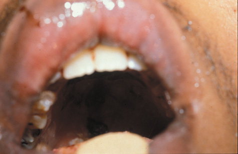
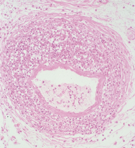
Biopsy of tissue within the orbit shows invasion of the mucosa, submucosa, and bone in cases of invasive fungal sinusitis or a granulomatous reaction without mucosal or bone invasion in the noninvasive form. The material within the sinuses may be yellowish, black, or friable. Hyphae are not seen within the mucosa or bone of patients with the noninvasive form but can be seen on routine stains of mucus. Multiple specimens should be examined116 in a search for septate fungal hyphae of relatively uniform width that are apparent on hematoxylin and eosin staining. The most common effects on the orbit are cellulitis, proptosis, ptosis, and diplopia. Invasion in both forms may produce pain. Optic nerve involvement is rare. The orbit may be affected by contiguous spread from the sinuses, usually the ethmoid. The treatment for aspergillosis in the immunosuppressed patient is both medical and surgical, with anjpgungal agents (usually amphotericin B) and radical debridement and sinus surgery.106,107,111 Flucytosine and rifampin with amphotericin B may be more effective than amphotericin alone.117–119 Conservative debridement, intravenous amphotericin, and local amphotericin irrigation have been described as an alternative to radical surgery.99 Five milliliters of amphotericin B is placed in sterile water, creating a concentration of 0.25 mg/mL. Irrigation is performed two times a day directly into the orbit through a site of entry into the orbit or sinus.116 MUCORMYCOSIS Mucormycosis is a fungal infection with the organisms from the genera Mucor, Absidia, and Rhizopus100–102 that normally are present in air, soil, vegetable matter, skin body orifices, manure, and bread mold.120 Most patients who develop mucormycosis have a predisposing systemic disease: Typically the infection occurs in the diabetic patient with ketoacidosis, but it can occur in patients with renal failure, gastroenteritis, or in those who are immunosuppressed.121 Rarely, it can occur in the normal host.124,125 An association between deferoxamine therapy for iron or aluminum excess and mucormycosis has also been described in hemodialysis patients with chronic renal failure, as well as in a variety of hematologic disorders.126 It is possible that the fungus uses the iron that is mobilized by deferoxamine to proliferate, thus facilitating invasive infection.126 Rhinocerebral mucormycosis originates as a rhinitis, parapharyngitis, or sinusitis, and spreads by invasion of blood vessel walls, causing a necrotizing vasculitis with thrombosis of the vascular lumina and resultant infarction. The patient typically presents with unilateral orbital apex syndrome, including severe pain, visual loss, total ophthalmoplegia, corneal anesthesia, and multiple cranial nerve palsies.127 Orbital cellulitis presenting with early visual loss is one of the hallmarks of mucormycosis.72 Gangrene may occur of external periorbital tissues as well as of the hard palate and nose, and eschar-like crusting may be observed within the nose or on the hard palate (Fig. 23). Obstruction of the central retinal artery, ciliary arteries, and choroidal circulation can also be seen.124,125 Brain damage may occur because of spread of infection or infarction or occlusion of affected intracranial vessels.72
CT shows sinusitis with or without bone destruction and is indistinguishable from other causes of orbital cellulitis.128 MRI may show carotid narrowing, occlusion, and absent flow in the superior ophthalmic vein (Fig. 24).72 Diagnosis is made by having a large index of suspicion and obtaining specimens of nasal turbinate, sinus, or infected orbital tissue. Large, branching nonseptate hyphae are readily apparent on hematoxylin and eosin staining or with methenamine silver staining (see Fig. 23). These hyphae can be grown on fungal culture. The treatment of mucormycosis involves the control of ketoacidosis or other systemic disease. Sinus drainage must be restored, and intravenous amphotericin B in doses of up to 1 mg/kg per day can be administered up to a total dose of 2 to 4 g. Radical and aggressive surgical debridement of infected tissue is advocated by most authors, including exeneration if necessary. Surgical debridement is important because local thrombosis caused by fungal invasion prevents amphotericin from reaching necrotic and infected tissue. In the patient with known infection and preserved vision, the need for exenteration and radical surgery is more controversial, particularly if orbital biopsy does not show orbital invasion with fungal elements. Successful outcomes have been achieved with debridement of infected tissue without exenteration.128 In one series intravenous amphotericin has been used successfully in conjunction with local irrigation of orbital and sinus tissues with amphotericin B.129 Hyperbaric oxygen has been used as an adjunctive therapy for rhinocerebral mucormycosis by treating patients with 100% oxygen at 2.5 atm for 90 minutes for approximately 6 weeks.130 In untreated patients, rhinocerebral mucormycosis is associated with a 14% survival rate. With amphotericin B and surgery, the survival rate improves to 57%.121 This implies nonetheless that a significant number of patients will die of this disease and emphasizes the need for early diagnosis and aggressive management. |