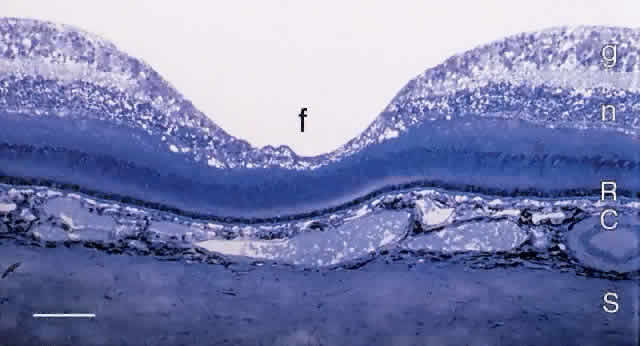
Fig. 21.
Light micrograph of human macula and fovea (f) stained with Richardson's methylene blue/azure II method. S, sclera; C, choroid; R, retinal pigment epithelium; n, inner nuclear layer; g, ganglion cell layer. Bar = 100 μm.