|
|
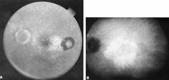
| Fig. 5 A. Ill-defined late leakage and mottled hyperfluorescence along with blockage of fluorescence from subretinal hemorrhage is evident temporal to the foveal center in this fluorescein angiogram of a patient with macular degeneration. B. The corresponding late-phase (22-minute) ICG angiogram shows a bright, well-delineated hyperfluorescent vascular-like structure in the central macula. (Courtesy of Dr. W. Benson) |