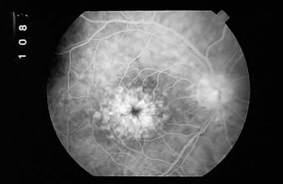
Fig. 10.
Fluorescein angiographic appearance of cystoid macular edema described as having a “petaloid pattern.”