|
|
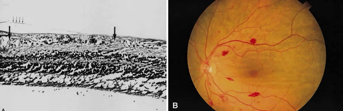
| Fig. 11. A. Section of retina with hemorrhage in the nerve fiber layer (between the two large arrows). Notice that the limits of the hemorrhage are not clearly defined, since scattered red blood cells can be seen to the right of the right-hand large arrow. This histologic picture corresponds to a clinically observed fame-shaped hemorrhage with an indistinct border. The detached posterior hyaloid is marked by four small arrows. B. Fundus photograph of nerve fiber layer hemorrhage. They are oriented parallel to the plane of the internal limiting membrane. Because of their dispersal within the ganglion cell layer, the borders are “feathery” (flame shaped). |