|
|
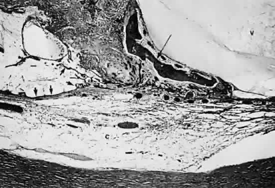
| Fig. 19. Histologic section demonstrating response of the pigment epithelium to injury. The sclera is at the bottom the micrograph; the choroid (c) contains scattered red blood cells and is edematous. The vitreous (v) also contains hemorrhage. Locate the pigment epithelium (short arrows) at the left third of the micrograph and notice the ribbons of pigment epithelium that proliferate into the membrane, partially formed by fibrous tissue (F). Bone (long arrow) also is present and probably was deposited by metaplastic pigment epithelium. |